Playing with Active Matter on ArXiv

One exemplar of the HEXBUGS used in the experiment. (Image by the Authors of the manuscript.)
Playing with Active Matter
Angelo Barona Balda, Aykut Argun, Agnese Callegari, Giovanni Volpe
arXiv: 2209.04168

In the last 20 years, active matter has been a very successful research field, bridging the fundamental physics of nonequilibrium thermodynamics with applications in robotics, biology, and medicine. This field deals with active particles, which, differently from passive Brownian particles, can harness energy to generate complex motions and emerging behaviors. Most active-matter experiments are performed with microscopic particles and require advanced microfabrication and microscopy techniques. Here, we propose some macroscopic experiments with active matter employing commercially available toy robots, i.e., the Hexbugs. We demonstrate how they can be easily modified to perform regular and chiral active Brownian motion. We also show that Hexbugs can interact with passive objects present in their environment and, depending on their shape, set them in motion and rotation. Furthermore, we show that, by introducing obstacles in the environment, we can sort the robots based on their motility and chirality. Finally, we demonstrate the emergence of Casimir-like activity-induced attraction between planar objects in the presence of active particles in the environment.

Invited Talk by G. Volpe at Fluctuations in small complex systems VI, Venice, 9 September 2022

Label-free measurement of biomolecules and their diffusion
Giovanni Volpe
9 September 2022, 16:45 (CEST)
Venice meeting on Fluctuations in small complex systems VI
Istituto Veneto di Scienze, Lettere ed Arti
Palazzo Franchetti, Venezia, 5-9 September 2022

An anomalous competition: assessment of methods for anomalous diffusion through a community effort

An anomalous competition: assessment of methods for anomalous diffusion through a community effort
Carlo Manzo, Giovanni Volpe
Submitted to SPIE-ETAI
Date: 25 August 2022
Time: 9:00 (PDT)

Deviations from the law of Brownian motion, typically referred to as anomalous diffusion, are ubiquitous in science and associated with non-equilibrium phenomena, flows of energy and information, and transport in living systems. In the last years, the booming of machine learning has boosted the development of new methods to detect and characterize anomalous diffusion from individual trajectories, going beyond classical calculations based on the mean squared displacement. We thus designed the AnDi challenge, an open community effort to objectively assess the performance of conventional and novel methods. We developed a python library for generating simulated datasets according to the most popular theoretical models of diffusion. We evaluated 16 methods over 3 different tasks and 3 different dimensions, involving anomalous exponent inference, model classification, and trajectory segmentation. Our analysis provides the first assessment of methods for anomalous diffusion in a variety of realistic conditions of trajectory length and noise. Furthermore, we compared the prediction provided by these methods for several experimental datasets. The results of this study further highlight the role that anomalous diffusion has in defining the biological function while revealing insight into the current state of the field and providing a benchmark for future developers.

Presenter: Giovanni Volpe

Soft Matter Lab members present at SPIE Optics+Photonics conference in San Diego, 21-25 August 2022

The Soft Matter Lab participates to the SPIE Optics+Photonics conference in San Diego, CA, USA, 21-25 August 2022, with the presentations listed below.

Giovanni Volpe is also co-author of the presentations:

Invited Talk by G. Volpe at UCLA, 19 August 2022

Quantitative Digital Microscopy with Deep Learning
Giovanni Volpe
19 August 2022, 14:40 (PDT)
At the intersection of Photonics, Neuroscience, and AI
Ozcan Lab, UCLA, 19 August 2022

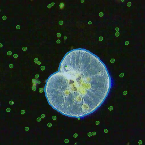
Video microscopy has a long history of providing insights and breakthroughs for a broad range of disciplines, from physics to biology. Image analysis to extract quantitative information from video microscopy data has traditionally relied on algorithmic approaches, which are often difficult to implement, time consuming, and computationally expensive. Recently, alternative data-driven approaches using deep learning have greatly improved quantitative digital microscopy, potentially offering automatized, accurate, and fast image analysis. However, the combination of deep learning and video microscopy remains underutilized primarily due to the steep learning curve involved in developing custom deep-learning solutions. To overcome this issue, we have introduced a software, currently at version DeepTrack 2.1, to design, train and validate deep-learning solutions for digital microscopy. We use it to exemplify how deep learning can be employed for a broad range of applications, from particle localization, tracking and characterization to cell counting and classification. Thanks to its user-friendly graphical interface, DeepTrack 2.1 can be easily customized for user-specific applications, and, thanks to its open-source object-oriented programming, it can be easily expanded to add features and functionalities, potentially introducing deep-learning-enhanced video microscopy to a far wider audience.

Multi-cohort and longitudinal Bayesian clustering study of stage and subtype in Alzheimer’s disease published in Nature Communications

Comparison of cluster-specific covariance matrixes with node strength. (Image by the Authors.)
Multi-cohort and longitudinal Bayesian clustering study of stage and subtype in Alzheimer’s disease
Konstantinos Poulakis, Joana B. Pereira, J.-Sebastian Muehlboeck, Lars-Olof Wahlund, Örjan Smedby, Giovanni Volpe, Colin L. Masters, David Ames, Yoshiki Niimi, Takeshi Iwatsubo, Daniel Ferreira, Eric Westman, Japanese Alzheimer’s Disease Neuroimaging Initiative & Australian Imaging, Biomarkers and Lifestyle study
Nature Communications 13, 4566 (2022)
doi: 10.1038/s41467-022-32202-6

Understanding Alzheimer’s disease (AD) heterogeneity is important for understanding the underlying pathophysiological mechanisms of AD. However, AD atrophy subtypes may reflect different disease stages or biologically distinct subtypes. Here we use longitudinal magnetic resonance imaging data (891 participants with AD dementia, 305 healthy control participants) from four international cohorts, and longitudinal clustering to estimate differential atrophy trajectories from the age of clinical disease onset. Our findings (in amyloid-β positive AD patients) show five distinct longitudinal patterns of atrophy with different demographical and cognitive characteristics. Some previously reported atrophy subtypes may reflect disease stages rather than distinct subtypes. The heterogeneity in atrophy rates and cognitive decline within the five longitudinal atrophy patterns, potentially expresses a complex combination of protective/risk factors and concomitant non-AD pathologies. By alternating between the cross-sectional and longitudinal understanding of AD subtypes these analyses may allow better understanding of disease heterogeneity.

Invited Talk by G. Volpe at Nordita, Stockholm, 2 August 2022

Interplay between active particles and their environment
Giovanni Volpe
2 August 2022, 10:30 (CEST)
Nordita workshop: Current and Future Themes in Soft and Biological Active Matter
Albano Building 3
Stockholm, 25 July-19 August 2022

In this seminar, I will present some examples of how the behaviour of active particles can be influenced by their environment. In particular, I’ll show the formation of active molecules and active droploids from passive colloidal building blocks; the emergence of non-Boltzmann statistics and active-depletion forces between plates in an active bath; and the environment topography alters the way to multicellularity in the bacterium Myxococcus xanthus.

Unraveling Parkinson’s disease heterogeneity using subtypes based on multimodal data published in Parkinsonism and Related Disorders

Particular of the brain in the group comparison analysis. (Image by the Authors.)
Unraveling Parkinson’s disease heterogeneity using subtypes based on multimodal data
Franziska Albrecht, Konstantinos Poulakis, Malin Freidle, Hanna Johansson, Urban Ekman, Giovanni Volpe, Eric Westman, Joana B. Pereira, Erika Franzén
Parkinsonism and Related Disorders 102, 19-29 (2022)
doi: 10.1016/j.parkreldis.2022.07.014

Background

Parkinson’s disease (PD) is a clinically and neuroanatomically heterogeneous neurodegenerative disease characterized by different subtypes. To this date, no studies have used multimodal data that combines clinical, motor, cognitive and neuroimaging assessments to identify these subtypes, which may provide complementary, clinically relevant information. To address this limitation, we subtyped participants with mild-moderate PD based on a rich, multimodal dataset of clinical, cognitive, motor, and neuroimaging variables.

Methods

Cross-sectional data from 95 PD participants from our randomized EXPANd (EXercise in PArkinson’s disease and Neuroplasticity) controlled trial were included. Participants were subtyped using clinical, motor, and cognitive assessments as well as structural and resting-state MRI data. Subtyping was done by random forest clustering. We extracted information about the subtypes by inspecting their neuroimaging profiles and descriptive statistics.

Results

Our multimodal subtyping analysis yielded three PD subtypes: a motor-cognitive subtype characterized by widespread alterations in brain structure and function as well as impairment in motor and cognitive abilities; a cognitive dominant subtype mainly impaired in cognitive function that showed frontoparietal structural and functional changes; and a motor dominant subtype impaired in motor variables without any brain alterations. Motor variables were most important for the subtyping, followed by gray matter volume in the right medial postcentral gyrus.

Conclusions

Three distinct PD subtypes were identified in our multimodal dataset. The most important features to subtype PD participants were motor variables in addition to structural MRI in the sensorimotor region. These findings have the potential to improve our understanding of PD heterogeneity, which in turn can lead to personalized interventions and rehabilitation.

Invited Talk by G. Volpe at MoLE Conference 2022, Donostia/San Sebastián, Spain, 27 July 2022

Artificial intelligence in microscopy, photonics, and active matter
Giovanni Volpe
27 July 2022, 12:40 (CEST)
MoLE Conference 2022
Donostia/San Sebastián, Spain, 25-29 July 2022

After a brief overview of artificial intelligence, machine learning and deep learning, I will present a series of recent works in which we have employed deep learning for applications in microscopy, optical tweezers, and active matter. In particular, I will explain how we employed deep learning to enhance digital video microscopy, to perform virtual staining of tissues, to estimate the properties of anomalous diffusion, to characterize microscopic force fields, to improve the calculation of optical forces, and to characterize nanoparticles. Finally, I will provide an outlook on the future for the application of deep learning in these fields.

Dynamic live/apoptotic cell assay using phase-contrast imaging and deep learning on bioRxiv

Phase-contrast image before virtual staining. (Image by the Authors.)
Dynamic live/apoptotic cell assay using phase-contrast imaging and deep learning
Zofia Korczak, Jesús Pineda, Saga Helgadottir, Benjamin Midtvedt, Mattias Goksör, Giovanni Volpe, Caroline B. Adiels
bioRxiv: https://doi.org/10.1101/2022.07.18.500422

Chemical live/dead assay has a long history of providing information about the viability of cells cultured in vitro. The standard methods rely on imaging chemically-stained cells using fluorescence microscopy and further analysis of the obtained images to retrieve the proportion of living cells in the sample. However, such a technique is not only time-consuming but also invasive. Due to the toxicity of chemical dyes, once a sample is stained, it is discarded, meaning that longitudinal studies are impossible using this approach. Further, information about when cells start programmed cell death (apoptosis) is more relevant for dynamic studies. Here, we present an alternative method where cell images from phase-contrast time-lapse microscopy are virtually-stained using deep learning. In this study, human endothelial cells are stained live or apoptotic and subsequently counted using the self-supervised single-shot deep-learning technique (LodeSTAR). Our approach is less labour-intensive than traditional chemical staining procedures and provides dynamic live/apoptotic cell ratios from a continuous cell population with minimal impact. Further, it can be used to extract data from dense cell samples, where manual counting is unfeasible.