Dual-Angle Interferometric Scattering Microscopy for Optical Multiparametric Particle Characterization published in Nano Letters

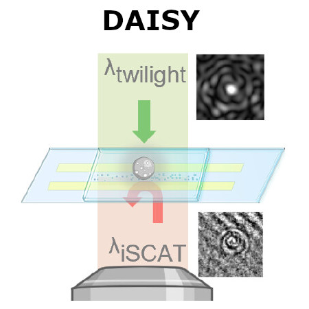
Conceptual schematic of dual-angle interferometric scattering microscopy (DAISY). (Image by the Authors of the manuscript.)
Dual-Angle Interferometric Scattering Microscopy for Optical Multiparametric Particle Characterization
Erik Olsén, Berenice García Rodríguez, Fredrik Skärberg, Petteri Parkkila, Giovanni Volpe, Fredrik Höök, and Daniel Sundås Midtvedt
Nano Letters (2024)
arXiv: 2309.07572
doi: 10.1021/acs.nanolett.3c03539

Traditional single-nanoparticle sizing using optical microscopy techniques assesses size via the diffusion constant, which requires suspended particles to be in a medium of known viscosity. However, these assumptions are typically not fulfilled in complex natural sample environments. Here, we introduce dual-angle interferometric scattering microscopy (DAISY), enabling optical quantification of both size and polarizability of individual nanoparticles (radius <170 nm) without requiring a priori information regarding the surrounding media or super-resolution imaging. DAISY achieves this by combining the information contained in concurrently measured forward and backward scattering images through twilight off-axis holography and interferometric scattering (iSCAT). Going beyond particle size and polarizability, single-particle morphology can be deduced from the fact that the hydrodynamic radius relates to the outer particle radius, while the scattering-based size estimate depends on the internal mass distribution of the particles. We demonstrate this by differentiating biomolecular fractal aggregates from spherical particles in fetal bovine serum at the single-particle level.
Fredrik Skärberg presented his half-time seminar on 10 January 2024

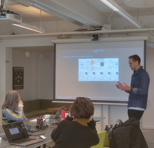
Fredrik Skärberg (right) and opponent Prof. Rebecka Jörnsten (left). (Photo by A. Ciarlo)
Fredrik Skärberg completed the first half of his doctoral studies and he defended his half-time on the 10th of January 2024.

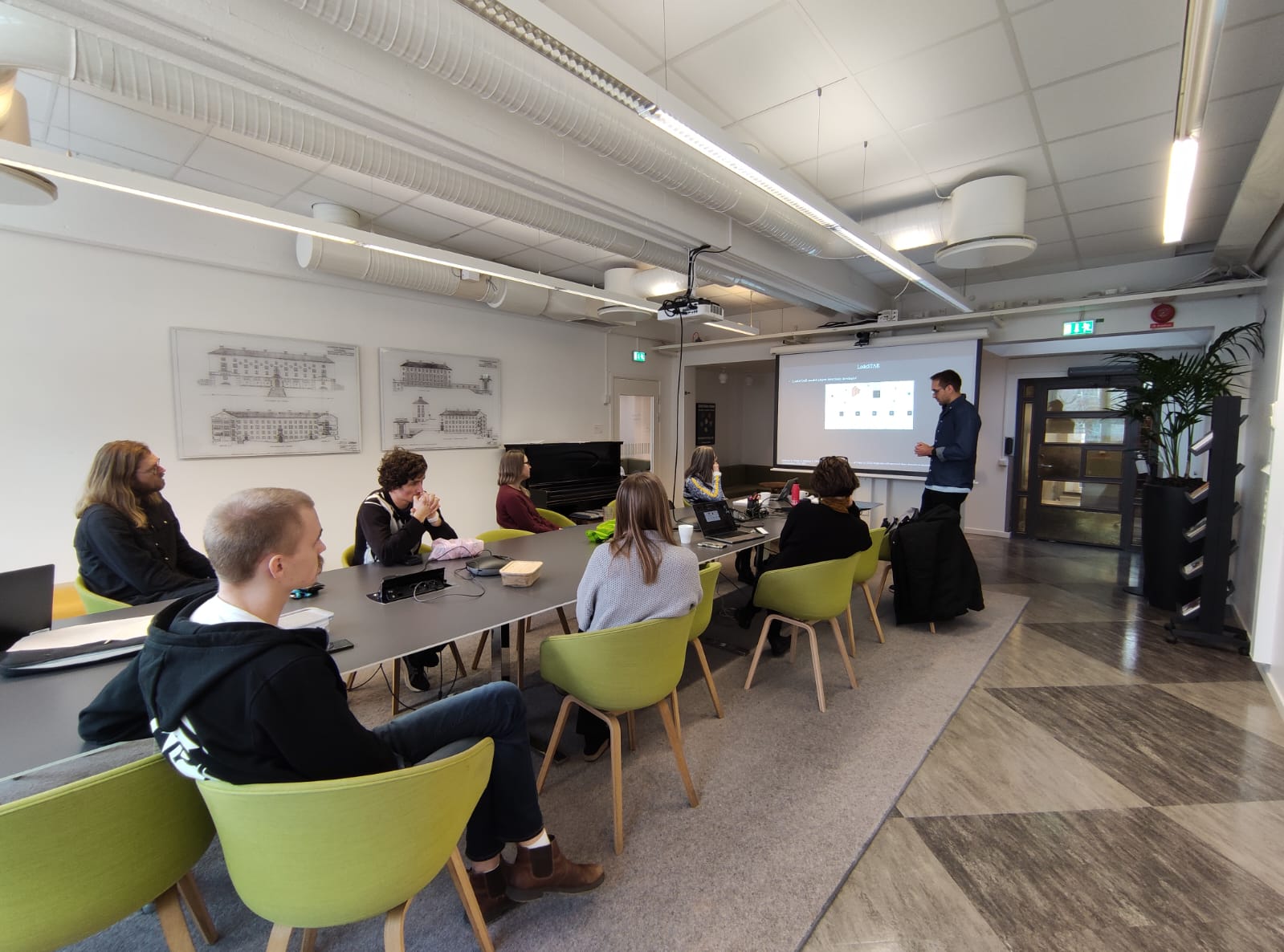
The presentation, with title: “Holographic characterization of biological nanoparticles using deep learning”, was held in hybrid format, with part of the audience in the Nexus room and the rest connected through zoom. The half-time consisted in a presentation about his past and planned projects and it was followed by a discussion and questions proposed by his opponent Prof. Rebecka Jörnsten.

The presentation started with a short background to characterization of biological particles inside cells and an introduction to the papers included in the half-time.

It continued with images and videos of various particle types inside cells, both tracked and characterized, followed by a description of the LodeSTAR-model.

In the last section, he outlined the proposed continuation of his PhD, with an ongoing project for monitoring lipid droplets during long timescales and a neural network for 3D rotation parameter estimation of rotating biological samples.

PhD Student: Fredrik Skärberg
Supervisor: Daniel Midtvedt
Co-supervisors: Giovanni Volpe, Fredrik Höök

Fredrik Skärberg and audience in Nexus. (Photo by A. Ciarlo.)

Single-shot self-supervised object detection in microscopy published in Nature Communications

LodeSTAR tracks the plankton Noctiluca scintillans. (Image by the Authors of the manuscript.)
Single-shot self-supervised particle tracking
Benjamin Midtvedt, Jesús Pineda, Fredrik Skärberg, Erik Olsén, Harshith Bachimanchi, Emelie Wesén, Elin K. Esbjörner, Erik Selander, Fredrik Höök, Daniel Midtvedt, Giovanni Volpe
Nature Communications 13, 7492 (2022)
arXiv: 2202.13546
doi: 10.1038/s41467-022-35004-y

Object detection is a fundamental task in digital microscopy, where machine learning has made great strides in overcoming the limitations of classical approaches. The training of state-of-the-art machine-learning methods almost universally relies on vast amounts of labeled experimental data or the ability to numerically simulate realistic datasets. However, experimental data are often challenging to label and cannot be easily reproduced numerically. Here, we propose a deep-learning method, named LodeSTAR (Localization and detection from Symmetries, Translations And Rotations), that learns to detect microscopic objects with sub-pixel accuracy from a single unlabeled experimental image by exploiting the inherent roto-translational symmetries of this task. We demonstrate that LodeSTAR outperforms traditional methods in terms of accuracy, also when analyzing challenging experimental data containing densely packed cells or noisy backgrounds. Furthermore, by exploiting additional symmetries we show that LodeSTAR can measure other properties, e.g., vertical position and polarizability in holographic microscopy.

Poster Presentation by F. Skärberg at SPIE-ETAI, San Diego, 22 August 2022

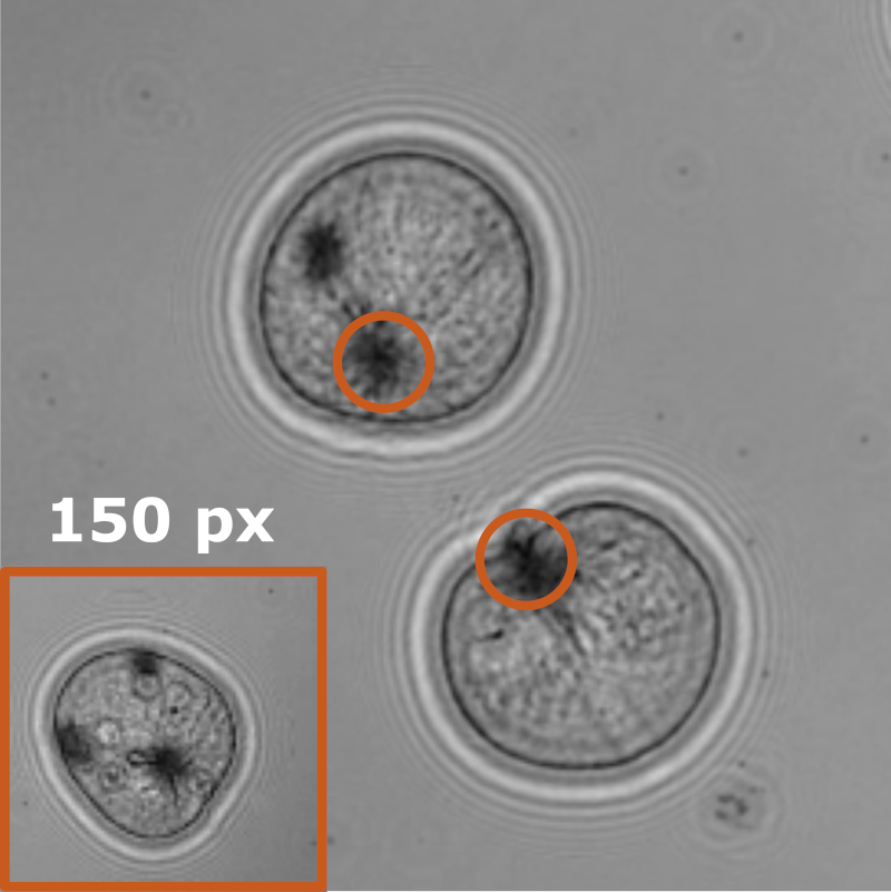
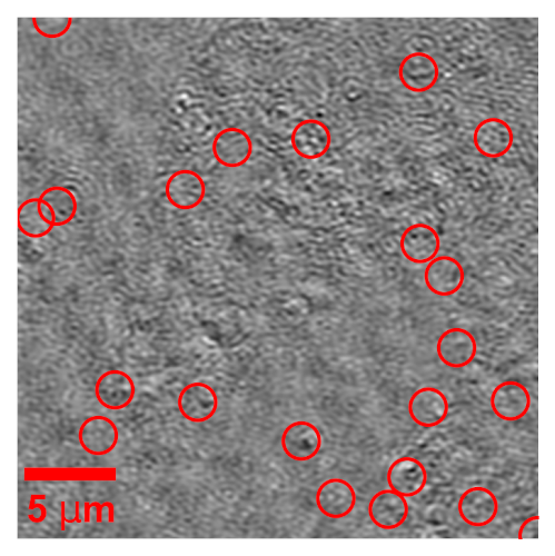
411nm Silica particles inside living cells. (Image by F. Skärberg.)
Holographic characterisation of biological nanoparticles using deep learning
Fredrik Skärberg, Erik Olsén, Benjamin Midtvedt, Emelie V. Wesén, Elin K. Esbjörner, Giovanni Volpe, Fredrik Höök, Daniel Midtvedt
Submitted to SPIE-ETAI
Date: 22 August 2022
Time: 17:30 (PDT)

Optical characterization of nanoparticles in/outside cells is a challenging task, as the scattering from the nanoparticles is distorted by the scattering from the cells. In this work, we demonstrate a framework for optical mass quantification of intra- and extracellular nanoparticles by leveraging a novel deep learning method, LodeSTAR, in combination with off-axis twilight holography. The result provides new means for the exploration of nanoparticle/cell interactions.

Soft Matter Lab members present at SPIE Optics+Photonics conference in San Diego, 21-25 August 2022

The Soft Matter Lab participates to the SPIE Optics+Photonics conference in San Diego, CA, USA, 21-25 August 2022, with the presentations listed below.

Giovanni Volpe is also co-author of the presentations: