Illustration of three different experiments autonomously performed by the SmartTrap system: DNA pulling experiments (top), red blood cell stretching (bottom left), and particle-particle interaction measurements (bottom right). (Image by M. Selin.)SmartTrap: automated precision experiments with optical tweezers
Martin Selin, Antonio Ciarlo, Giuseppe Pesce, Lars Bengtsson, Joan Camunas-Soler, Vinoth Sundar Rajan, Fredrik Westerlund, L. Marcus Wilhelmsson, Isabel Pastor, Felix Ritort, Steven B. Smith, Carlos Bustamante, Giovanni Volpe
Nature Methods (2026)
arXiv: 2505.05290
doi: 10.1038/s41592-026-03129-3
Optical tweezers are widely used in single-molecule biophysics, cell biomechanics and soft matter physics, but require a human operator, limiting throughput and repeatability. Here we present a smart optical tweezers platform, named SmartTrap, capable of performing complex experiments autonomously by integrating real-time three-dimensional particle tracking, custom electronics and a microfluidics system. Through a series of experiments, we demonstrate it can operate continuously, acquiring high-precision data over extended periods of time. By bridging the gap between manual experimentation and autonomous operation, SmartTrap establishes a robust and open-source framework for the next generation of optical tweezers research, capable of performing large-scale studies in single-molecule biophysics, cell mechanics and colloidal science with minimal experimental overhead and operator bias.
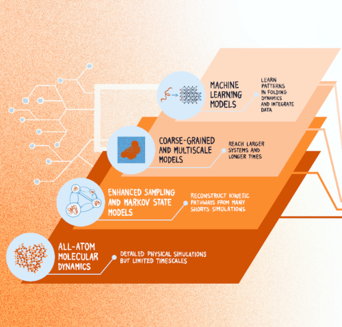
Computational advances in protein folding studies. Current approaches address multiple levels of resolution and methodological frameworks, however, none of the existing methods provides quantitative and dynamic information of the relationship between protein sequence and folding mechanism at all-atom resolution and at scale. (Graphics by J. Sacquegno.)Protein Dynamics Beyond Structure Prediction
Juliette Griffié, Sviatlana Shashkova, Antonio Ciarlo, Sreekanth K. Manikandan, Claes Andréasson, Malin Bäckström, Tristan Bereau, Hjalmar Brismar, Carlos Bustamante, Marta Carroni, Roberto Covino, Andreas Dahlin, Sebastian Deindl, Lucie Delemotte, Arne Elofsson, John Eriksson, Giovanna Fragneto, Anders Gunnarsson, Per Hammarström, Caroline Ingre, Christian Kaiser, Petronella Kettunen, Mark C. Leake, Benjamin Loos, Anna Månberg, Antonia S. J. S. Mey, Richard Neutze, Thomas Nyström, Karl Palmås, Charley Schaefer, Markus J. Tamás, Nicola Ticozzi, Tomás S. Pilvelic, Jacopo Sacquegno, B.M. (Betty)Tijms, Gunnar von Heijne, Björn Wallner, Vitali Zhaunerchyk, Simon Olsson, Joana B. Pereira, Julia Fernandez-Rodriguez, Fredrik Westerlund, Giovanni Volpe
arXiv: 2606.08647
How microorganisms respond to and interact with their environment can vary significantly from individual to individual, which can have important microbiological and ecological implications. However, most microscopy techniques can only observe motile microorganisms for short times because of their limited fields of view. Using Lagrangian tracking, a single microorganism can be followed in 3D, potentially indefinitely, allowing to decipher individual phenotypical traits. Current Lagrangian tracking methods use the fluorescence signal emitted by the microorganism as feedback to keep it in focus. However, over long times, epifluorescent imaging can induce photobleaching and photodamage, and importantly, not all microorganisms can easily be made fluorescent. Additionally, traditional algorithms used in feedback loops to determine microorganism position are prone to errors, especially in optically complex media. Here, we present a faster, more reliable, and versatile Lagrangian tracking method that uses deep learning to determine the 3D position of the microorganism. This new method demonstrates enhanced accuracy and speed in tracking fluorescent bacteria with fluorescence microscopy also in optically complex media. Furthermore, we track bacteria with other microscopy modalities, such as brightfield microscopy — for example, this enables us to track magnetotactic bacteria, which cannot be made fluorescent without degrading their magnetotactic properties. These novel capabilities allow to extract previously inaccessible quantitative information, significantly advancing the study of microorganism behavior — and thus opening new avenues for research in complex biological and ecological systems.
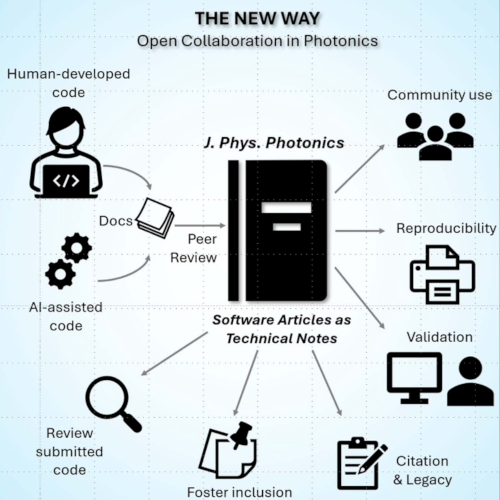
A centralized, peer-reviewed ecosystem in J. Phys. Photonics where human agents and/or AI agents co-develop validated, documented software. (Image from the manuscript.)Beyond the black box: towards an open and citable software ecosystem in photonics
Anoop C Patil, Maciej Trusiak, Fei Xia, Liangcai Cao, Carlo Manzo and Giovanni Volpe
Journal of Physics: Photonics 8, 020201 (2026)
doi: 10.1088/2515-7647/ae68cd
This editorial draws attention to a major issue in photonics: important research software is often hidden, undocumented, and lost over time, making results hard to reproduce. The rise of AI-generated code increases this problem by adding more ‘black box’ systems. To address this, J. Phys. Photonics is introducing Software Articles, a new article type allowing researchers to publish, validate, and share their code as formal scientific outputs. This initiative aims to promote transparency, reproducibility, and proper credit for developers. Open and peer-reviewed software helps the community verify results, reduce duplication of effort, and build lasting tools, ensuring that computational methods become reliable, accessible, and integral to scientific progress.
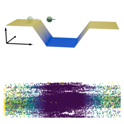
Experimental setup. (Top) Thermophoretic microswimmer undergoes active Brownian motion in a spatially-varying laser intensity profile that controls the self-thermophoretic propulsion of the swimmer using a feedback loop. (Bottom) Sample trajectory of the microswimmer over 15 minutes in a chamber. Colors indicate instantaneous velocity. (Image from the manuscript.)Delayed Active Swimmer in a Velocity Landscape
Viktor Holubec, Alexander Fischer, Giovanni Volpe, Frank Cichos
Physical Review Research 8, L022017 (2026)
arXiv: 2505.11042
doi: 10.1103/xn9x-ppjx
Active systems in nature and synthetic environments commonly exhibit spatially heterogeneous activity patterns and time-delayed responses from internal feedback mechanisms, exemplified by bacterial chemotaxis. We study an idealized active gas where particles modulate their self-propulsion based on local environmental conditions with such delays. Through integrated theoretical, computational, and experimental approaches, we demonstrate that steady-state density distributions and collective polarization exhibit characteristic peaks and valleys as functions of response delay time. We find that delays can amplify polarization by nearly an order of magnitude and trigger complete polarization reversal when particle displacement during the delay period surpasses the persistence length. Multiparticle simulations incorporating interparticle interactions validate that these phenomena remain robust in sufficiently dilute collective systems. Since density and polarization determine the current in active matter, our findings show that temporally programming the delay time allows control over both static and dynamic states in active systems, with implications for biological microswimmers and engineered microrobots.
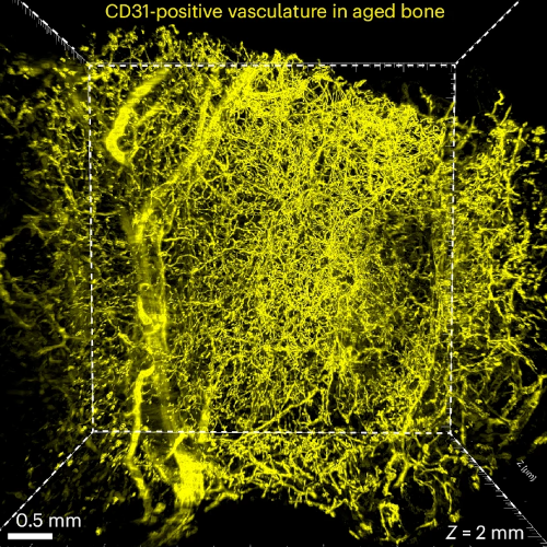
Visualization of the vasculature within human bone from a 75-year-old patient by immunostaining with antibodies against CD31. (Image from the manuscript.)Three-dimensional quantitative tissue clearing reveals differences in osteovascular niche of aged and young human mesenchymal stromal cells
Nelson Tsz Long Chu, Ostap Dregval, Yu-Wei Chang, Emil Kriukov, Xin Tian, Xin Liu, Dana Trompet, Misty Shuo Zhang, Lei Li, Zhong Li, Emiliano Gomez Ruiz, Joana B. Pereira, Mats Brittberg, Björn Barenius, Lars Sävendahl, Ralf H. Adams, Inger Gjertsson, Claes Ohlsson, Giovanni Volpe & Andrei S. Chagin
Nature Biomedical Engineering (2026)
bioRxiv: 10.1101/2025.10.07.680053
doi: 10.1038/s41551-026-01645-3
Human bone marrow mesenchymal stromal/stem cells (BM-MSCs) are widely used in clinical trials and tissue engineering, yet their native microenvironment remains poorly understood. Here we introduce a tissue-clearing protocol, DeepBone, for human bones and integrate it with simultaneous mRNA and protein detection. Using this protocol, we spatially map BM-MSCs relative to key bone microenvironment components, including human blood capillaries, adipocytes, sinusoids and bony trabeculae. Quantitative analysis reveals that the native microenvironment of human BM-MSCs in young bone is enriched in vasculature, sinusoids, bone matrix and adipocytes. In contrast, in aged bone, BM-MSCs show no preferential association with bone or adipocytes. Proliferative BM-MSCs are predominantly found along blood vessels. Moreover, we identify a specialized microenvironment for BM-MSCs in young bone, characterized by sinusoids coiled around trabeculae and enriched by R-type vessels. These findings provide insights into the native niches of BM-MSCs, offering a foundation for the development of tissue engineering strategies that mimic their physiological context.
Automated segnmentation of bacterial structures within a droplet. The image shows a bright-field microscopy view where a large biofilm region (green, outlined in blue) has been segmented from surrounding features. Small aggregates (yellow contours) are also highlighted. This segmentation enables structural differentiation of biofilm components for downstream quantitative analysis. (Image by D. Pérez Guerrero.)Latent space-driven quantification of biofilm formation using time-resolved droplet microfluidics
Daniela Pérez Guerrero, Jesús Manuel Antúnez Domínguez, Aurélie Vigne, Daniel Midtvedt, Wylie Ahmed, Lisa D. Muiznieks, Giovanni Volpe, Caroline Beck Adiels
Microchemical Journal 225, 117685 (2026)
arXiv: 2507.07632
DOI: 10.1016/j.microc.2026.117685
Bacterial biofilms play crucial roles across diverse contexts, from public health risks to beneficial applications in bioremediation, biodegradation, and wastewater treatment. However, tools that enable high-resolution, dynamic analysis of their responses to environmental cues and collective cellular behaviors remain limited. Here, we present a droplet-based microfluidic platform that combines continuous in situ microscopy with subsequent unsupervised deep learning for quantitative analysis of biofilm development. In our setup, Bacillus subtilis cells are encapsulated in monodisperse aqueous microdroplets containing Lysogeny Broth, suspended in an oil phase and immobilized within microfabricated traps, providing continuous optical access throughout biofilm formation at the water–oil interface. The platform supports both fluorescence and bright-field imaging, enabling high-throughput, time-resolved monitoring of thousands of droplets under controlled conditions. To extract quantitative information from these large datasets, we developed an automated analysis pipeline based on a Variational Autoencoder (VAE) trained directly on microscopy images from our experiments. This unsupervised model enables segmentation and latent-space representation of bacterial structures without manual annotation or synthetic training data. Post-segmentation size thresholding enables classification of bacterial aggregates and larger biofilm-like clusters, including quantification of biofilm porosity, thereby supporting detailed morphological and temporal analyses across droplets and conditions. By integrating droplet microfluidics with unsupervised deep learning, our platform provides a scalable, robust, and rapid approach for high-throughput quantitative studies of biofilm behavior. It resolves complex structural biofilm patterns, bypasses the need for manual annotation, and opens new opportunities to probe environmental determinants of biofilm formation. Departing from earlier methods, our framework fuses biological training data with unsupervised models to quantify microbial community dynamics across scales, offering a generalizable platform for future high-resolution microbiology.
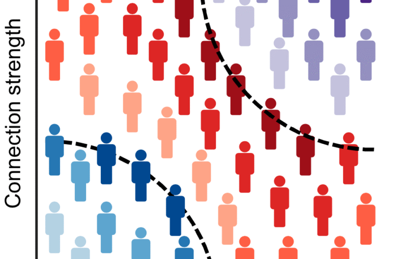
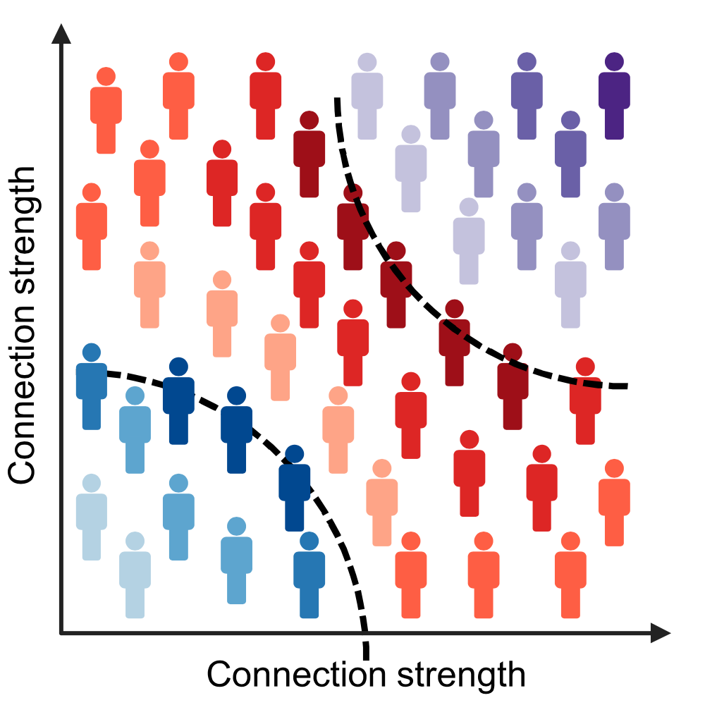
Diagnostic classification. (Image from the article.)Mapping individual molecular connectomes in Alzheimer’s disease
Zhilei Xu, Mite Mijalkov, Jiawei Sun, Yu-Wei Chang, Arianna Sala, Giovanni Volpe, Mario Severino, Mattia Veronese, Sara Garcia-Ptacek, Joana B. Pereira, for the Alzheimer’s Disease Neuroimaging Initiative
Alzheimer’s & Dementia 22, e71310 (2026)
DOI: 10.1002/alz.71310
INTRODUCTION
Mapping individual differences is crucial to improve personalized medicine approaches in Alzheimer’s disease (AD), which is characterized by strong inter-individual variability in the accumulation patterns of tau and amyloid beta pathology.
METHODS
We assess the progression of AD across the disease continuum by building individual molecular connectomes using longitudinal positron emission tomography (PET) data.
RESULTS
We demonstrate that these connectomes constitute a unique fingerprint, capable of identifying a single individual from a large group of subjects. Alterations in the connectomes discriminate different diagnostic groups and predict cognitive decline to a higher extent than conventional PET measures. We introduce a novel gene-specific transcription network analysis that linked individual tau and amyloid connectomes to a common transcriptomic profile of apoptosis, with the tau connectome being specifically related to pyrimidine metabolism, and the amyloid connectome to histone acetylation.
DISCUSSION
Individual molecular connectome mapping provides a novel and sensitive framework to monitor AD progression.
Highlights
Individual molecular connectomes constitute a unique fingerprint, capable of identifying a single individual from a large group of subjects.
Alterations in individual molecular connectomes significantly increase both across the Alzheimer’s disease (AD) continuum and over time.
Alterations in individual molecular connectomes discriminate different diagnostic groups and predict cognitive decline to a higher extent than conventional positron emission tomography measures.
Susceptibilities of individual tau and amyloid connectomes to AD are linked to a common transcriptomic profile of apoptosis, with the tau connectome being specifically related to pyrimidine metabolism, and the amyloid connectome to histone acetylation.
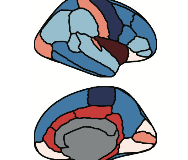
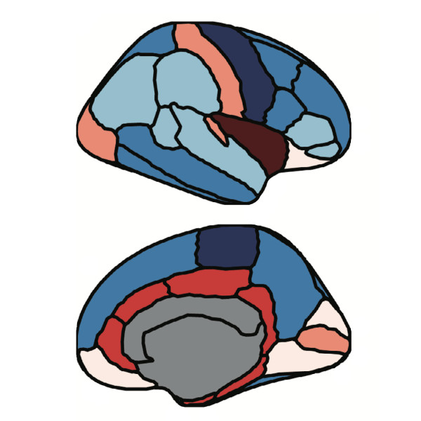
Parcellation of the brain cortex. (Image from the article.)Tracking early cognitive decline in preclinical AD with brain MRI similarity
Jiawei Sun, Blanca Zufiria-Gerbolés, Massimiliano Passaretti, Giovanni Volpe, Mite Mijalkov, Joana B. Pereira, for the Alzheimer’s Disease Neuroimaging Initiative
Alzheimer’s & Dementia 22, e71170 (2026)
DOI: 10.1002/alz.71170
INTRODUCTION
Early detection of neuroanatomical changes in preclinical Alzheimer’s disease (AD) is critical for timely intervention. However, conventional magnetic resonance imaging (MRI) and fluid biomarkers often lack sensitivity to subtle structural alterations in early disease stages.
METHODS
To identify early brain alterations, we applied a perturbation-based brain similarity approach to cognitively normal participants from Alzheimer’s Disease Neuroimaging Initiative (ADNI) and Open Access Series of Imaging Studies (OASIS), stratified by amyloid status. We evaluated its predictive performance for cognition and diagnostic conversion against cortical thickness, volumetric MRI, and fluid biomarkers.
RESULTS
In both cohorts, brain similarity consistently outperformed other biomarkers across cognitive domains and amyloid groups. It also achieved superior accuracy in predicting clinical conversion and exhibited associations with cytoarchitectural organization.
DISCUSSION
These findings highlight brain similarity as a sensitive marker of early neuroanatomical disruption in AD. Its ability to detect subtle structural changes before overt atrophy underscores its potential for early disease monitoring and treatment assessment in preclinical AD trials.
Highlights
Brain similarity captures early brain changes in preclinical Alzheimer’s disease (AD).
Brain similarity outperforms conventional biomarkers such as cortical thickness, volume measures, and fluid biomarkers in predicting cognitive decline.
Brain similarity predicts conversion to mild cognitive impairment and AD more accurately than traditional imaging markers, and its predictive performance is further improved when combined with fluid biomarkers.
Brain similarity captures structural disruptions associated with cortical layer II of the cytoarchitectonic lamina of human neocortex.
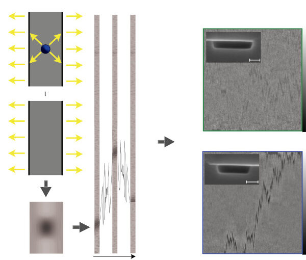
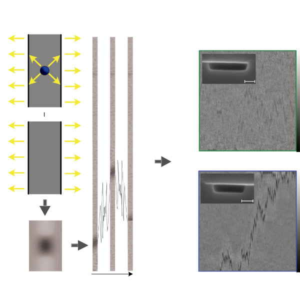
The principle of differential imaging in NSM, in which we subtract the light scattered (yellow arrows indicate the scattered-light direction) by an empty nanochannel from the light scattered by the same channel with a molecule inside. A sequence of differential images of a nanochannel containing a diffusing single molecule obtained in this way is combined into a kymograph, which then contains the full molecular trajectory. (Image from the article.)Label-free mass and size characterization of few-kDa biomolecules by hierarchical vision transformer augmented nanofluidic scattering microscopy
Henrik K. Moberg, Bohdan Yeroshenko, Joachim Fritzsche, David Albinsson, Barbora Spackova, Daniel Midtvedt, Giovanni Volpe, Christoph Langhammer
Nature Communications 17, 2533 (2026)
DOI: 10.1038/s41467-026-70514-z
Nanofluidic scattering microscopy characterizes single molecules in subwavelength nanofluidic channels label-free, using the interference of visible light scattered by the molecule and nanochannel. It determines a molecule’s hydrodynamic radius by tracking its diffusion trajectory and its molecular weight by analyzing its scattering intensity along that trajectory. However, using standard analysis algorithms, it is limited to characterization of proteins larger than ≈ 60 kDa. Here, we push this limit by one order of magnitude to below ≈ 6 kDa molecular weight and ≈ 1.5 nm hydrodynamic radius — as we exemplify on the peptide hormone insulin — by using ultrasmall nanofluidic channels and by analyzing the data with a hierarchical vision transformer. When we benchmark this approach against the theoretical limit set by the Cramér–Rao Lower Bound, we find that it can be approached with sufficiently long molecular trajectories. This enables quantitative label-free single-molecule microscopy for biologically relevant families of sub-10-kDa molecules, such as cytokines, chemokines and peptide hormones.
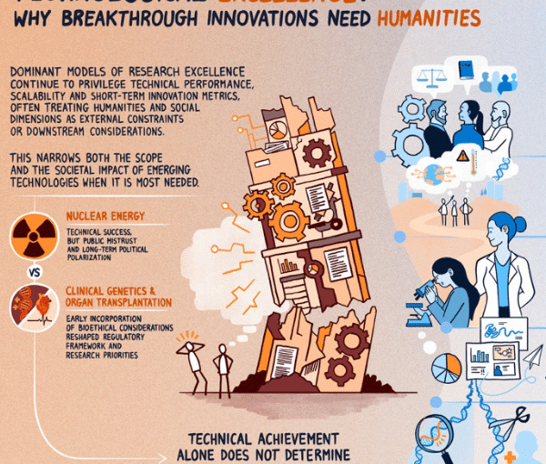
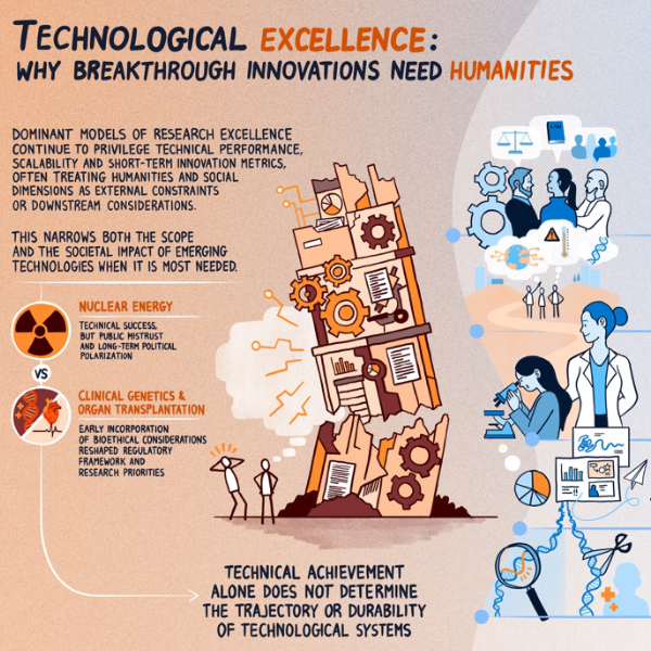
Why breakthrough research needs humanities and social sciences. (From an artwork by Jacopo Sacquegno.)Technological Excellence Requires Human and Social Context
Karl Palmås, Mats Benner, Monica Billger, Ben Clarke, Raimund Feifel, Julia Fernandez-Rodriguez, Anna Foka, Juliette Griffié, Claes Gustafsson, Kerstin Hamilton, Johan Holmén, Kristina Lindström, Tobias Olofsson, Joana B. Pereira, Marisa Ponti, Julia Ravanis, Sviatlana Shashkova, Emma Sparr, Pontus Strimling, Fredrik Höök, Giovanni Volpe
arXiv: 2603.10653
Breakthrough technologies increasingly shape social institutions, economic systems, and political futures. Yet models of research excellence associated with such technologies often prioritize technical performance, scalability, and short-term innovation metrics while treating ethical, social, and cultural dimensions as secondary considerations. This perspective article argues that such separation is no longer tenable. We propose a broader understanding of excellence that combines technical rigor with ethical robustness, social intelligibility, and long-term relevance. The rapid emergence of generative and agentic artificial intelligence further underscores this argument. As technological systems increasingly operate through language, interpretation, and normative alignment, expertise traditionally cultivated in the humanities and social sciences becomes integral to the design, governance, and responsible deployment of such systems. Drawing on historical examples and contemporary research practices, this article examines five interconnected domains where the humanities and social sciences, treated as integrated dimensions of research practice, can strengthen technological development: (1) ethical, legal, and social integration in agenda-setting and research design; (2) plural and reflexive foresight practices that shape technological futures; (3) graduate education as a leverage point for cross-disciplinary literacy; (4) visualization and communication as epistemic and civic practices; and (5) institutional frameworks that move beyond rigid distinctions between basic and applied research. Across these dimensions, we propose practical strategies for embedding interdisciplinary collaboration structurally rather than symbolically.