Poster presentation by M. Granfors at Optics and Photonics in Sweden, Norrköping, 7 October, 2026

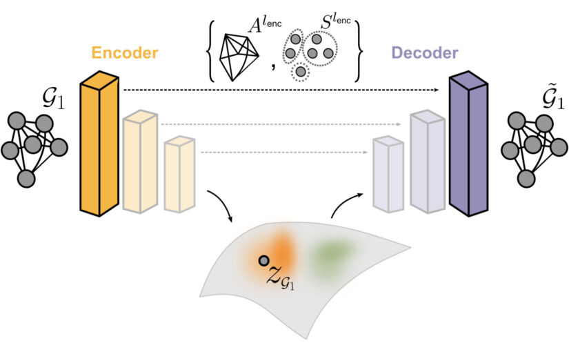
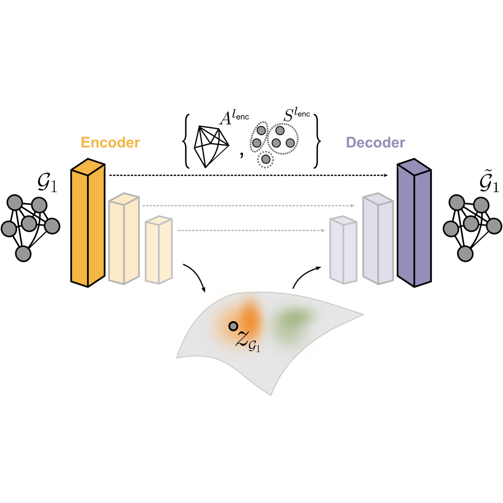
GAUDI leverages a hierarchical graph-convolutional variational autoencoder architecture, where an encoder progressively compresses the graph into a low-dimensional latent space, and a decoder reconstructs the graph from the latent embedding. (Image by M. Granfors and J. Pineda.)
Unsupervised Graph Deep Learning Uncovers Protein Organization in Super-Resolution Microscopy
Mirja Granfors, Jesús Pineda, Blanca Zufiria Gerbolés, Joana B. Pereira, Carlo Manzo, and Giovanni Volpe
Date: 7 October 2026
Time: 11:45 – 13:00 and 17:20 – 19:00
Place: Biblioteket, Arbetets museum, Norrköping

Modern optical techniques such as super-resolution microscopy generate rich, spatially resolved data that can be naturally represented as graphs. However, the structural variability of these graphs poses significant challenges for their analysis and classification, particularly when similar underlying processes can produce substantially different graph realizations.

To address this challenge, we introduce Graph Autoencoder Uncovering Descriptive Information (GAUDI), an unsupervised graph deep learning framework designed to capture both local details and global structure. GAUDI employs a hierarchical graph-convolutional variational autoencoder architecture with pooling and upsampling layers connected through skip connections, preserving essential connectivity information throughout the encoding–decoding process.

Despite substantial variability between graph realizations generated from identical or highly similar underlying parameters, GAUDI consistently maps them to nearby regions of a structured, continuous latent space. This enables the identification of invariant process-level features while separating them from stochastic variability.

We demonstrate this capability using super-resolution microscopy, where GAUDI characterizes protein assemblies and organizes structurally variable graphs according to their underlying state. The framework is further demonstrated on small-world network models, collective motion in the Vicsek model, and age-related changes in brain connectivity. These results highlight GAUDI as a broadly applicable approach for extracting meaningful information from complex graph-structured data across microscopy and other fields.
Poster presentation by M. Granfors at Optics and Photonics in Sweden, Norrköping, 7 October, 2026

DeepTrack2 Logo. (Image by J. Pineda.)
DeepTrack2: Physics-Based Microscopy Simulations for Deep Learning
Mirja Granfors, Alex Lech, Jiacheng Huang, Eduard Duta, Guillem Guigo i Corominas, Harshith Bachimanchi, Benjamin Midtvedt, Jesús Pineda, Carlo Manzo, and Giovanni Volpe
Date: 7 October 2026
Time: 11:45 – 13:00 and 17:20 – 19:00
Place: Biblioteket, Arbetets museum, Norrköping

Deep learning has become an increasingly powerful tool for microscopy image analysis, but training robust models often requires large and diverse datasets that are difficult and time-consuming to acquire experimentally. Physics-based simulations provide a way to generate synthetic microscopy data while retaining control over the optical and physical properties of the imaging process.

DeepTrack2 is a flexible and scalable Python library designed to generate physics-based synthetic microscopy datasets for training deep learning models. It supports a wide range of imaging modalities, including brightfield, fluorescence, darkfield, and holography, and allows users to customize optical systems, incorporate optical aberrations and noise, simulate diverse objects and imaging conditions, and apply image augmentations.

Its modular architecture also enables simulations to be executed using a PyTorch backend, providing GPU acceleration and allowing backpropagation through the optical system. This makes it possible to optimize the optical setup and the deep learning model jointly. The DeepTrack2 repository further provides tutorials covering the complete workflow, from physics-based simulation to deep-learning-based analysis using its companion library, deeplay.

Together, these capabilities provide a flexible framework for developing and benchmarking deep learning methods for microscopy while reducing the need for large experimentally acquired training datasets.

Global graph features unveiled by unsupervised deep learning published in Machine Learning: Science and Technology

GAUDI leverages a hierarchical graph-convolutional variational autoencoder architecture, where an encoder progressively compresses the graph into a low-dimensional latent space, and a decoder reconstructs the graph from the latent embedding. (Image by M. Granfors and J. Pineda.)
Global graph features unveiled by unsupervised deep learning
Mirja Granfors, Jesús Pineda, Blanca Zufiria Gerbolés, Joana B. Pereira, Carlo Manzo, Giovanni Volpe
Machine Learning: Science and Technology, 7, 045054 (2026)
arXiv: 2503.05560
doi: 10.1088/2632-2153/ae8d7f

Graphs provide a powerful framework for modeling complex systems, but their structural variability poses significant challenges for analysis and classification. To address these challenges, we introduce Graph Autoencoder Uncovering Descriptive Information (GAUDI), an unsupervised graph deep learning framework designed to capture both local details and global structure. GAUDI employs an hourglass architecture with hierarchical pooling and upsampling layers linked through skip connections, which preserve essential connectivity information throughout the encoding–decoding process. Even though identical or highly similar underlying parameters describing a system’s state can lead to significant variability in graph realizations, GAUDI consistently maps them into nearby regions of a structured and continuous latent space, effectively disentangling invariant process-level features from stochastic noise. We demonstrate GAUDI’s versatility across multiple applications, including small-world networks modeling, characterization of protein assemblies from super-resolution microscopy, analysis of collective motion in the Vicsek model, and identification of age-related changes in brain connectivity. Comparison with related approaches highlights GAUDI’s superior performance in analyzing complex graphs, providing new insights into emergent phenomena across diverse scientific domains.

Beyond the black box: towards an open and citable software ecosystem in photonics published in Journal of Physics: Photonics

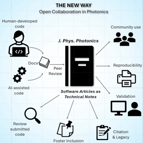
A centralized, peer-reviewed ecosystem in J. Phys. Photonics where human agents and/or AI agents co-develop validated, documented software. (Image from the manuscript.)
Beyond the black box: towards an open and citable software ecosystem in photonics
Anoop C Patil, Maciej Trusiak, Fei Xia, Liangcai Cao, Carlo Manzo and Giovanni Volpe
Journal of Physics: Photonics 8, 020201 (2026)
doi: 10.1088/2515-7647/ae68cd

This editorial draws attention to a major issue in photonics: important research software is often hidden, undocumented, and lost over time, making results hard to reproduce. The rise of AI-generated code increases this problem by adding more ‘black box’ systems. To address this, J. Phys. Photonics is introducing Software Articles, a new article type allowing researchers to publish, validate, and share their code as formal scientific outputs. This initiative aims to promote transparency, reproducibility, and proper credit for developers. Open and peer-reviewed software helps the community verify results, reduce duplication of effort, and build lasting tools, ensuring that computational methods become reliable, accessible, and integral to scientific progress.

Roadmap on Deep Learning for Microscopy published in Journal of Physics: Photonics

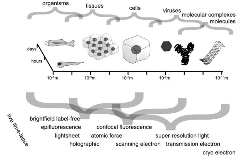
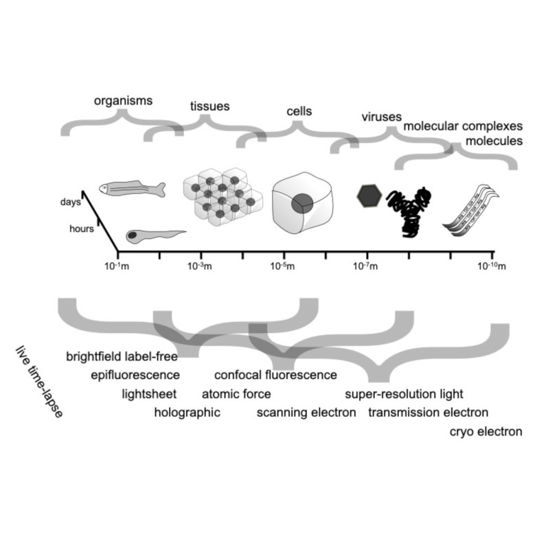
Spatio-temporal spectrum diagram of microscopy techniques and their applications. (Image by the Authors of the manuscript.)
Roadmap on Deep Learning for Microscopy
Giovanni Volpe, Carolina Wählby, Lei Tian, Michael Hecht, Artur Yakimovich, Kristina Monakhova, Laura Waller, Ivo F. Sbalzarini, Christopher A. Metzler, Mingyang Xie, Kevin Zhang, Isaac C.D. Lenton, Halina Rubinsztein-Dunlop, Daniel Brunner, Bijie Bai, Aydogan Ozcan, Daniel Midtvedt, Hao Wang, Nataša Sladoje, Joakim Lindblad, Jason T. Smith, Marien Ochoa, Margarida Barroso, Xavier Intes, Tong Qiu, Li-Yu Yu, Sixian You, Yongtao Liu, Maxim A. Ziatdinov, Sergei V. Kalinin, Arlo Sheridan, Uri Manor, Elias Nehme, Ofri Goldenberg, Yoav Shechtman, Henrik K. Moberg, Christoph Langhammer, Barbora Špačková, Saga Helgadottir, Benjamin Midtvedt, Aykut Argun, Tobias Thalheim, Frank Cichos, Stefano Bo, Lars Hubatsch, Jesus Pineda, Carlo Manzo, Harshith Bachimanchi, Erik Selander, Antoni Homs-Corbera, Martin Fränzl, Kevin de Haan, Yair Rivenson, Zofia Korczak, Caroline Beck Adiels, Mite Mijalkov, Dániel Veréb, Yu-Wei Chang, Joana B. Pereira, Damian Matuszewski, Gustaf Kylberg, Ida-Maria Sintorn, Juan C. Caicedo, Beth A Cimini, Muyinatu A. Lediju Bell, Bruno M. Saraiva, Guillaume Jacquemet, Ricardo Henriques, Wei Ouyang, Trang Le, Estibaliz Gómez-de-Mariscal, Daniel Sage, Arrate Muñoz-Barrutia, Ebba Josefson Lindqvist, Johanna Bergman
Journal of Physics: Photonics 8, 012501 (2026)
arXiv: 2303.03793
doi: 10.1088/2515-7647/ae0fd1

Through digital imaging, microscopy has evolved from primarily being a means for visual observation of life at the micro- and nano-scale, to a quantitative tool with ever-increasing resolution and throughput. Artificial intelligence, deep neural networks, and machine learning (ML) are all niche terms describing computational methods that have gained a pivotal role in microscopy-based research over the past decade. This Roadmap encompasses key aspects of how ML is applied to microscopy image data, with the aim of gaining scientific knowledge by improved image quality, automated detection, segmentation, classification and tracking of objects, and efficient merging of information from multiple imaging modalities. We aim to give the reader an overview of the key developments and an understanding of possibilities and limitations of ML for microscopy. It will be of interest to a wide cross-disciplinary audience in the physical sciences and life sciences.

Enhanced spatial clustering of single-molecule localizations with graph neural networks published in Nature Communications

MIRO employs a recurrent graph neural network to refine SMLM point clouds by compressing clusters around their center, enhancing inter-cluster distinction and background separation for efficient clustering. (Image by J. Pineda.)
Enhanced spatial clustering of single-molecule localizations with graph neural networks
Jesús Pineda, Sergi Masó-Orriols, Montse Masoliver, Joan Bertran, Mattias Goksör, Giovanni Volpe and Carlo Manzo
Nature Communications 16, 9693 (2025)
arXiv: 2412.00173
doi: 10.1038/s41467-025-65557-7

Single-molecule localization microscopy generates point clouds corresponding to fluorophore localizations. Spatial cluster identification and analysis of these point clouds are crucial for extracting insights about molecular organization. However, this task becomes challenging in the presence of localization noise, high point density, or complex biological structures. Here, we introduce MIRO (Multifunctional Integration through Relational Optimization), an algorithm that uses recurrent graph neural networks to transform the point clouds in order to improve clustering efficiency when applying conventional clustering techniques. We show that MIRO supports simultaneous processing of clusters of different shapes and at multiple scales, demonstrating improved performance across varied datasets. Our comprehensive evaluation demonstrates MIRO’s transformative potential for single-molecule localization applications, showcasing its capability to revolutionize cluster analysis and provide accurate, reliable details of molecular architecture. In addition, MIRO’s robust clustering capabilities hold promise for applications in various fields such as neuroscience, for the analysis of neural connectivity patterns, and environmental science, for studying spatial distributions of ecological data.

Quantitative evaluation of methods to analyze motion changes in single-particle experiments published on Nature Communications

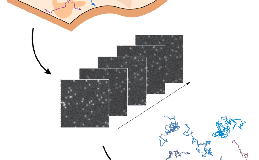
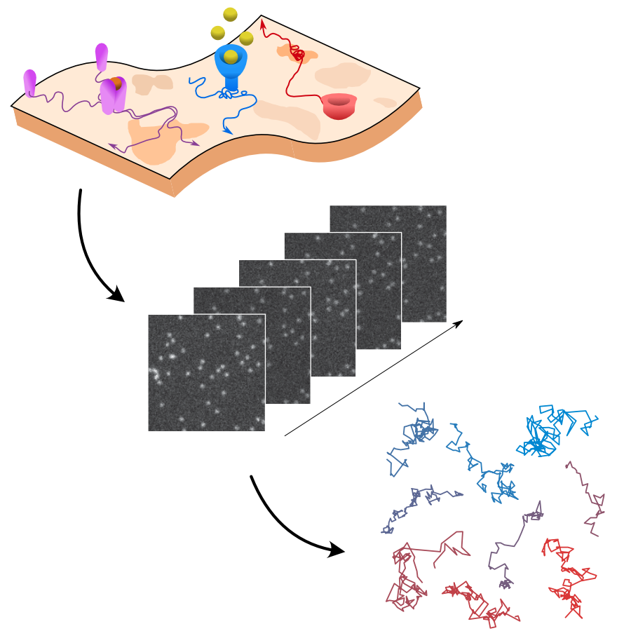
Rationale for the challenge organization. The interactions of biomolecules in complex environments, such as the cell membrane, regulate physiological processes in living systems. These interactions produce changes in molecular motion that can be used as a proxy to measure interaction parameters. Time-lapse single-molecule imaging allows us to visualize these processes with high spatiotemporal resolution and, in combination with single-particle tracking methods, provide trajectories of individual molecules. (Image by the Authors of the manuscript.)
Quantitative evaluation of methods to analyze motion changes in single-particle experiments
Gorka Muñoz-Gil, Harshith Bachimanchi, Jesús Pineda, Benjamin Midtvedt, Gabriel Fernández-Fernández, Borja Requena, Yusef Ahsini, Solomon Asghar, Jaeyong Bae, Francisco J. Barrantes, Steen W. B. Bender, Clément Cabriel, J. Alberto Conejero, Marc Escoto, Xiaochen Feng, Rasched Haidari, Nikos S. Hatzakis, Zihan Huang, Ignacio Izeddin, Hawoong Jeong, Yuan Jiang, Jacob Kæstel-Hansen, Judith Miné-Hattab, Ran Ni, Junwoo Park, Xiang Qu, Lucas A. Saavedra, Hao Sha, Nataliya Sokolovska, Yongbing Zhang, Giorgio Volpe, Maciej Lewenstein, Ralf Metzler, Diego Krapf, Giovanni Volpe, Carlo Manzo
Nature Communications 16, 6749 (2025)
arXiv: 2311.18100
doi: https://doi.org/10.1038/s41467-025-61949-x

The analysis of live-cell single-molecule imaging experiments can reveal valuable information about the heterogeneity of transport processes and interactions between cell components. These characteristics are seen as motion changes in the particle trajectories. Despite the existence of multiple approaches to carry out this type of analysis, no objective assessment of these methods has been performed so far. Here, we report the results of a competition to characterize and rank the performance of these methods when analyzing the dynamic behavior of single molecules. To run this competition, we implemented a software library that simulates realistic data corresponding to widespread diffusion and interaction models, both in the form of trajectories and videos obtained in typical experimental conditions. The competition constitutes the first assessment of these methods, providing insights into the current limitations of the field, fostering the development of new approaches, and guiding researchers to identify optimal tools for analyzing their experiments.

Seminar by G. Volpe and C. Manzo at CIG, Makerere University, Kampala, Uganda, 3 July 2025 (Online)

GAUDI leverages a hierarchical graph-convolutional variational autoencoder architecture, where an encoder progressively compresses the graph into a low-dimensional latent space, and a decoder reconstructs the graph from the latent embedding. (Image by M. Granfors and J. Pineda.)
Cutting Training Data Needs through Inductive Bias & Unsupervised Learning
Giovanni Volpe and Carlo Manzo
Computational Intelligence Group (CIG), Weekly Reading Session
Date: 3 July 2025
Time: 17:00
Place: Makerere University, Kampala, Uganda (Online)

Graphs provide a powerful framework for modeling complex systems, but their structural variability makes analysis and classification challenging. To address this, we introduce GAUDI (Graph Autoencoder Uncovering Descriptive Information), a novel unsupervised geometric deep learning framework that captures both local details and global structure. GAUDI employs an innovative hourglass architecture with hierarchical pooling and upsampling layers, linked through skip connections to preserve essential connectivity information throughout the encoding–decoding process. By mapping different realizations of a system — generated from the same underlying parameters — into a continuous, structured latent space, GAUDI disentangles invariant process-level features from stochastic noise. We demonstrate its power across multiple applications, including modeling small-world networks, characterizing protein assemblies from super-resolution microscopy, analyzing collective motion in the Vicsek model, and capturing age-related changes in brain connectivity. This approach not only improves the analysis of complex graphs but also provides new insights into emergent phenomena across diverse scientific domains.

Youtube: Global graph features unveiled by unsupervised geometric deep learning

Book “Deep Learning Crash Course” published at No Starch Press

The book Deep Learning Crash Course, authored by Giovanni Volpe, Benjamin Midtvedt, Jesús Pineda, Henrik Klein Moberg, Harshith Bachimanchi, Joana B. Pereira, and Carlo Manzo, has been published online by No Starch Press in July 2024.

Preorder Discount
A preorder discount is available: preorders with coupon code PREORDER will receive 25% off. Link: Preorder @ No Starch Press | Deep Learning Crash Course

Links
@ No Starch Press

Citation 
Giovanni Volpe, Benjamin Midtvedt, Jesús Pineda, Henrik Klein Moberg, Harshith Bachimanchi, Joana B. Pereira, and Carlo Manzo. Deep Learning Crash Course. No Starch Press.
ISBN-13: 9781718503922

Geometric deep learning reveals the spatiotemporal fingerprint of microscopic motion published in Nature Machine Intelligence

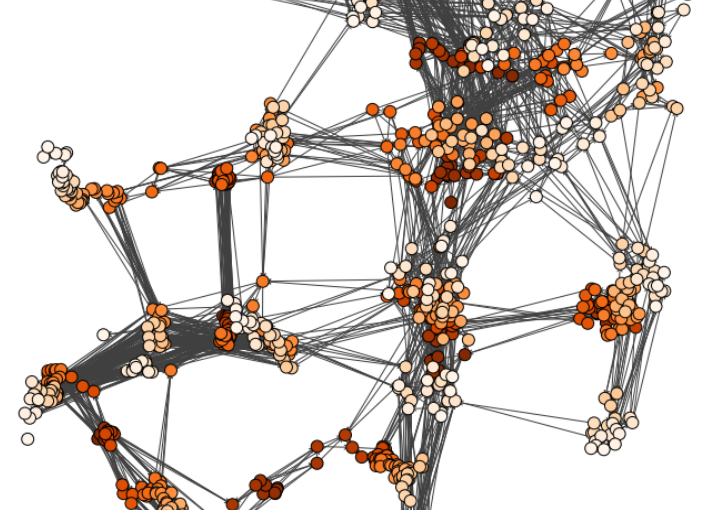
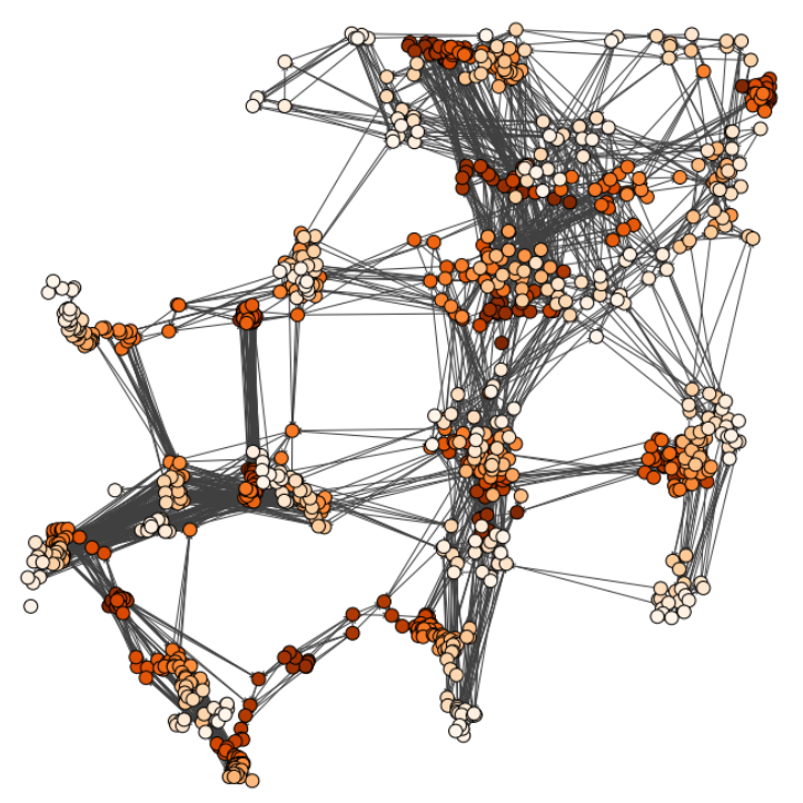
Input graph structure including a redundant number of edges. (Image by J. Pineda.)
Geometric deep learning reveals the spatiotemporal fingerprint of microscopic motion
Jesús Pineda, Benjamin Midtvedt, Harshith Bachimanchi, Sergio Noé, Daniel Midtvedt, Giovanni Volpe, Carlo Manzo
Nature Machine Intelligence 5, 71–82 (2023)
arXiv: 2202.06355
doi: 10.1038/s42256-022-00595-0

The characterization of dynamical processes in living systems provides important clues for their mechanistic interpretation and link to biological functions. Thanks to recent advances in microscopy techniques, it is now possible to routinely record the motion of cells, organelles, and individual molecules at multiple spatiotemporal scales in physiological conditions. However, the automated analysis of dynamics occurring in crowded and complex environments still lags behind the acquisition of microscopic image sequences. Here, we present a framework based on geometric deep learning that achieves the accurate estimation of dynamical properties in various biologically-relevant scenarios. This deep-learning approach relies on a graph neural network enhanced by attention-based components. By processing object features with geometric priors, the network is capable of performing multiple tasks, from linking coordinates into trajectories to inferring local and global dynamic properties. We demonstrate the flexibility and reliability of this approach by applying it to real and simulated data corresponding to a broad range of biological experiments.