Optimal calibration of optical tweezers with arbitrary integration time and sampling frequencies – A general framework published in Biomedical Optics Express

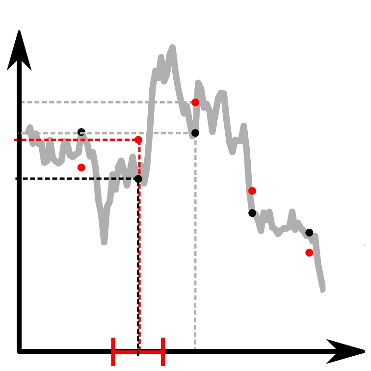
Different sampling methods for the trajectory of a particle. (Adapted from the manuscript.)
Optimal calibration of optical tweezers with arbitrary integration time and sampling frequencies — A general framework
Laura Pérez-García, Martin Selin, Antonio Ciarlo, Alessandro Magazzù, Giuseppe Pesce, Antonio Sasso, Giovanni Volpe, Isaac Pérez Castillo, Alejandro V. Arzola
Biomedical Optics Express, 14, 6442-6469 (2023)
doi: 10.1364/BOE.495468
arXiv: 2305.07245

Optical tweezers (OT) have become an essential technique in several fields of physics, chemistry, and biology as precise micromanipulation tools and microscopic force transducers. Quantitative measurements require the accurate calibration of the trap stiffness of the optical trap and the diffusion constant of the optically trapped particle. This is typically done by statistical estimators constructed from the position signal of the particle, which is recorded by a digital camera or a quadrant photodiode. The finite integration time and sampling frequency of the detector need to be properly taken into account. Here, we present a general approach based on the joint probability density function of the sampled trajectory that corrects exactly the biases due to the detector’s finite integration time and limited sampling frequency, providing theoretical formulas for the most widely employed calibration methods: equipartition, mean squared displacement, autocorrelation, power spectral density, and force reconstruction via maximum-likelihood-estimator analysis (FORMA). Our results, tested with experiments and Monte Carlo simulations, will permit users of OT to confidently estimate the trap stiffness and diffusion constant, extending their use to a broader set of experimental conditions.
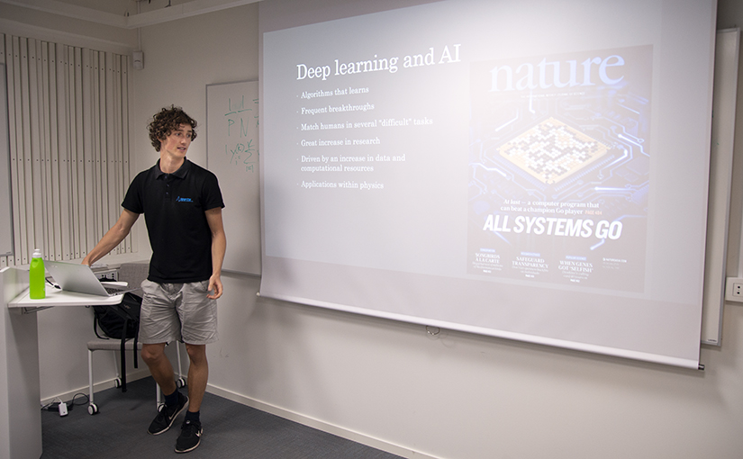
Presentation by M.Selin at S3IC, Barcelona, 23 November 2023

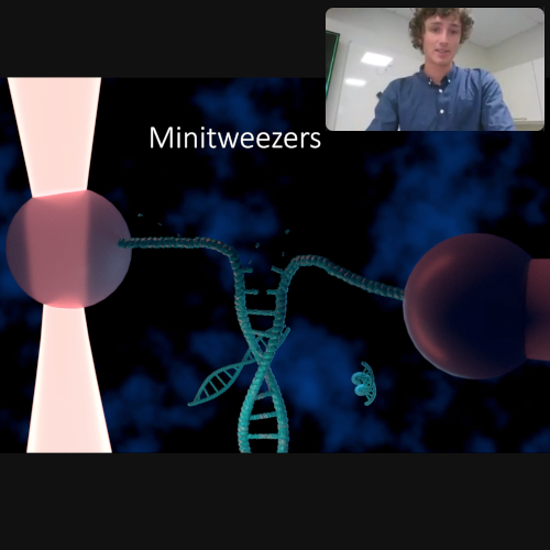
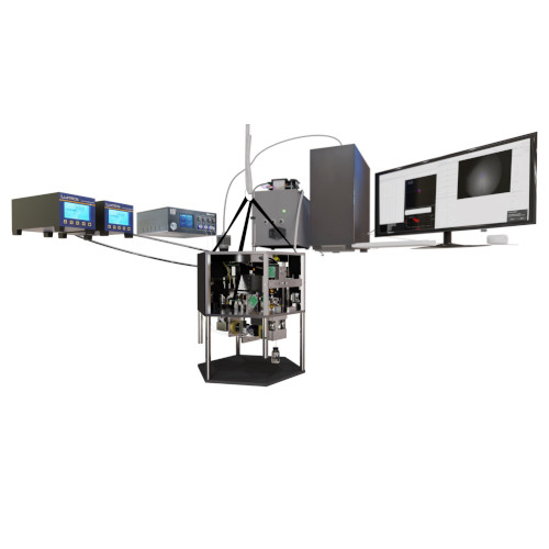
3d Visualization of the full Minitweezers 2.0 system. (Illustration by M. Selin.)
Minitweezers 2.0, Paving the way for fully autonomous optical tweezers experiments.
Martin Selin
Single-Molecule Sensors and NanoSystems International Conference – S3IC 2023
23 November 2023, 11:51 (CET)

Since their invention by Ashkin et al. in the 1980s, optical tweezers have evolved into an indispensable tool in physics, especially in biophysics, with applications spanning from cell sorting to stretching single DNA strands. By the 2000s, commercial systems became available. Nevertheless, owing to their unique requirements, many labs prefer to construct their own, often drawing inspiration from existing designs.

A prominent optical tweezers design is the “miniTweezers” system, pioneered by Bustamante’s group in the late 1990s. This system has been widely adopted globally for force spectroscopy experiments on single molecules, including DNA, proteins, and RNA.

In this presentation, we unveil an advanced iteration of the miniTweezers. By enhancing its control and acquisition capabilities, we’ve augmented its versatility, enabling new experiment types. A significant breakthrough is the integration of real-time image feedback, which paves the way for automated procedures via deep learning-based image analysis, the first of which we demonstrate in this presentation.

We showcase this system’s capabilities through three distinct experiments:

  1. A pulling experiment on a λ-DNA strand. By tethering DNA between two polystyrene beads – one anchored in a micropipette and the other manipulated by the tweezer – we illustrate near-complete automation, with the system autonomously handling bead trapping, attachment of the DNA and the pulling procedure.
  2. An exploration of Coulomb interactions between charged particles. Here, one particle remains in a micropipette, while the other orbits the stationary bead, providing a 3D map of the interaction.
  3. A non-contact stretching experiment on red blood cells is conducted under low osmotic pressure conditions. Modulating the laser power induces cell elongation along the laser’s propagation direction. By correlating this elongation with the optical force exerted by the lasers, we present a simple and non-invasive method to measure membrane rigidity.

In summary, these advancements mark a significant leap in the capabilities and applications of optical tweezers in biophysics. As we push the boundaries of automation and precision, we envision a future where such instruments can unravel even more intricate molecular interactions and cellular mechanics, setting the stage for groundbreaking discoveries.

Presentation by M. Selin at SBE congress, 30 June 2023

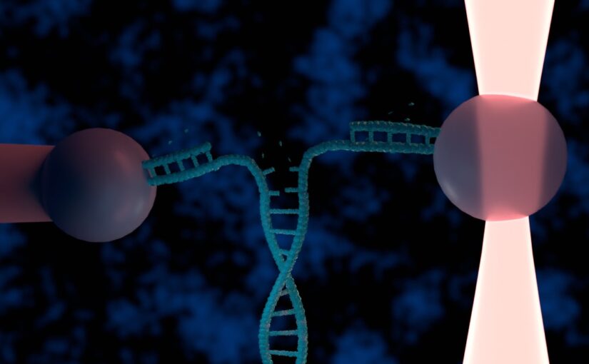
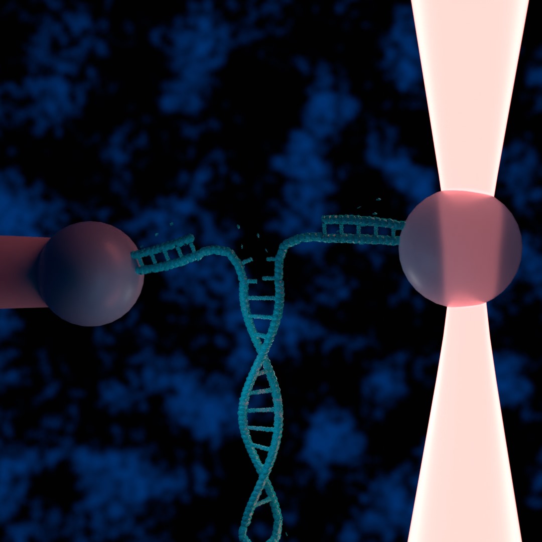
Illustration of a DNA hairpin being unzipped by an optical tweezers. (Illustration by M. Selin.)
Automating optical tweezers experiments using deep learning and custom electronics
Martin Selin
30 June 2023, 13:00 CEST

Optical tweezers are powerful tools for manipulating and studying the mechanical properties of single biomolecules, such as DNA. However, conducting such experiments manually is both time-consuming and labor-intensive limiting the amount of data collectable. In this work, we present a method to automate optical tweezers with the use of deep learning applying it to DNA pulling experiments.

A typical DNA pulling experiment can be divided into three main steps, each of which we have automated. The first is positioning of a bead in a micropipette(or secondary optical trap), second is connecting DNA of a another optically trapped bead with the bead in the micropipette and lastly the stretching of the DNA by moving the trapped bead while monitoring the force.

We have used deep learning, in particular a unet, to track beads and identify important features in the sample such as the micropipette. Combining this with realtime feedback allows the system to both trap beads and carefully position trap beads.

We demonstrate the viability of our method by stretching lambda DNA, showing human like reliability in performing the experiments. We expect our method to find use in the study of small biomolecules enabling more and faster data collection as well as longer running experiments.

Martin Selin presented his half-time seminar on 2 September 2022

Martin Selin’s half-time seminar: Opponent Dag Hanstorp (left), Martin Selin (right). (Photo by H. P. Tanabalan.)
Martin Selin completed the first half of his doctoral studies and defended his half-time on the 2nd of September 2022.

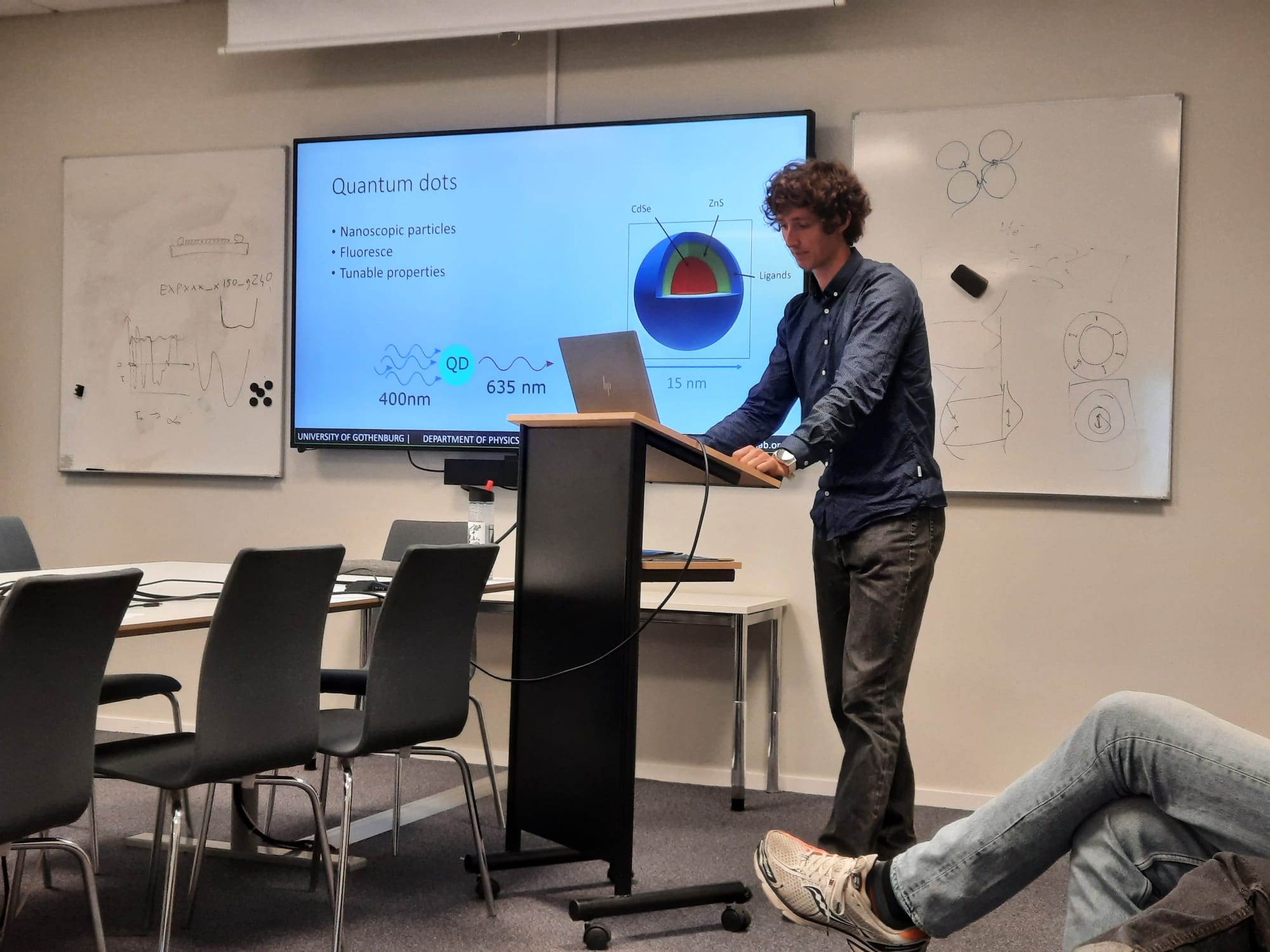
The presentation was held in hybrid format, with part of the audience in the Von Bahr room and the rest connected through zoom. The half-time consisted of a presentation of Martins two main projects followed by a discussion and questions proposed by Martins opponent Dag Hanstorp.

The presentation started providing a background on optical tweezers and continued with the ongoing project of positioning quantum dots using optical tweezers. Thereafter the presentation continued with the Minitweezers project. Data on DNA stretching was presented and shown to be in good agreement with results found in literature. Lastly the future of the two projects were outlined. Specifically, how to address the challenging task of detecting moving quantum dots and how to improve on the Minitweezers system through automation.

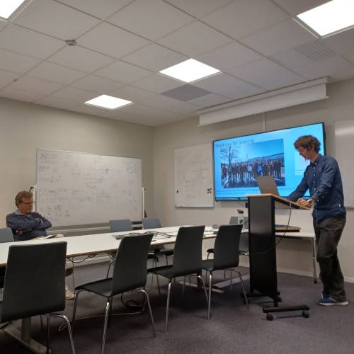
Martin Selin during his half-time seminar. (Photo by L. Natali.)

Soft Matter Lab members present at SPIE Optics+Photonics conference in San Diego, 21-25 August 2022

The Soft Matter Lab participates to the SPIE Optics+Photonics conference in San Diego, CA, USA, 21-25 August 2022, with the presentations listed below.

Giovanni Volpe is also co-author of the presentations:

Presentation by L. Pérez García at OSA-OMA-2021

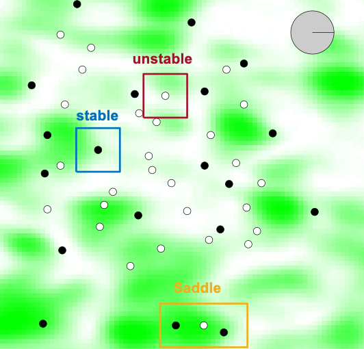
FORMA allows to identify and characterize all the equilibrium points in a force field generated by a speckle pattern.
FORMA and BEFORE: Expanding Applications of Optical Tweezers. Laura Pérez Garcia, Martin Selin, Alejandro V. Arzola, Giovanni Volpe, Alessandro Magazzù, Isaac Pérez Castillo.
Submitted to OSA-OMA 2021,  ATh1D.5
Date: 15 April
Time: 15:45 (CEST)

Abstract: 
FORMA (force reconstruction via maximum-likelihood-estimator analysis) addresses the need to measure the force fields acting on microscopic particles. Compared to alternative established methods, FORMA is faster, simpler, more accurate, and more precise. Furthermore, FORMA can also measure non-conservative and out-of-equilibrium force fields. Here, after a brief introduction to FORMA, I will present its use, advantages, and limitations. I will conclude with the most recent work where we exploit Bayesian inference to expand FORMA’s scope of application.

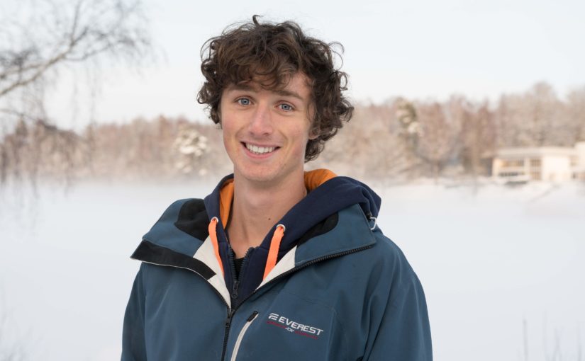
Martin Selin joins the Soft Matter Lab

Martin Selin starts his PhD at the Physics Department of the University of Gothenburg on 16th March 2020.

Martin has a Master degree in Applied Physics at Chalmers University of Technology, Gothenburg, Sweden.

In his PhD, he will focus on automating particle trapping using optical tweezers and machine learning.

Influence of Sensorial Delay on Clustering and Swarming published in Phys. Rev. E

Influence of Sensorial Delay on Clustering and Swarming

Influence of Sensorial Delay on Clustering and Swarming
Rafal Piwowarczyk, Martin Selin, Thomas Ihle & Giovanni Volpe
Physical Review E 100(1), 012607 (2019)
doi: 10.1103/PhysRevE.100.012607
arXiv:  1803.06026

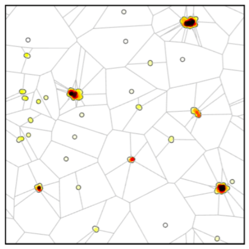
We show that sensorial delay alters the collective motion of self-propelling agents with aligning interactions: In a two-dimensional Vicsek model, short delays enhance the emergence of clusters and swarms, while long or negative delays prevent their formation. In order to quantify this phenomenon, we introduce a global clustering parameter based on the Voronoi tessellation, which permits us to efficiently measure the formation of clusters. Thanks to its simplicity, sensorial delay might already play a role in the organization of living organisms and can provide a powerful tool to engineer and dynamically tune the behavior of large ensembles of autonomous robots.

Martin Selin defended his Master Thesis. Congrats!

Martin Selin defended his Master thesis in Physics at Chalmers University of Technology on 5 June 2019

Title: Growing Artificial Neural Networks. Novel approaches to Deep Learning for Image Analysis and Particle Tracking

Deep-learning has recently emerged as one of the most successful methods for an- alyzing large amounts of data and constructing models from it. It has virtually revolutionized the field of image analysis and the algorithms are now being employed in research field outside of computer science. The methods do however suffer from several drawbacks such as large computational costs.

In this thesis alternative methods for training the networks underlying networks are evaluated based on gradually growing networks during training using layer-by- layer training as well as a method based on increasing network width dubbed breadth training.

These training methods lends themselves to easily implementing networks of tune- able size allowing for choice between high accuracy or fast execution or the construc- tion of modular network in which one can chose to execute only a small part of the network to get a very fast prediction at the cost of some accuracy. The layer-by-layer method is applied to multiple different image analysis tasks and the performance is evaluated and compared to that of regular training. Both the layer by layer training and the breadth training comparable to normal training in performance and in some cases slightly superior while in others slightly inferior. The modular nature of the networks make them suitable for applications within multi-particle tracking.

​Name of the master programme: MPPHYS – Physics
Supervisor: Giovanni Volpe, Department of Physics, University of Gothenburg
Examiner: Giovanni Volpe, Department of Physics, University of Gothenburg
Opponent: Henry Yang, MP Complex Adaptive Systems, Department of Physics, Chalmers University of Technology

Place: Raven & Fox room
Time: 5 June, 2019, 15:00