Illustration of three different experiments autonomously performed by the SmartTrap system: DNA pulling experiments (top), red blood cell stretching (bottom left), and particle-particle interaction measurements (bottom right). (Image by M. Selin.)Smart Machines and Optical Manipulation at the Microscale
Giovanni Volpe 5th Münster Symposium on Intelligent Matter (MüSIM) (Flyer) Date: 24 June 2026 Time: 15:00 Place: Center for Soft Nanoscience (SoN), Münster, Germany
Microscale systems offer a unique opportunity to engineer machines whose function emerges from the interplay of geometry, interactions, and fluctuations. In this talk, I will present our work on smart machines at the microscale, combining nanofabrication, programmable interactions, and advanced optical methods to design and control colloidal micromechanisms and metamaterials.
I will first introduce how nanotechnology enables the realization of colloidal metamachines and microscopic mechanisms, where shape and mechanical constraints are engineered to produce targeted motion and response in fluid environments. I will then show how smart microscopy and optical manipulation—including high-resolution imaging, automated tracking, and light-based control—allow us to probe these machines in real time and quantify their dynamics. This approach enables precision measurements of effective interaction landscapes, including critical Casimir forces and their relation to fluctuation-induced forces known from QED Casimir physics.
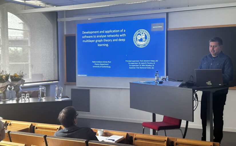
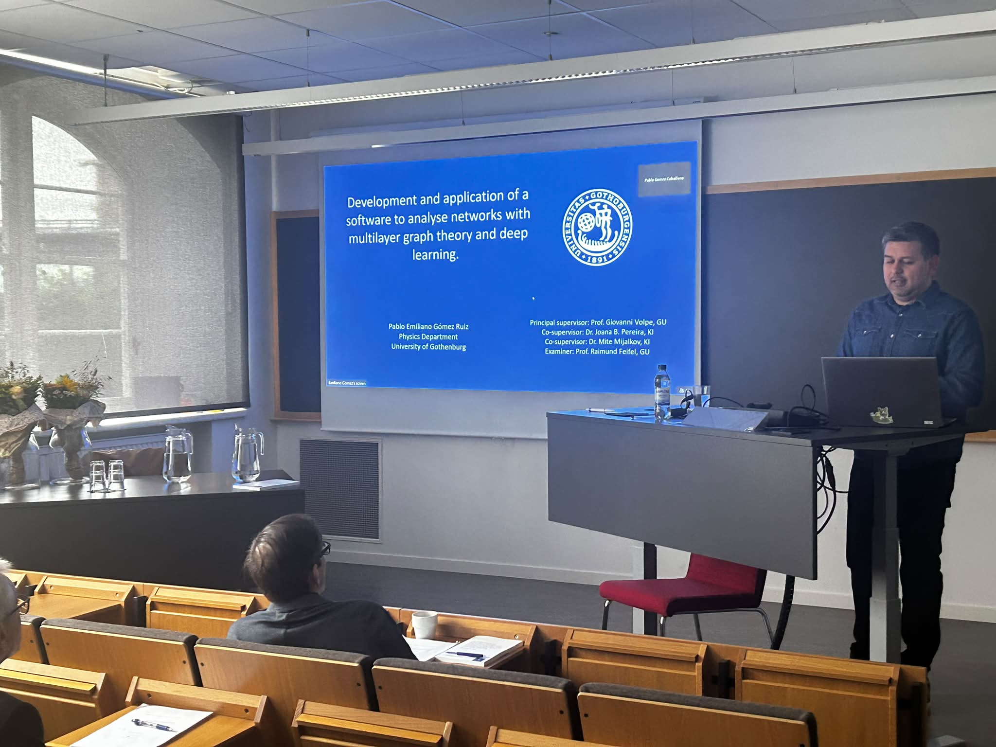
PhD defense of Emiliano Gomez-Ruiz. (Photo by H. Zhao.)Pablo Emiliano Gomez Ruiz defended his PhD thesis on June 15, 2026. Congrats!
The defense took place in PJ Salen lecture hall, Institutionen för fysik, Johanneberg Campus, Göteborg, at 14:00.
Title: Development and application of software to analyze networks with multilayer graph theory and deep learning.
Abstract:
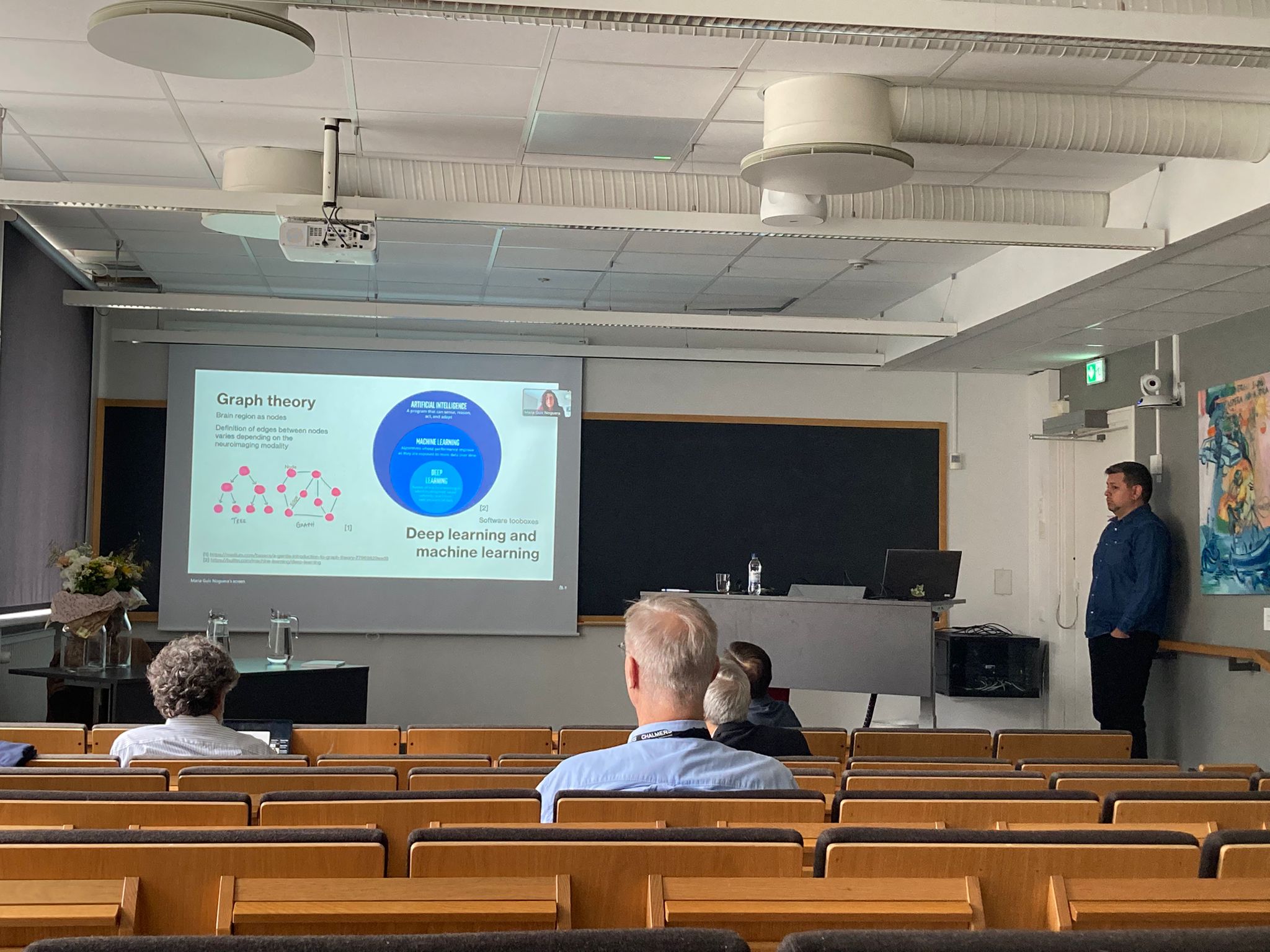
Understanding how the brain is wired is essential, it gives us a new level of insight of its functionality. By modeling the brain as a complex intercon- nected network, the connectome, researchers can abstract biological com- plexity into a mathematical framework suitable for analysis. The connec- tome can be understood by it’s structural links such as neuron’s synapses or by the functional links such as a statistical relationships between neu- ral activity between the brain’s regions. The mapping of these networks is achieved with neuroimaging, while their analysis is driven by the integration of graph theory and deep learning architectures.
In this work, we present a software “Brain Analysis using Graph Theory 2” (BRAPH 2.0), which is a direct solution of the need for a toolbox de- signed for both complex graph theory and deep learning analyses. Central to the software’s architecture is the “Genesis” pseudo-language, which allows researchers to bridge human-readable properties with computer code, facilitating the modular expansion of multilayer graph theory and deep learning pipelines of the software.
The capabilities of this framework are demonstrated through large-scale clinical applications. We analyze sex-related differences in the aging brain using a cohort of 37,543 participants from the UK Biobank. Our results reveal that multilayer metrics, which capture the dynamic interplay between positive and negative functional connections, are significantly more sensitive to sex-related topological changes than traditional unilayer measures.
Furthermore, we implement a Reservoir Computing (RC) pipeline to define computational “Memory Capacity” (MC) as a physical indicator of biological aging. Using the Cam-CAN and LEMON cohorts, we demonstrate that MC reliably predicts age-related decline, particularly within the frontal and parietal regions, and reflects the underlying integrity of white matter tracts and the locus coeruleus.
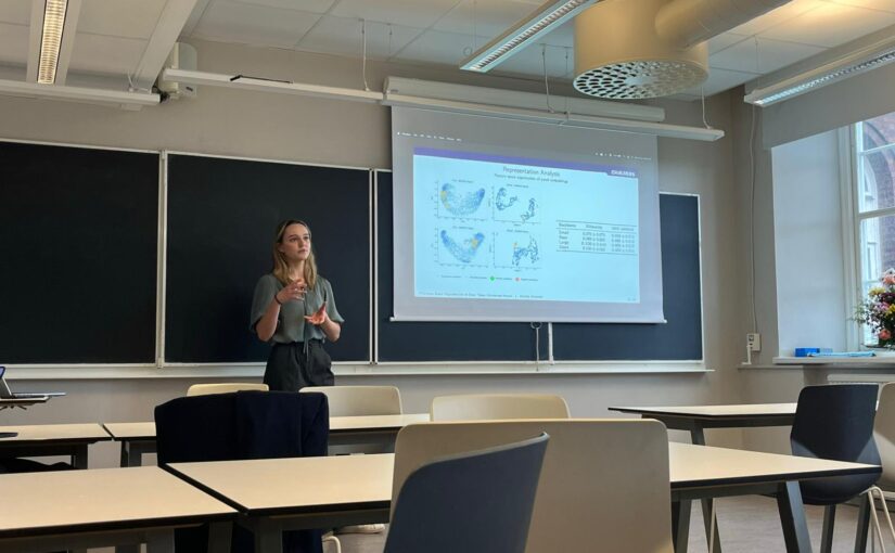
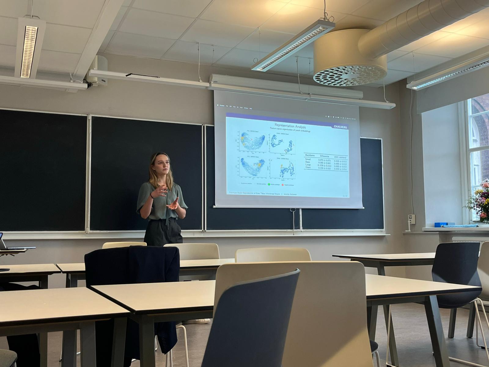
Matilda presenting her thesis. (Photo by M. Granfors.)Matilda Hellström, master student in the program of Complex Adaptive Systems at Chalmers University of Technology, defended her Master thesis on June 15 2026. Congrats!
Title: Prototype Based Segmentation of Bone Tissue Microscopy Images Using Self-Supervised Vision Transformers and Feature Space Similarity
Abstract:
Segmentation of microscopy images constitutes a fundamental task in biomedical research and clinical analysis. However, many segmentation methods rely on large annotated datasets. As the creation of labeled datasets tend to be highly time consuming and difficult to scale, there exists a need for finding alternative segmentation methods that can use unlabeled data directly.
This thesis investigates whether pretrained self-supervised Vision Transformers can be used for prototype based segmentation of bone tissue microscopy images. A segmentation framework based on pretrained DINOv2 backbones was developed, in which positive and negative reference points are used to construct prototype embeddings that guide similarity based segmentation in the learned feature space. The framework was evaluated using multiple DINOv2 backbone variants, feature space analysis and prototype transfer experiments.
The results demonstrated the potential of using pretrained self-supervised Vision Transformers for microscopy image segmentation by showing that the models produce feature representations in which tissue and background regions become partially separable. Despite being trained on natural RGB images rather than microscopy data, the pretrained backbones enabled segmentation of bone structures using the proposed similarity based segmentation framework.
Supervisor: Mirja Granfors Pineda Examiner: Giovanni Volpe Opponent: Patrik Dennis
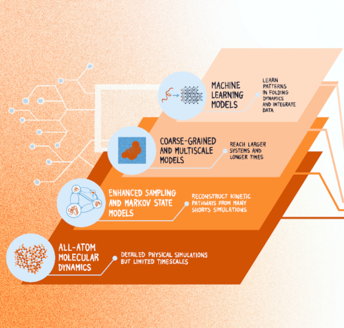
Computational advances in protein folding studies. Current approaches address multiple levels of resolution and methodological frameworks, however, none of the existing methods provides quantitative and dynamic information of the relationship between protein sequence and folding mechanism at all-atom resolution and at scale. (Graphics by J. Sacquegno.)Protein Dynamics Beyond Structure Prediction
Juliette Griffié, Sviatlana Shashkova, Antonio Ciarlo, Sreekanth K. Manikandan, Claes Andréasson, Malin Bäckström, Tristan Bereau, Hjalmar Brismar, Carlos Bustamante, Marta Carroni, Roberto Covino, Andreas Dahlin, Sebastian Deindl, Lucie Delemotte, Arne Elofsson, John Eriksson, Giovanna Fragneto, Anders Gunnarsson, Per Hammarström, Caroline Ingre, Christian Kaiser, Petronella Kettunen, Mark C. Leake, Benjamin Loos, Anna Månberg, Antonia S. J. S. Mey, Richard Neutze, Thomas Nyström, Karl Palmås, Charley Schaefer, Markus J. Tamás, Nicola Ticozzi, Tomás S. Pilvelic, Jacopo Sacquegno, B.M. (Betty)Tijms, Gunnar von Heijne, Björn Wallner, Vitali Zhaunerchyk, Simon Olsson, Joana B. Pereira, Julia Fernandez-Rodriguez, Fredrik Westerlund, Giovanni Volpe
arXiv: 2606.08647
How microorganisms respond to and interact with their environment can vary significantly from individual to individual, which can have important microbiological and ecological implications. However, most microscopy techniques can only observe motile microorganisms for short times because of their limited fields of view. Using Lagrangian tracking, a single microorganism can be followed in 3D, potentially indefinitely, allowing to decipher individual phenotypical traits. Current Lagrangian tracking methods use the fluorescence signal emitted by the microorganism as feedback to keep it in focus. However, over long times, epifluorescent imaging can induce photobleaching and photodamage, and importantly, not all microorganisms can easily be made fluorescent. Additionally, traditional algorithms used in feedback loops to determine microorganism position are prone to errors, especially in optically complex media. Here, we present a faster, more reliable, and versatile Lagrangian tracking method that uses deep learning to determine the 3D position of the microorganism. This new method demonstrates enhanced accuracy and speed in tracking fluorescent bacteria with fluorescence microscopy also in optically complex media. Furthermore, we track bacteria with other microscopy modalities, such as brightfield microscopy — for example, this enables us to track magnetotactic bacteria, which cannot be made fluorescent without degrading their magnetotactic properties. These novel capabilities allow to extract previously inaccessible quantitative information, significantly advancing the study of microorganism behavior — and thus opening new avenues for research in complex biological and ecological systems.
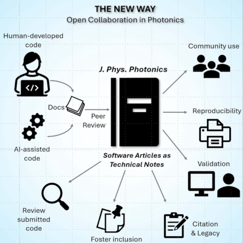
A centralized, peer-reviewed ecosystem in J. Phys. Photonics where human agents and/or AI agents co-develop validated, documented software. (Image from the manuscript.)Beyond the black box: towards an open and citable software ecosystem in photonics
Anoop C Patil, Maciej Trusiak, Fei Xia, Liangcai Cao, Carlo Manzo and Giovanni Volpe
Journal of Physics: Photonics 8, 020201 (2026)
doi: 10.1088/2515-7647/ae68cd
This editorial draws attention to a major issue in photonics: important research software is often hidden, undocumented, and lost over time, making results hard to reproduce. The rise of AI-generated code increases this problem by adding more ‘black box’ systems. To address this, J. Phys. Photonics is introducing Software Articles, a new article type allowing researchers to publish, validate, and share their code as formal scientific outputs. This initiative aims to promote transparency, reproducibility, and proper credit for developers. Open and peer-reviewed software helps the community verify results, reduce duplication of effort, and build lasting tools, ensuring that computational methods become reliable, accessible, and integral to scientific progress.
(Image created by G. Volpe with the assistance of DALL·E 2)What remain for physicists to do in the age of AI?
Giovanni Volpe
QT (Quantum Technology Division of MC2, Chalmers University of Technology) community building activity 2026 Date: 11 May 2026 Place: Stenungsbaden Yacht club
In recent years, the rapid growth of artificial intelligence, particularly deep learning, has transformed fields from natural sciences to technology. While deep learning is often viewed as a glorified form of curve fitting, its advancement to multi-layered, deep neural networks has resulted in unprecedented performance improvements, often surprising experts. As AI models grow larger and more complex, many wonder whether AI will eventually take over the world and what role remains for physicists and, more broadly, humans.
A critical, yet underappreciated fact is that these AI systems rely heavily on vast amounts of training data, most of which are generated and annotated by humans. This dependency raises an intriguing issue: what happens when human-generated data is no longer available, or when AI begins to train on AI-generated data? The phenomenon of AI poisoning, where the quality of AI outputs declines due to self-referencing, demonstrates the limitations of current AI models. For example, in image recognition tasks, such as those involving the MNIST dataset, AI tends to gravitate towards ‘safe’ or average outputs, diminishing originality and accuracy.
In this context, the unique role of humans becomes clear. Physicists, with their capacity for originality, deep understanding of physical phenomena, and the ability to exploit fundamental symmetries in nature, bring invaluable perspectives to the development of AI. By incorporating physics-informed training architectures and embracing the human drive for meaning and discovery, we can guide the future of AI in truly innovative directions. The message is clear: physicists must remain original, pursue their passions, and continue searching for the hidden laws that govern the world and society.
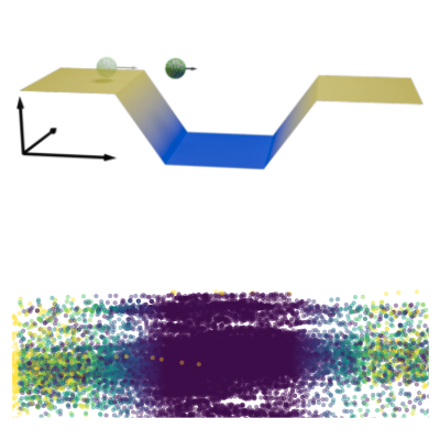
Experimental setup. (Top) Thermophoretic microswimmer undergoes active Brownian motion in a spatially-varying laser intensity profile that controls the self-thermophoretic propulsion of the swimmer using a feedback loop. (Bottom) Sample trajectory of the microswimmer over 15 minutes in a chamber. Colors indicate instantaneous velocity. (Image from the manuscript.)Delayed Active Swimmer in a Velocity Landscape
Viktor Holubec, Alexander Fischer, Giovanni Volpe, Frank Cichos
Physical Review Research 8, L022017 (2026)
arXiv: 2505.11042
doi: 10.1103/xn9x-ppjx
Active systems in nature and synthetic environments commonly exhibit spatially heterogeneous activity patterns and time-delayed responses from internal feedback mechanisms, exemplified by bacterial chemotaxis. We study an idealized active gas where particles modulate their self-propulsion based on local environmental conditions with such delays. Through integrated theoretical, computational, and experimental approaches, we demonstrate that steady-state density distributions and collective polarization exhibit characteristic peaks and valleys as functions of response delay time. We find that delays can amplify polarization by nearly an order of magnitude and trigger complete polarization reversal when particle displacement during the delay period surpasses the persistence length. Multiparticle simulations incorporating interparticle interactions validate that these phenomena remain robust in sufficiently dilute collective systems. Since density and polarization determine the current in active matter, our findings show that temporally programming the delay time allows control over both static and dynamic states in active systems, with implications for biological microswimmers and engineered microrobots.
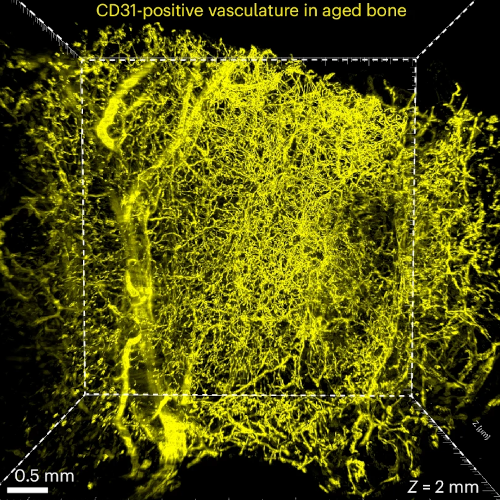
Visualization of the vasculature within human bone from a 75-year-old patient by immunostaining with antibodies against CD31. (Image from the manuscript.)Three-dimensional quantitative tissue clearing reveals differences in osteovascular niche of aged and young human mesenchymal stromal cells
Nelson Tsz Long Chu, Ostap Dregval, Yu-Wei Chang, Emil Kriukov, Xin Tian, Xin Liu, Dana Trompet, Misty Shuo Zhang, Lei Li, Zhong Li, Emiliano Gomez Ruiz, Joana B. Pereira, Mats Brittberg, Björn Barenius, Lars Sävendahl, Ralf H. Adams, Inger Gjertsson, Claes Ohlsson, Giovanni Volpe & Andrei S. Chagin
Nature Biomedical Engineering (2026)
bioRxiv: 10.1101/2025.10.07.680053
doi: 10.1038/s41551-026-01645-3
Human bone marrow mesenchymal stromal/stem cells (BM-MSCs) are widely used in clinical trials and tissue engineering, yet their native microenvironment remains poorly understood. Here we introduce a tissue-clearing protocol, DeepBone, for human bones and integrate it with simultaneous mRNA and protein detection. Using this protocol, we spatially map BM-MSCs relative to key bone microenvironment components, including human blood capillaries, adipocytes, sinusoids and bony trabeculae. Quantitative analysis reveals that the native microenvironment of human BM-MSCs in young bone is enriched in vasculature, sinusoids, bone matrix and adipocytes. In contrast, in aged bone, BM-MSCs show no preferential association with bone or adipocytes. Proliferative BM-MSCs are predominantly found along blood vessels. Moreover, we identify a specialized microenvironment for BM-MSCs in young bone, characterized by sinusoids coiled around trabeculae and enriched by R-type vessels. These findings provide insights into the native niches of BM-MSCs, offering a foundation for the development of tissue engineering strategies that mimic their physiological context.
DeepTrack 2 Logo. (Image from DeepTrack 2 Project)Artificial Intelligence for Microscopy: From Pixels to Physical Insight
Giovanni Volpe DINAMO 2026
Franschhoek, South Africa
7 April 2026
Diagnostic classification. (Image from the article.)Mapping individual molecular connectomes in Alzheimer’s disease
Zhilei Xu, Mite Mijalkov, Jiawei Sun, Yu-Wei Chang, Arianna Sala, Giovanni Volpe, Mario Severino, Mattia Veronese, Sara Garcia-Ptacek, Joana B. Pereira, for the Alzheimer’s Disease Neuroimaging Initiative
Alzheimer’s & Dementia 22, e71310 (2026)
DOI: 10.1002/alz.71310
INTRODUCTION
Mapping individual differences is crucial to improve personalized medicine approaches in Alzheimer’s disease (AD), which is characterized by strong inter-individual variability in the accumulation patterns of tau and amyloid beta pathology.
METHODS
We assess the progression of AD across the disease continuum by building individual molecular connectomes using longitudinal positron emission tomography (PET) data.
RESULTS
We demonstrate that these connectomes constitute a unique fingerprint, capable of identifying a single individual from a large group of subjects. Alterations in the connectomes discriminate different diagnostic groups and predict cognitive decline to a higher extent than conventional PET measures. We introduce a novel gene-specific transcription network analysis that linked individual tau and amyloid connectomes to a common transcriptomic profile of apoptosis, with the tau connectome being specifically related to pyrimidine metabolism, and the amyloid connectome to histone acetylation.
DISCUSSION
Individual molecular connectome mapping provides a novel and sensitive framework to monitor AD progression.
Highlights
Individual molecular connectomes constitute a unique fingerprint, capable of identifying a single individual from a large group of subjects.
Alterations in individual molecular connectomes significantly increase both across the Alzheimer’s disease (AD) continuum and over time.
Alterations in individual molecular connectomes discriminate different diagnostic groups and predict cognitive decline to a higher extent than conventional positron emission tomography measures.
Susceptibilities of individual tau and amyloid connectomes to AD are linked to a common transcriptomic profile of apoptosis, with the tau connectome being specifically related to pyrimidine metabolism, and the amyloid connectome to histone acetylation.