Myxococcus xanthus colonies develop different strategies to adapt to their environment, leading to the formation of macroscopic patterns from microscopic entities. (Image by the Authors of the manuscript.)Tutorial for the growth and development of Myxococcus xanthus as a Model System at the Intersection of Biology and Physics
Jesus Manuel Antúnez Domínguez, Laura Pérez García, Natsuko Rivera-Yoshida, Jasmin Di Franco, David Steiner, Alejandro V. Arzola, Mariana Benítez, Charlotte Hamngren Blomqvist, Roberto Cerbino, Caroline Beck Adiels, Giovanni Volpe
Soft Matter 21, 8602-8623 (2025)
arXiv: 2407.18714
doi: 10.1039/d5sm00284b
Myxococcus xanthus is a unicellular organism known for its capacity to move and communicate, giving rise to complex collective properties, structures and behaviors. These characteristics have contributed to position M. xanthus as a valuable model organism for exploring emergent collective phenomena at the interface of biology and physics, particularly within the growing domain of active matter research. Yet, researchers frequently encounter difficulties in establishing reproducible and reliable culturing protocols. This tutorial provides a detailed and accessible guide to the culture, growth, development, and experimental sample preparation of M. xanthus. In addition, it presents several exemplary experiments that can be conducted using these samples, including motility assays, fruiting body formation, predation, and elasticotaxis—phenomena of direct relevance for active matter studies.
High-resolution display of “The Kiss” on Retina E-Paper vs. iPhone 15: Photographs comparing the display of “The Kiss” on an iPhone 15 and Retina E-paper. The surface area of the Retina E-paper is ~ 1/4000 times smaller than the iPhone 15. (Image by the Authors of the manuscript.)Video‐rate tunable colour electronic paper with human resolution
Ade Satria Saloka Santosa, Yu-Wei Chang, Andreas B. Dahlin, Lars Osterlund, Giovanni Volpe, Kunli Xiong
Nature 646, 1089-1095 (2025)
arXiv: 2502.03580
doi: 10.1038/s41586-025-09642-3
As demand for immersive experiences grows, displays are moving closer to the eye with smaller sizes and higher resolutions. However, shrinking pixel emitters reduce intensity, making them harder to perceive. Electronic Papers utilize ambient light for visibility, maintaining optical contrast regardless of pixel size, but cannot achieve high resolution. We show electrically tunable meta-pixels down to ~560 nm in size (>45,000 PPI) consisting of WO3 nanodiscs, allowing one-to-one pixel-photodetector mapping on the retina when the display size matches the pupil diameter, which we call Retina Electronic Paper. Our technology also supports video display (25 Hz), high reflectance (~80%), and optical contrast (~50%), which will help create the ultimate virtual reality display.
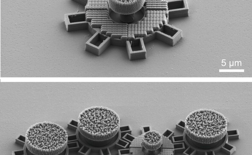
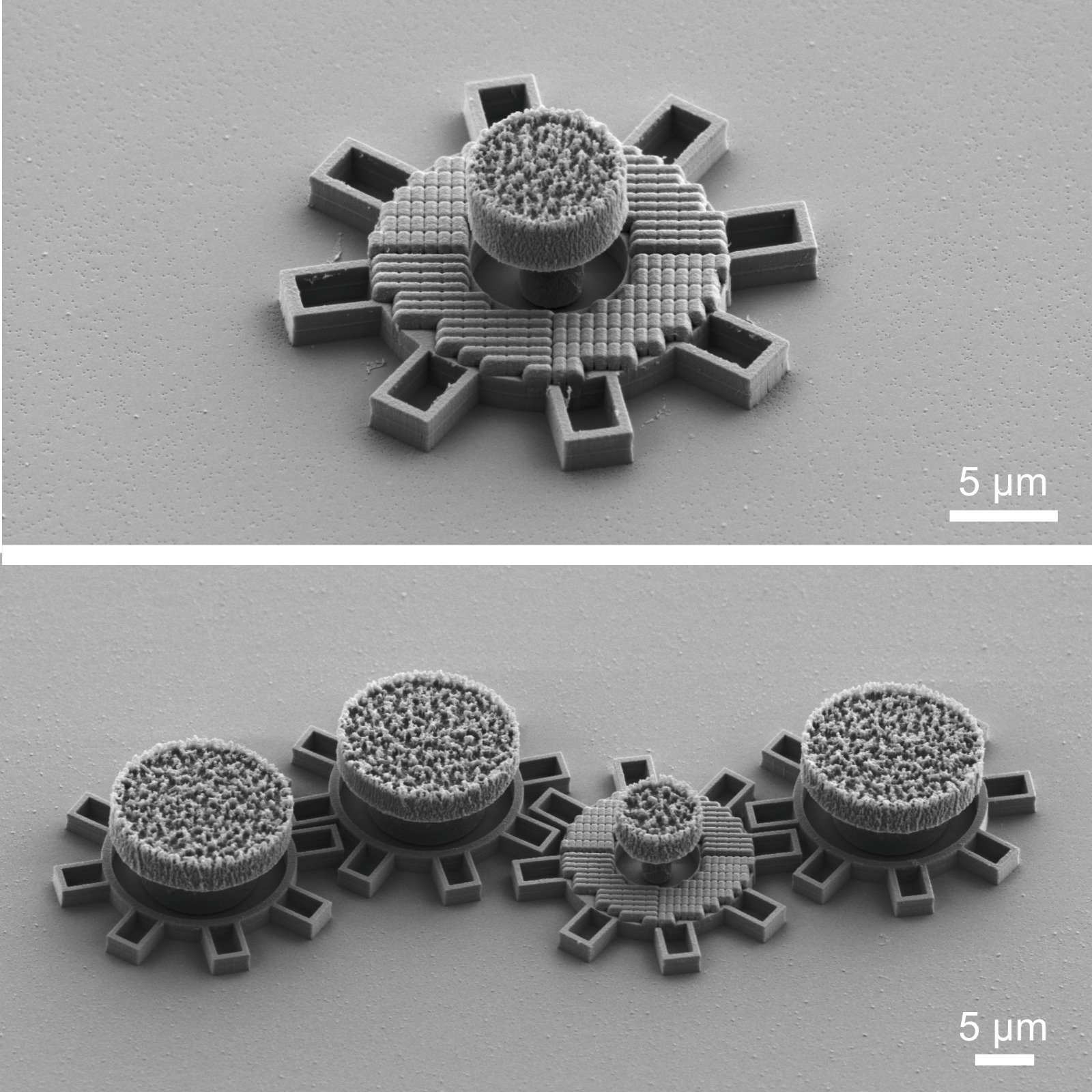
Top: single gear; Bottom: the second gear from the right has an optical metamaterial that react to laserlight and makes the gear move. All gears are made in silica directly on a chip. Each gear is about 0.016 mm in diameter. (Image by G. Wang)
Microscopic Geared Metamachines
Gan Wang, Marcel Rey, Antonio Ciarlo, Mohanmmad Mahdi Shanei, Kunli Xiong, Giuseppe Pesce, Mikael Käll and Giovanni Volpe
Nature Communications 16, 7767 (2025)
doi: 10.1038/s41467-025-62869-6
arXiv: 2409.17284
The miniaturization of mechanical machines is critical for advancing nanotechnology and reducing device footprints. Traditional efforts to downsize gears and micromotors have faced limitations at around 0.1 mm for over thirty years due to the complexities of constructing drives and coupling systems at such scales. Here, we present an alternative approach utilizing optical metasurfaces to locally drive microscopic machines, which can then be fabricated using standard lithography techniques and seamlessly integrated on the chip, achieving sizes down to tens of micrometers with movements precise to the sub-micrometer scale. As a proof of principle, we demonstrate the construction of microscopic gear trains powered by a single driving gear with a metasurface activated by a plane light wave. Additionally, we develop a versatile pinion and rack micromachine capable of transducing rotational motion, performing periodic motion, and controlling microscopic mirrors for light deflection. Our on-chip fabrication process allows for straightforward parallelization and integration. Using light as a widely available and easily controllable energy source, these miniaturized metamachines offer precise control and movement, unlocking new possibilities for micro- and nanoscale systems.
After the article was published, it was reported by many media outlets, University of Gothenburg, New Scientist, Optics.org, Phys.org, ScienceDaily, Discover Magazine, among others.
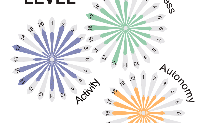
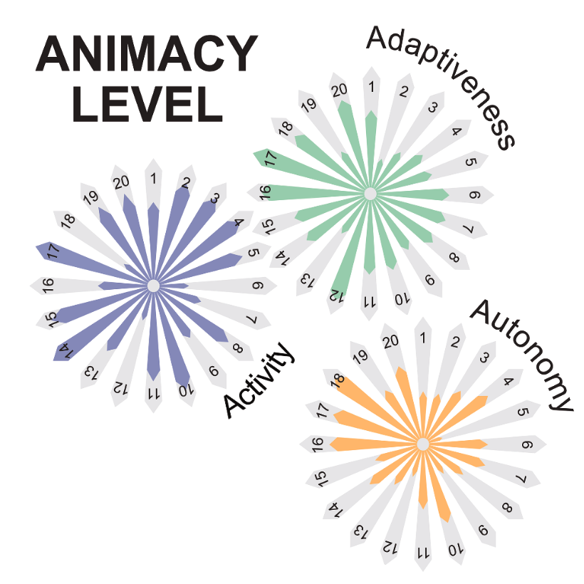
The three properties of animacy. The three polar plots sketch our jointly perceived level of development for each principle of animacy (i.e. activity, adaptiveness and autonomy) for each system discussed in this roadmap. The polar coordinate represents the various systems, while the radial coordinate represents the level of development (from low to high) that each system shows in the principle of each polar plot. Ideally, within a generation, all systems will fill these polar plots to show high levels in each of the three attributes of animacy. For now, only biological materials (not represented here) can be considered fully animated. (Image from the manuscript, adapted.)Roadmap for animate matter
Giorgio Volpe, Nuno A M Araújo, Maria Guix, Mark Miodownik, Nicolas Martin, Laura Alvarez, Juliane Simmchen, Roberto Di Leonardo, Nicola Pellicciotta, Quentin Martinet, Jérémie Palacci, Wai Kit Ng, Dhruv Saxena, Riccardo Sapienza, Sara Nadine, João F Mano, Reza Mahdavi, Caroline Beck Adiels, Joe Forth, Christian Santangelo, Stefano Palagi, Ji Min Seok, Victoria A Webster-Wood, Shuhong Wang, Lining Yao, Amirreza Aghakhani, Thomas Barois, Hamid Kellay, Corentin Coulais, Martin van Hecke, Christopher J Pierce, Tianyu Wang, Baxi Chong, Daniel I Goldman, Andreagiovanni Reina, Vito Trianni, Giovanni Volpe, Richard Beckett, Sean P Nair, Rachel Armstrong
Journal of Physics: Condensed Matter 37, 333501 (2025)
arXiv: 2407.10623
doi: 10.1088/1361-648X/adebd3
Humanity has long sought inspiration from nature to innovate materials and devices. As science advances, nature-inspired materials are becoming part of our lives. Animate materials, characterized by their activity, adaptability, and autonomy, emulate properties of living systems. While only biological materials fully embody these principles, artificial versions are advancing rapidly, promising transformative impacts in the circular economy, health and climate resilience within a generation. This roadmap presents authoritative perspectives on animate materials across different disciplines and scales, highlighting their interdisciplinary nature and potential applications in diverse fields including nanotechnology, robotics and the built environment. It underscores the need for concerted efforts to address shared challenges such as complexity management, scalability, evolvability, interdisciplinary collaboration, and ethical and environmental considerations. The framework defined by classifying materials based on their level of animacy can guide this emerging field to encourage cooperation and responsible development. By unravelling the mysteries of living matter and leveraging its principles, we can design materials and systems that will transform our world in a more sustainable manner.
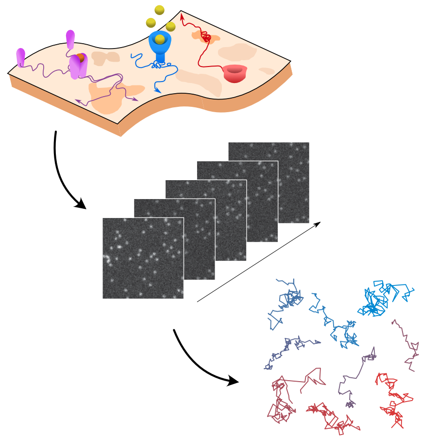
Rationale for the challenge organization. The interactions of biomolecules in complex environments, such as the cell membrane, regulate physiological processes in living systems. These interactions produce changes in molecular motion that can be used as a proxy to measure interaction parameters. Time-lapse single-molecule imaging allows us to visualize these processes with high spatiotemporal resolution and, in combination with single-particle tracking methods, provide trajectories of individual molecules. (Image by the Authors of the manuscript.)Quantitative evaluation of methods to analyze motion changes in single-particle experiments
Gorka Muñoz-Gil, Harshith Bachimanchi, Jesús Pineda, Benjamin Midtvedt, Gabriel Fernández-Fernández, Borja Requena, Yusef Ahsini, Solomon Asghar, Jaeyong Bae, Francisco J. Barrantes, Steen W. B. Bender, Clément Cabriel, J. Alberto Conejero, Marc Escoto, Xiaochen Feng, Rasched Haidari, Nikos S. Hatzakis, Zihan Huang, Ignacio Izeddin, Hawoong Jeong, Yuan Jiang, Jacob Kæstel-Hansen, Judith Miné-Hattab, Ran Ni, Junwoo Park, Xiang Qu, Lucas A. Saavedra, Hao Sha, Nataliya Sokolovska, Yongbing Zhang, Giorgio Volpe, Maciej Lewenstein, Ralf Metzler, Diego Krapf, Giovanni Volpe, Carlo Manzo
Nature Communications 16, 6749 (2025)
arXiv: 2311.18100
doi: https://doi.org/10.1038/s41467-025-61949-x
The analysis of live-cell single-molecule imaging experiments can reveal valuable information about the heterogeneity of transport processes and interactions between cell components. These characteristics are seen as motion changes in the particle trajectories. Despite the existence of multiple approaches to carry out this type of analysis, no objective assessment of these methods has been performed so far. Here, we report the results of a competition to characterize and rank the performance of these methods when analyzing the dynamic behavior of single molecules. To run this competition, we implemented a software library that simulates realistic data corresponding to widespread diffusion and interaction models, both in the form of trajectories and videos obtained in typical experimental conditions. The competition constitutes the first assessment of these methods, providing insights into the current limitations of the field, fostering the development of new approaches, and guiding researchers to identify optimal tools for analyzing their experiments.
In this work, we present an unsupervised deep learning framework using Variational Autoencoders (VAEs) to decode stress-specific biomolecular fingerprints directly from Raman spectral data across multiple plant species and genotypes. (Image by the Authors of the manuscript. A part of the image was designed using Biorender.com.)From Spectra to Stress: Unsupervised Deep Learning for Plant Health Monitoring
Anoop C. Patil, Benny Jian Rong Sng, Yu-Wei Chang, Joana B. Pereira, Chua Nam-Hai, Rajani Sarojam, Gajendra Pratap Singh, In-Cheol Jang, and Giovanni Volpe
ArXiv: 2507.15772
Detecting stress in plants is crucial for both open-farm and controlled-environment agriculture. Biomolecules within plants serve as key stress indicators, offering vital markers for continuous health monitoring and early disease detection. Raman spectroscopy provides a powerful, non-invasive means to quantify these biomolecules through their molecular vibrational signatures. However, traditional Raman analysis relies on customized data-processing workflows that require fluorescence background removal and prior identification of Raman peaks of interest-introducing potential biases and inconsistencies. Here, we introduce DIVA (Deep-learning-based Investigation of Vibrational Raman spectra for plant-stress Analysis), a fully automated workflow based on a variational autoencoder. Unlike conventional approaches, DIVA processes native Raman spectra-including fluorescence backgrounds-without manual preprocessing, identifying and quantifying significant spectral features in an unbiased manner. We applied DIVA to detect a range of plant stresses, including abiotic (shading, high light intensity, high temperature) and biotic stressors (bacterial infections). By integrating deep learning with vibrational spectroscopy, DIVA paves the way for AI-driven plant health assessment, fostering more resilient and sustainable agricultural practices.
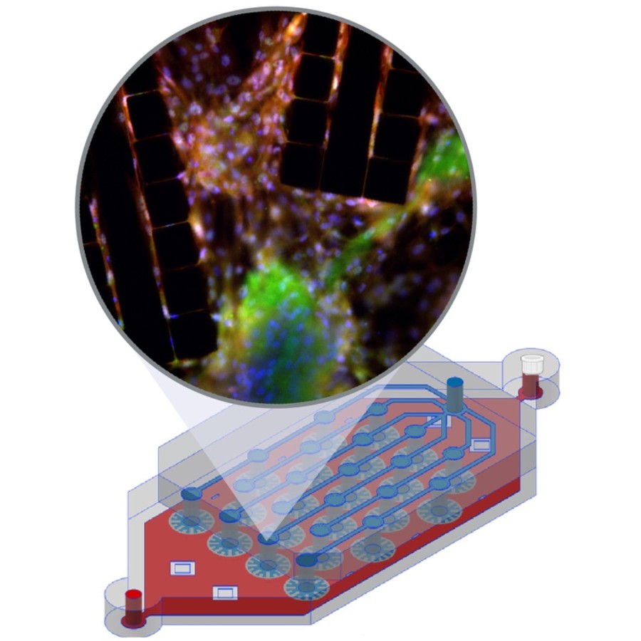
The image shows a liver-lobule-chip (LLoC) with 21 artificial lobules mimicking liver microarchitecture. Its PDMS design supports diffusion-based perfusion, shear stress, and nutrient gradients and enables iPSC-derived hepatic maturation and spatially organized, zonated function in 3D. (Image by C. Beck Adiels)An in vivo mimetic liver-lobule-chip (LLoC) for stem cell maturation, and zonation of hepatocyte-like cells on chip
Philip Dalsbecker, Siiri Suominen, Muhammad Asim Faridi, Reza Mahdavi, Julia Johansson, Charlotte Hamngren Blomqvist, Mattias Goksör, Katriina Aalto-Setälä, Leena E. Viiri and Caroline B. Adiels
Lab on a Chip 25, 4328 – 4344 (2025)
doi: 10.1039/D4LC00509K
In vitro cell culture models play a crucial role in preclinical drug discovery. To achieve optimal culturing environments and establish physiologically relevant organ-specific conditions, it is imperative to replicate in vivo scenarios when working with primary or induced pluripotent cell types. However, current approaches to recreating in vivo conditions and generating relevant 3D cell cultures still fall short. In this study, we validate a liver-lobule-chip (LLoC) containing 21 artificial liver lobules, each representing the smallest functional unit of the human liver. The LLoC facilitates diffusion-based perfusion via sinusoid-mimetic structures, providing physiologically relevant shear stress exposure and radial nutrient concentration gradients within each lobule. We demonstrate the feasibility of long term cultures (up to 14 days) of viable and functional HepG2 cells in a 3D discoid tissue structure, serving as initial proof of concept. Thereafter, we successfully differentiate sensitive, human induced pluripotent stem cell (iPSC)-derived cells into hepatocyte-like cells over a period of 20 days on-chip, exhibiting advancements in maturity compared to traditional 2D cultures. Further, hepatocyte-like cells cultured in the LLoC exhibit zonated protein expression profiles, indicating the presence of metabolic gradients characteristic of liver lobules. Our results highlight the suitability of the LLoC for long-term discoid tissue cultures, specifically for iPSCs, and their differentiation in a perfused environment. We envision the LLoC as a starting point for more advanced in vitro models, allowing for the combination of multiple liver cell types to create a comprehensive liver model for disease-onchip studies. Ultimately, when combined with stem cell technology, the LLoC offers a promising and robust on-chip liver model that serves as a viable alternative to primary hepatocyte cultures—ideally suited for preclinical drug screening and personalized medicine applications.
BRAPH 2 Genesis enables swift creation of custom, reproducible software distributions—tackling the growing complexity of neuroscience by streamlining analysis across diverse data types and workflows. (Image by B. Zufiria-Gerbolés and Y.-W. Chang.)BRAPH 2: a flexible, open-source, reproducible, community-oriented, easy-to-use framework for network analyses in neurosciences
Yu-Wei Chang, Blanca Zufiria-Gerbolés, Pablo Emiliano Gómez-Ruiz, Anna Canal-Garcia, Hang Zhao, Mite Mijalkov, Joana Braga Pereira, Giovanni Volpe
bioRxiv: 10.1101/2025.04.11.648455
As network analyses in neuroscience continue to grow in both complexity and size, flexible methods are urgently needed to provide unbiased, reproducible insights into brain function. BRAPH 2 is a versatile, open-source framework that meets this challenge by offering streamlined workflows for advanced statistical models and deep learning in a community-oriented environment. Through its Genesis compiler, users can build specialized distributions with custom pipelines, ensuring flexibility and scalability across diverse research domains. These powerful capabilities will ensure reproducibility and accelerate discoveries in neuroscience.
Memory capacity in aging. A Brain reservoir computing architecture with uniform random signals applied to all nodes. (Image from the article.)Computational memory capacity predicts aging and cognitive decline
Mite Mijalkov, Ludvig Storm, Blanca Zufiria-Gerbolés, Dániel Veréb, Zhilei Xu, Anna Canal-Garcia, Jiawei Sun, Yu-Wei Chang, Hang Zhao, Emiliano Gómez-Ruiz, Massimiliano Passaretti, Sara Garcia-Ptacek, Miia Kivipelto, Per Svenningsson, Henrik Zetterberg, Heidi Jacobs, Kathy Lüdge, Daniel Brunner, Bernhard Mehlig, Giovanni Volpe, Joana B. Pereira
Nature Communications 16, 2748 (2025)
doi: 10.1038/s41467-025-57995-0
Memory is a crucial cognitive function that deteriorates with age. However, this ability is normally assessed using cognitive tests instead of the architecture of brain networks. Here, we use reservoir computing, a recurrent neural network computing paradigm, to assess the linear memory capacities of neural-network reservoirs extracted from brain anatomical connectivity data in a lifespan cohort of 636 individuals. The computational memory capacity emerges as a robust marker of aging, being associated with resting-state functional activity, white matter integrity, locus coeruleus signal intensity, and cognitive performance. We replicate our findings in an independent cohort of 154 young and 72 old individuals. By linking the computational memory capacity of the brain network with cognition, brain function and integrity, our findings open new pathways to employ reservoir computing to investigate aging and age-related disorders.
Propagation of scattered light through a scattering microscope, illustrating typical nanoparticles studied. (Image by B. García Rodriguez.)Optical Label-Free Microscopy Characterization of Dielectric Nanoparticles
Berenice Garcia Rodriguez, Erik Olsén, Fredrik Skärberg, Giovanni Volpe, Fredrik Höök, Daniel Sundås Midtvedt
Nanoscale, 17, 8336-8362 (2025)
arXiv: 2409.11810
doi: 10.1039/D4NR03860F
In order to relate nanoparticle properties to function, fast and detailed particle characterization, is needed. The ability to characterize nanoparticle samples using optical microscopy techniques has drastically improved over the past few decades; consequently, there are now numerous microscopy methods available for detailed characterization of particles with nanometric size. However, there is currently no “one size fits all” solution to the problem of nanoparticle characterization. Instead, since the available techniques have different detection limits and deliver related but different quantitative information, the measurement and analysis approaches need to be selected and adapted for the sample at hand. In this tutorial, we review the optical theory of single particle scattering and how it relates to the differences and similarities in the quantitative particle information obtained from commonly used microscopy techniques, with an emphasis on nanometric (submicron) sized dielectric particles. Particular emphasis is placed on how the optical signal relates to mass, size, structure, and material properties of the detected particles and to its combination with diffusivity-based particle sizing. We also discuss emerging opportunities in the wake of new technology development, with the ambition to guide the choice of measurement strategy based on various challenges related to different types of nanoparticle samples and associated analytical demands.