Optical Label-Free Microscopy Characterization of Dielectric Nanoparticles: A tutorial on ArXiv

Propagation of scattered light through a scattering microscope, illustrating typical nanoparticles studied. (Image by B. García Rodriguez.)
Optical Label-Free Microscopy Characterization of Dielectric Nanoparticles: A tutorial
Berenice Garcia Rodriguez, Erik Olsén, Fredrik Skärberg, Giovanni Volpe, Fredrik Höök, Daniel Sundås Midtvedt
arXiv: 2409.11810

In order to relate nanoparticle properties to function, fast and detailed particle characterization, is needed. The ability to characterize nanoparticle samples using optical microscopy techniques has drastically improved over the past few decades; consequently, there are now numerous microscopy methods available for detailed characterization of particles with nanometric size. However, there is currently no “one size fits all” solution to the problem of nanoparticle characterization. Instead, since the available techniques have different detection limits and deliver related but different quantitative information, the measurement and analysis approaches need to be selected and adapted for the sample at hand. In this tutorial, we review the optical theory of single particle scattering and how it relates to the differences and similarities in the quantitative particle information obtained from commonly used microscopy techniques, with an emphasis on nanometric (submicron) sized dielectric particles. Particular emphasis is placed on how the optical signal relates to mass, size, structure, and material properties of the detected particles and to its combination with diffusivity-based particle sizing. We also discuss emerging opportunities in the wake of new technology development, with the ambition to guide the choice of measurement strategy based on various challenges related to different types of nanoparticle samples and associated analytical demands.

Dual-Angle Interferometric Scattering Microscopy for Optical Multiparametric Particle Characterization published in Nano Letters

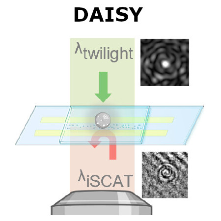
Conceptual schematic of dual-angle interferometric scattering microscopy (DAISY). (Image by the Authors of the manuscript.)
Dual-Angle Interferometric Scattering Microscopy for Optical Multiparametric Particle Characterization
Erik Olsén, Berenice García Rodríguez, Fredrik Skärberg, Petteri Parkkila, Giovanni Volpe, Fredrik Höök, and Daniel Sundås Midtvedt
Nano Letters, 24(6), 1874-1881 (2024)
doi: 10.1021/acs.nanolett.3c03539
arXiv: 2309.07572

Traditional single-nanoparticle sizing using optical microscopy techniques assesses size via the diffusion constant, which requires suspended particles to be in a medium of known viscosity. However, these assumptions are typically not fulfilled in complex natural sample environments. Here, we introduce dual-angle interferometric scattering microscopy (DAISY), enabling optical quantification of both size and polarizability of individual nanoparticles (radius <170 nm) without requiring a priori information regarding the surrounding media or super-resolution imaging. DAISY achieves this by combining the information contained in concurrently measured forward and backward scattering images through twilight off-axis holography and interferometric scattering (iSCAT). Going beyond particle size and polarizability, single-particle morphology can be deduced from the fact that the hydrodynamic radius relates to the outer particle radius, while the scattering-based size estimate depends on the internal mass distribution of the particles. We demonstrate this by differentiating biomolecular fractal aggregates from spherical particles in fetal bovine serum at the single-particle level.
Single-shot self-supervised object detection in microscopy published in Nature Communications

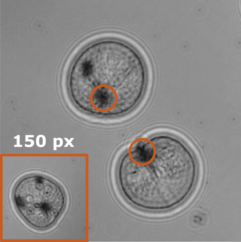
LodeSTAR tracks the plankton Noctiluca scintillans. (Image by the Authors of the manuscript.)
Single-shot self-supervised particle tracking
Benjamin Midtvedt, Jesús Pineda, Fredrik Skärberg, Erik Olsén, Harshith Bachimanchi, Emelie Wesén, Elin K. Esbjörner, Erik Selander, Fredrik Höök, Daniel Midtvedt, Giovanni Volpe
Nature Communications 13, 7492 (2022)
arXiv: 2202.13546
doi: 10.1038/s41467-022-35004-y

Object detection is a fundamental task in digital microscopy, where machine learning has made great strides in overcoming the limitations of classical approaches. The training of state-of-the-art machine-learning methods almost universally relies on vast amounts of labeled experimental data or the ability to numerically simulate realistic datasets. However, experimental data are often challenging to label and cannot be easily reproduced numerically. Here, we propose a deep-learning method, named LodeSTAR (Localization and detection from Symmetries, Translations And Rotations), that learns to detect microscopic objects with sub-pixel accuracy from a single unlabeled experimental image by exploiting the inherent roto-translational symmetries of this task. We demonstrate that LodeSTAR outperforms traditional methods in terms of accuracy, also when analyzing challenging experimental data containing densely packed cells or noisy backgrounds. Furthermore, by exploiting additional symmetries we show that LodeSTAR can measure other properties, e.g., vertical position and polarizability in holographic microscopy.

Fast and Accurate Nanoparticle Characterization Using Deep-Learning-Enhanced Off-Axis Holography published in ACS Nano

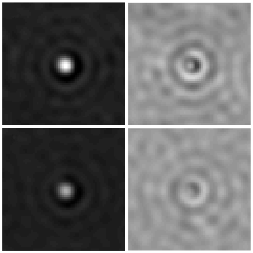
Phase and amplitude signals from representative particles for testing the performance of the Deep-learning approach

Fast and Accurate Nanoparticle Characterization Using Deep-Learning-Enhanced Off-Axis Holography
Benjamin Midtvedt, Erik Olsén, Fredrik Eklund, Fredrik Höök, Caroline Beck Adiels, Giovanni Volpe, Daniel Midtvedt
ACS Nano 15(2), 2240–2250 (2021)
doi: 10.1021/acsnano.0c06902
arXiv: 2006.11154

The characterisation of the physical properties of nanoparticles in their native environment plays a central role in a wide range of fields, from nanoparticle-enhanced drug delivery to environmental nanopollution assessment. Standard optical approaches require long trajectories of nanoparticles dispersed in a medium with known viscosity to characterise their diffusion constant and, thus, their size. However, often only short trajectories are available, while the medium viscosity is unknown, e.g., in most biomedical applications. In this work, we demonstrate a label-free method to quantify size and refractive index of individual subwavelength particles using two orders of magnitude shorter trajectories than required by standard methods, and without assumptions about the physicochemical properties of the medium. We achieve this by developing a weighted average convolutional neural network to analyse the holographic images of the particles. As a proof of principle, we distinguish and quantify size and refractive index of silica and polystyrene particles without prior knowledge of solute viscosity or refractive index. As an example of an application beyond the state of the art, we demonstrate how this technique can monitor the aggregation of polystyrene nanoparticles, revealing the time-resolved dynamics of the monomer number and fractal dimension of individual subwavelength aggregates. This technique opens new possibilities for nanoparticle characterisation with a broad range of applications from biomedicine to environmental monitoring.