Illustration of three different experiments autonomously performed by the SmartTrap system: DNA pulling experiments (top), red blood cell stretching (bottom left), and particle-particle interaction measurements (bottom right). (Image by M. Selin.)SmartTrap: automated precision experiments with optical tweezers
Martin Selin, Antonio Ciarlo, Giuseppe Pesce, Lars Bengtsson, Joan Camunas-Soler, Vinoth Sundar Rajan, Fredrik Westerlund, L. Marcus Wilhelmsson, Isabel Pastor, Felix Ritort, Steven B. Smith, Carlos Bustamante, Giovanni Volpe
Nature Methods (2026)
arXiv: 2505.05290
doi: 10.1038/s41592-026-03129-3
Optical tweezers are widely used in single-molecule biophysics, cell biomechanics and soft matter physics, but require a human operator, limiting throughput and repeatability. Here we present a smart optical tweezers platform, named SmartTrap, capable of performing complex experiments autonomously by integrating real-time three-dimensional particle tracking, custom electronics and a microfluidics system. Through a series of experiments, we demonstrate it can operate continuously, acquiring high-precision data over extended periods of time. By bridging the gap between manual experimentation and autonomous operation, SmartTrap establishes a robust and open-source framework for the next generation of optical tweezers research, capable of performing large-scale studies in single-molecule biophysics, cell mechanics and colloidal science with minimal experimental overhead and operator bias.
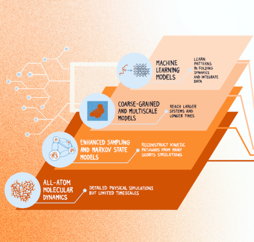
Computational advances in protein folding studies. Current approaches address multiple levels of resolution and methodological frameworks, however, none of the existing methods provides quantitative and dynamic information of the relationship between protein sequence and folding mechanism at all-atom resolution and at scale. (Graphics by J. Sacquegno.)Protein Dynamics Beyond Structure Prediction
Juliette Griffié, Sviatlana Shashkova, Antonio Ciarlo, Sreekanth K. Manikandan, Claes Andréasson, Malin Bäckström, Tristan Bereau, Hjalmar Brismar, Carlos Bustamante, Marta Carroni, Roberto Covino, Andreas Dahlin, Sebastian Deindl, Lucie Delemotte, Arne Elofsson, John Eriksson, Giovanna Fragneto, Anders Gunnarsson, Per Hammarström, Caroline Ingre, Christian Kaiser, Petronella Kettunen, Mark C. Leake, Benjamin Loos, Anna Månberg, Antonia S. J. S. Mey, Richard Neutze, Thomas Nyström, Karl Palmås, Charley Schaefer, Markus J. Tamás, Nicola Ticozzi, Tomás S. Pilvelic, Jacopo Sacquegno, B.M. (Betty)Tijms, Gunnar von Heijne, Björn Wallner, Vitali Zhaunerchyk, Simon Olsson, Joana B. Pereira, Julia Fernandez-Rodriguez, Fredrik Westerlund, Giovanni Volpe
arXiv: 2606.08647
How microorganisms respond to and interact with their environment can vary significantly from individual to individual, which can have important microbiological and ecological implications. However, most microscopy techniques can only observe motile microorganisms for short times because of their limited fields of view. Using Lagrangian tracking, a single microorganism can be followed in 3D, potentially indefinitely, allowing to decipher individual phenotypical traits. Current Lagrangian tracking methods use the fluorescence signal emitted by the microorganism as feedback to keep it in focus. However, over long times, epifluorescent imaging can induce photobleaching and photodamage, and importantly, not all microorganisms can easily be made fluorescent. Additionally, traditional algorithms used in feedback loops to determine microorganism position are prone to errors, especially in optically complex media. Here, we present a faster, more reliable, and versatile Lagrangian tracking method that uses deep learning to determine the 3D position of the microorganism. This new method demonstrates enhanced accuracy and speed in tracking fluorescent bacteria with fluorescence microscopy also in optically complex media. Furthermore, we track bacteria with other microscopy modalities, such as brightfield microscopy — for example, this enables us to track magnetotactic bacteria, which cannot be made fluorescent without degrading their magnetotactic properties. These novel capabilities allow to extract previously inaccessible quantitative information, significantly advancing the study of microorganism behavior — and thus opening new avenues for research in complex biological and ecological systems.
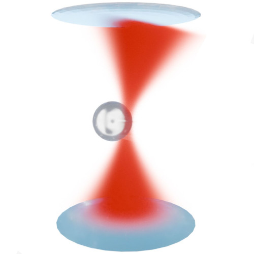
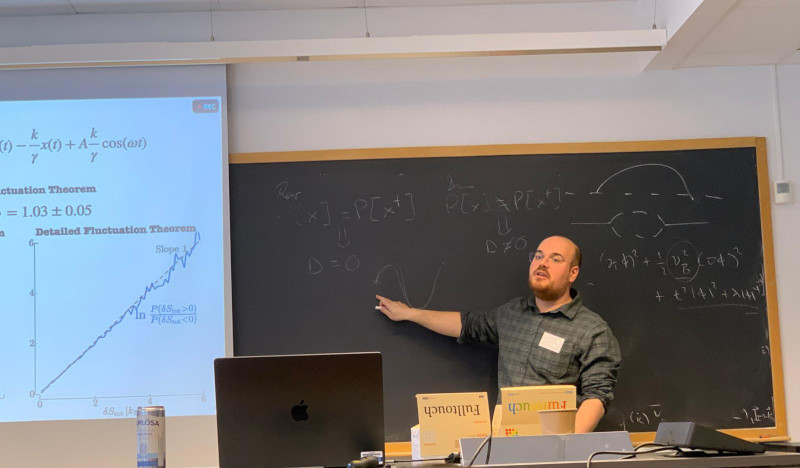
Schematic illustration of the light-momentum detection principle underlying SmartTrap. The momentum change of the trapping laser, induced by its interaction with the trapped particle, is measured to directly quantify optical forces with high precision, enabling real-time feedback and autonomous control in non-equilibrium experiments. (Figure by A. Ciarlo.)SmartTrap: Autonomous Optical Tweezers for Statistical Physics of Non-Equilibrium Systems
Antonio Ciarlo Date: 26th February 2026 Time: 13.30 Place: NORDITA, Stockholm, Sweden The 15th Nordic Workshop on Statistical Physics: Biological, Complex and Non-equilibrium Systems
Optical tweezers are a key tool in non-equilibrium statistical physics, allowing direct measurements of forces, work, and fluctuations in single-molecule and soft matter systems. However, manual operation limits throughput and the systematic study of rare events.
In this talk, Antonio Ciarlo will present SmartTrap, a fully autonomous optical tweezers platform integrating deep learning–based 3D tracking, adaptive feedback control, and automated microfluidics. The system operates without human intervention, executing complete force spectroscopy protocols.
Demonstrated with high-throughput DNA pulling experiments on λ-DNA, SmartTrap enables precise measurements of force–extension curves and folding kinetics. The platform also opens new possibilities for studies of colloids, single cells, and quantitative tests of non-equilibrium statistical physics.
Active Matter: Model Systems and Experimental Tests
Agnese Callegari, Antonio Ciarlo, Sreekanth Manikandan Dates and times:
23 Feb 14:00-15:00 (Agnese)
24 Feb 11:30-12:30 (Antonio)
24 Feb 14:00-15:00 (Sreekanth) Place: PJ Winter school on Geometry of nonequilibrium critical phenomena
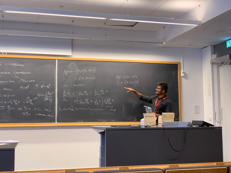
Active matter is a broad class of systems that operate intrinsically out of equilibrium. It spans multiple length scales—from macroscopic to micro- and nanoscopic—and includes both biological and artificial realizations, often displaying rich and emerging collective behaviors. The study of active matter aims to explain and interpret these phenomena using concepts and tools from physics. As such, understanding active and non-equilibrium systems requires a combination of theoretical, computational, and experimental approaches.
In the first part of the lecture, we introduce the concept of an active particle and demonstrate how it can be embodied in a macroscopic, self-propelled toy robot (a Hexbug). Despite their simplicity, such systems reproduce characteristic—and sometimes counterintuitive—features of microscopic active matter. These experiments have a strong pedagogical value and are designed to help bridge a gap in traditional physics curricula at the primary and secondary education levels.
The second part of the lecture focuses on active matter and non-equilibrium phenomena at the microscopic scale, where advanced experimental tools are essential. Optical tweezers provide precise control over microscopic systems and access to key physical observables. We introduce their operating principles and illustrate how they can be used to construct a minimal, well-controlled experimental model for studying non-equilibrium dynamics at the single-particle level.
In the final part of the lecture, we turn to the theoretical and computational tools required to analyze active matter systems. We discuss how non-equilibrium dynamics can be quantitatively characterized directly from experimental data in a model-independent framework. This naturally leads to an introduction to machine-learning–based inference techniques, which extract dynamical and thermodynamic information from data without relying on a priori assumptions about the underlying physical model.
References:
[1] A. Barona Balda, A. Argun, A. Callegari, G. Volpe. Playing with Active Matter, Am. J. Phys. 92, 847–858 (2024). https://doi.org/10.1119/5.0125111
[2] Martins, T.T., Malavazi, A.H.A., Kamizaki, L.P. et al. Fluctuation theorems with optical tweezers: theory and practice. Eur. Phys. J. Plus 141, 71 (2026). https://doi.org/10.1140/epjp/s13360-025-07181-4
[3] Manikandan, Sreekanth K. and Ghosh, T. and Mandal, T. and Biswas, A. and Sinha, B. and Mitra, D. Estimate of entropy production rate can spatiotemporally resolve the active nature of cell flickering. Phys. Rev. Res. 6, 023310 (2024). https://doi.org/10.1103/PhysRevResearch.6.023310
Photos
Antonio, presenting. (Photo by M. Orsino)Sreekanth, presenting. (Photo by A. Ciarlo)
(Image from the manuscript.)Directional flows using capillary assembly of photo-deformable colloidal particles at water-air interfaces
David Urban, Marcel Rey, Antonio Ciarlo, Marie Friederike Schulte, Emiliano Descrovi & Giovanni Volpe
Nature Communications 17, 1004 (2026)
doi: 10.1038/s41467-025-67739-9
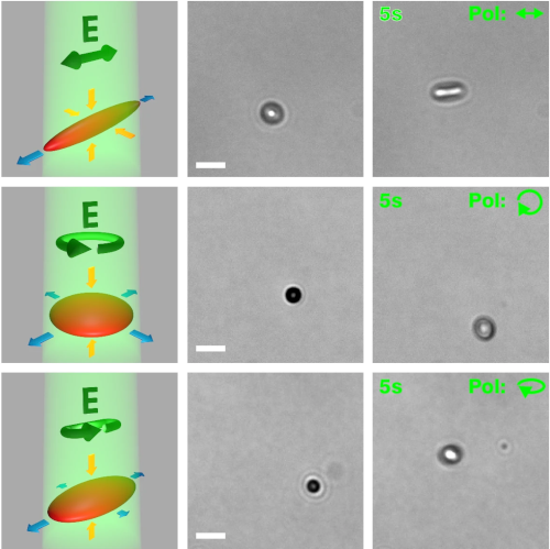
Colloidal particles at liquid interfaces experience long-ranged capillary interactions, whose magnitude and directionality depend on the particle shapes. When particle shapes are determined by fabrication or synthesis, the resulting shape-mediated interactions are predefined and often lead to the formation of persistent interfacial structures. Here, we introduce polymer particles at water-air interfaces whose shape and, therefore, interactions can be altered by illumination with polarized light. Specifically, we selectively trigger capillary self-assembly by anisotropically deforming the particles at the interface. Intriguingly, further deformation of already assembled particles induces sustained interfacial flows with velocities of up to 90 μm/s. Benefitting from polarization-defined deformation directions, we create flow-patterns that do not simply follow the illumination intensity pattern, such as shear flows along a single rectangular illumination stripe. We anticipate that this interplay between photo-deformation and capillary interactions of particles will enable various forms of mixing, manipulation, and assembly of soft matter at liquid interfaces.
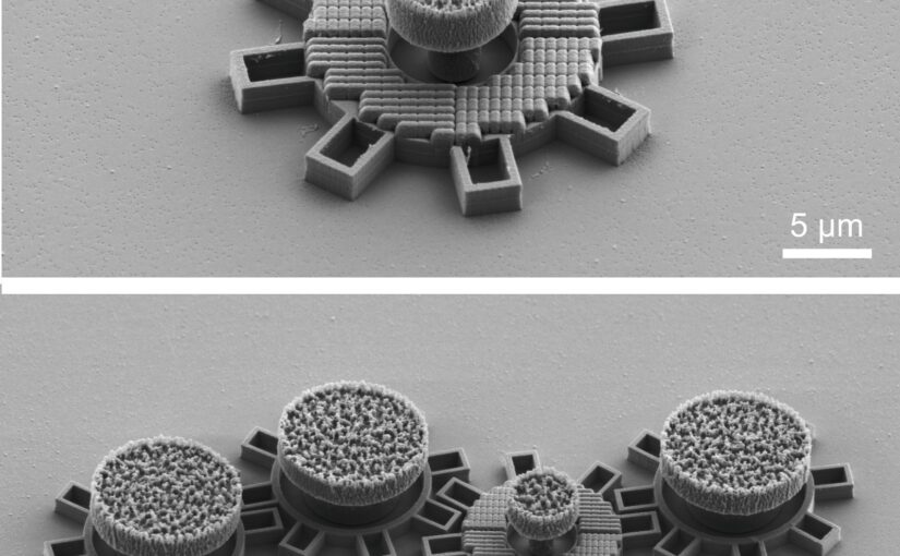
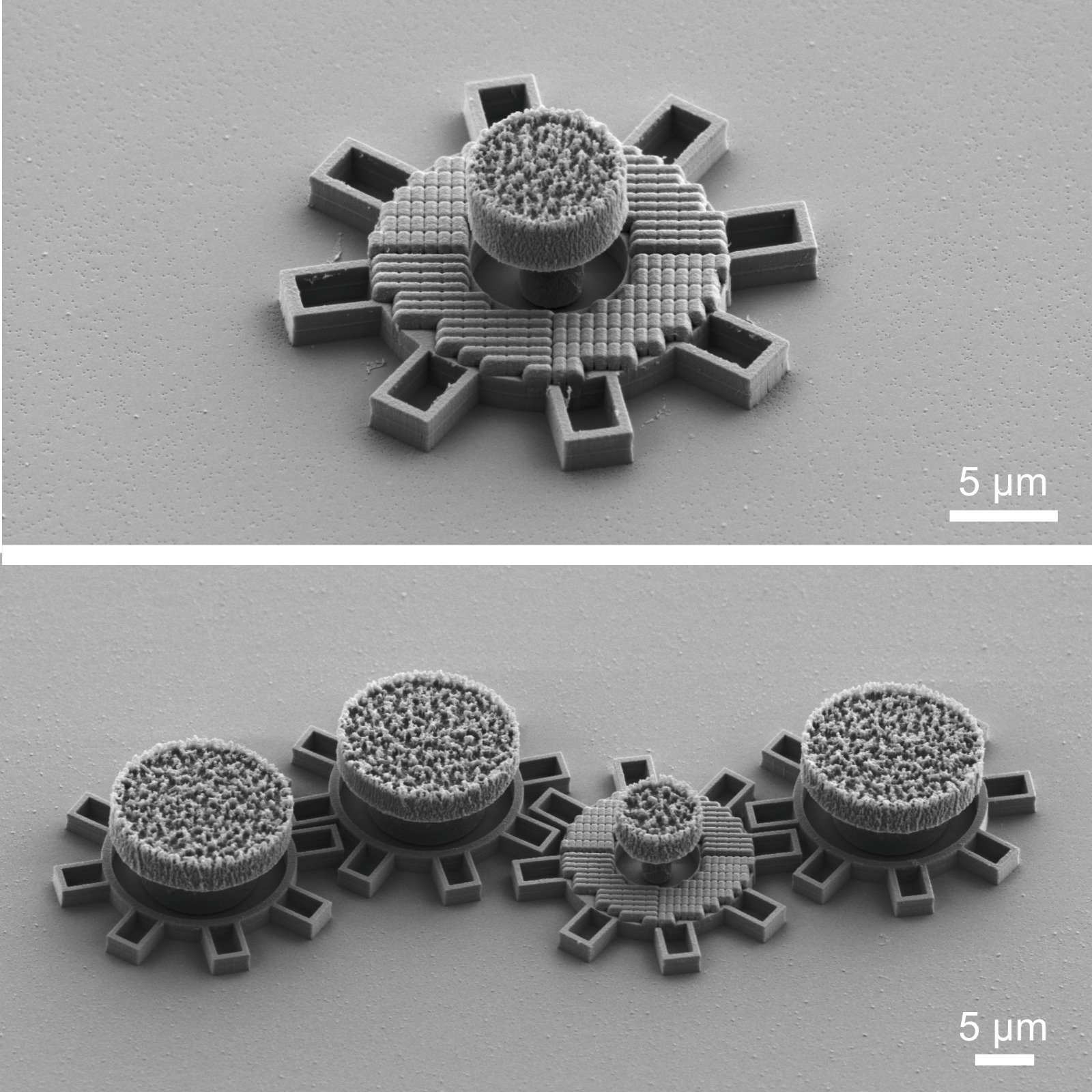
Top: single gear; Bottom: the second gear from the right has an optical metamaterial that react to laserlight and makes the gear move. All gears are made in silica directly on a chip. Each gear is about 0.016 mm in diameter. (Image by G. Wang)
Microscopic Geared Metamachines
Gan Wang, Marcel Rey, Antonio Ciarlo, Mohanmmad Mahdi Shanei, Kunli Xiong, Giuseppe Pesce, Mikael Käll and Giovanni Volpe
Nature Communications 16, 7767 (2025)
doi: 10.1038/s41467-025-62869-6
arXiv: 2409.17284
The miniaturization of mechanical machines is critical for advancing nanotechnology and reducing device footprints. Traditional efforts to downsize gears and micromotors have faced limitations at around 0.1 mm for over thirty years due to the complexities of constructing drives and coupling systems at such scales. Here, we present an alternative approach utilizing optical metasurfaces to locally drive microscopic machines, which can then be fabricated using standard lithography techniques and seamlessly integrated on the chip, achieving sizes down to tens of micrometers with movements precise to the sub-micrometer scale. As a proof of principle, we demonstrate the construction of microscopic gear trains powered by a single driving gear with a metasurface activated by a plane light wave. Additionally, we develop a versatile pinion and rack micromachine capable of transducing rotational motion, performing periodic motion, and controlling microscopic mirrors for light deflection. Our on-chip fabrication process allows for straightforward parallelization and integration. Using light as a widely available and easily controllable energy source, these miniaturized metamachines offer precise control and movement, unlocking new possibilities for micro- and nanoscale systems.
After the article was published, it was reported by many media outlets, University of Gothenburg, New Scientist, Optics.org, Phys.org, ScienceDaily, Discover Magazine, among others.
Graphical representation of colloidal interaction measurements using the automated miniTweezer. (Image by A. Ciarlo.)miniTweezer: an autonomous high-throughput optical tweezers platform for force spectroscopy Antonio Ciarlo, Martin Selin, Giuseppe Pesce, Carlos Bustamante, and Giovanni Volpe Date: 5 August 2025 Time: 9:45 AM – 10:15 AM Place: Conv. Ctr. Room 4
Optical tweezers are essential for single-molecule studies, providing direct access to the forces underlying biological processes such as protein folding, DNA transcription, and replication. However, manual experiments are labor-intensive, costly, and slow, especially when large data sets are required. Here we present the miniTweezer, a fully autonomous force spectroscopy platform that integrates optical tweezers with real-time image analysis and adaptive control. Once configured, it operates independently to perform high-throughput trapping, molecular attachment, and force measurements. Its robust design allows for extended unattended operation, significantly increasing the efficiency of data acquisition. We demonstrate its capabilities through DNA pulling experiments and highlight its broader applicability to microparticle interactions, colloidal assembly, and soft matter mechanics. By automating force spectroscopy, the miniTweezer enables large-scale, high-precision studies in biophysics, materials science, and nanotechnology.
Experimental trajectory (blue) of a particle trapped in air when the laser rotates at 1 Hz. The orange line represents the experimental laser trajectory. (Image by A. Ciarlo.)Probing fluid dynamics inertial effects of particles using optical tweezers Antonio Ciarlo, Giuseppe Pesce, Bernhard Mehlig, Antonio Sasso, and Giovanni Volpe Date: 4 August 2025 Time: 11:45 AM – 12:00 PM Place: Conv. Ctr. Room 3
Many natural phenomena involve dense particles suspended in a moving fluid, such as water droplets in clouds or dust grains in circumstellar disks. Studying these systems at the single particle level is challenging and requires precise control of flow and particle motion. Optical tweezers provide a powerful method for studying inertial effects in such environments. Here, we trap micrometer-sized particles in air and induce controlled dynamics by moving the trapping laser. We show that inertia becomes significant when the trap motion frequency is less than the harmonic trapping frequency, while at much higher motion frequencies, inertia has no effect. These results demonstrate the potential of trapping particles in air for studying inertial phenomena in fluids.
Representation of DNA stretching experiment with the miniTweezer. (Image by A. Ciarlo)miniTweezers2.0: smart optical tweezers for health and life sciences
Antonio Ciarlo Italy-Sweden bilateral workshop on smart sensor technologies and applications Date: 1 October 2024 Time: 14:40-15:05 Place: Meeting Room Kronan, Studenthuset, Linköping University, Campus Valla
Optical tweezers have become indispensable tools in various scientific fields such as biology, physics, chemistry, and materials science. Their wide range of applications has attracted the interest of scientists with limited expertise in optics and physics. Therefore, it is crucial to have a system that is accessible to non-experts. In this study, we present miniTweezers2.0, a highly versatile and user-friendly instrument enhanced by artificial intelligence. We demonstrate the capabilities of the system through three autonomous case study experiments. The first is DNA stretching, a fundamental experiment in single-molecule force spectroscopy. The second experiment focuses on stretching red blood cells, providing insight into their membrane stiffness. The final experiment examines the electrostatic interactions between microparticles in different environments. Our results highlight the potential of automated, versatile optical tweezers to advance our understanding of nanoscale and microscale systems by enabling high-throughput, unbiased measurements. The miniTweezers2.0 system successfully demonstrates the integration of artificial intelligence and automation to make optical tweezers more accessible and versatile, especially for health and life sciences. The adaptability of miniTweezers2.0 underscores its potential as a powerful tool for future scientific exploration across multiple disciplines.