Label-free mass and size characterization of few-kDa biomolecules by hierarchical vision transformer augmented nanofluidic scattering microscopy published in Nature Communications

The principle of differential imaging in NSM, in which we subtract the light scattered (yellow arrows indicate the scattered-light direction) by an empty nanochannel from the light scattered by the same channel with a molecule inside. A sequence of differential images of a nanochannel containing a diffusing single molecule obtained in this way is combined into a kymograph, which then contains the full molecular trajectory. (Image from the article.)
Label-free mass and size characterization of few-kDa biomolecules by hierarchical vision transformer augmented nanofluidic scattering microscopy
Henrik K. Moberg, Bohdan Yeroshenko, Joachim Fritzsche, David Albinsson, Barbora Spackova, Daniel Midtvedt, Giovanni Volpe, Christoph Langhammer
Nature Communications 17, 2533 (2026)
DOI: 10.1038/s41467-026-70514-z

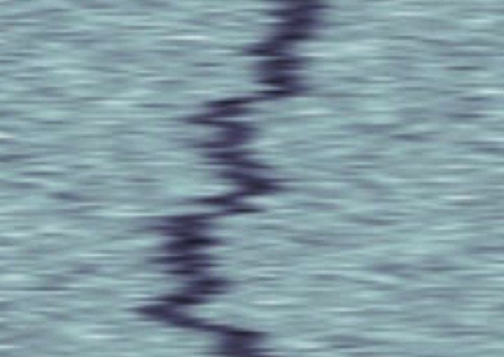
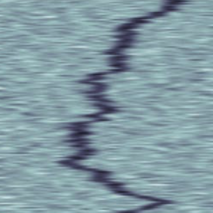
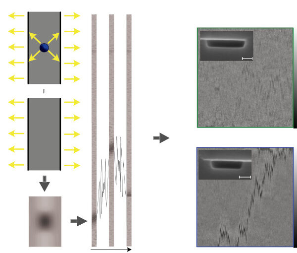
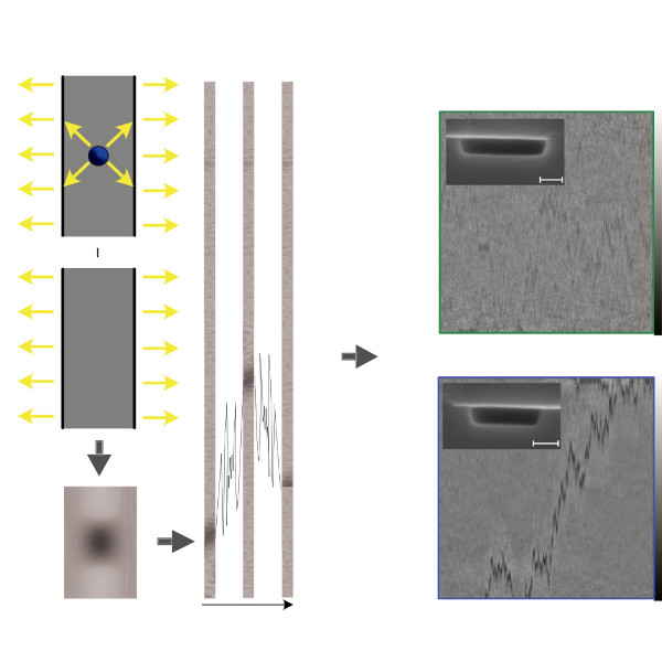
Nanofluidic scattering microscopy characterizes single molecules in subwavelength nanofluidic channels label-free, using the interference of visible light scattered by the molecule and nanochannel. It determines a molecule’s hydrodynamic radius by tracking its diffusion trajectory and its molecular weight by analyzing its scattering intensity along that trajectory. However, using standard analysis algorithms, it is limited to characterization of proteins larger than ≈ 60 kDa. Here, we push this limit by one order of magnitude to below ≈ 6 kDa molecular weight and ≈ 1.5 nm hydrodynamic radius — as we exemplify on the peptide hormone insulin — by using ultrasmall nanofluidic channels and by analyzing the data with a hierarchical vision transformer. When we benchmark this approach against the theoretical limit set by the Cramér–Rao Lower Bound, we find that it can be approached with sufficiently long molecular trajectories. This enables quantitative label-free single-molecule microscopy for biologically relevant families of sub-10-kDa molecules, such as cytokines, chemokines and peptide hormones.

Roadmap on Deep Learning for Microscopy published in Journal of Physics: Photonics

Spatio-temporal spectrum diagram of microscopy techniques and their applications. (Image by the Authors of the manuscript.)
Roadmap on Deep Learning for Microscopy
Giovanni Volpe, Carolina Wählby, Lei Tian, Michael Hecht, Artur Yakimovich, Kristina Monakhova, Laura Waller, Ivo F. Sbalzarini, Christopher A. Metzler, Mingyang Xie, Kevin Zhang, Isaac C.D. Lenton, Halina Rubinsztein-Dunlop, Daniel Brunner, Bijie Bai, Aydogan Ozcan, Daniel Midtvedt, Hao Wang, Nataša Sladoje, Joakim Lindblad, Jason T. Smith, Marien Ochoa, Margarida Barroso, Xavier Intes, Tong Qiu, Li-Yu Yu, Sixian You, Yongtao Liu, Maxim A. Ziatdinov, Sergei V. Kalinin, Arlo Sheridan, Uri Manor, Elias Nehme, Ofri Goldenberg, Yoav Shechtman, Henrik K. Moberg, Christoph Langhammer, Barbora Špačková, Saga Helgadottir, Benjamin Midtvedt, Aykut Argun, Tobias Thalheim, Frank Cichos, Stefano Bo, Lars Hubatsch, Jesus Pineda, Carlo Manzo, Harshith Bachimanchi, Erik Selander, Antoni Homs-Corbera, Martin Fränzl, Kevin de Haan, Yair Rivenson, Zofia Korczak, Caroline Beck Adiels, Mite Mijalkov, Dániel Veréb, Yu-Wei Chang, Joana B. Pereira, Damian Matuszewski, Gustaf Kylberg, Ida-Maria Sintorn, Juan C. Caicedo, Beth A Cimini, Muyinatu A. Lediju Bell, Bruno M. Saraiva, Guillaume Jacquemet, Ricardo Henriques, Wei Ouyang, Trang Le, Estibaliz Gómez-de-Mariscal, Daniel Sage, Arrate Muñoz-Barrutia, Ebba Josefson Lindqvist, Johanna Bergman
Journal of Physics: Photonics 8, 012501 (2026)
arXiv: 2303.03793
doi: 10.1088/2515-7647/ae0fd1

Through digital imaging, microscopy has evolved from primarily being a means for visual observation of life at the micro- and nano-scale, to a quantitative tool with ever-increasing resolution and throughput. Artificial intelligence, deep neural networks, and machine learning (ML) are all niche terms describing computational methods that have gained a pivotal role in microscopy-based research over the past decade. This Roadmap encompasses key aspects of how ML is applied to microscopy image data, with the aim of gaining scientific knowledge by improved image quality, automated detection, segmentation, classification and tracking of objects, and efficient merging of information from multiple imaging modalities. We aim to give the reader an overview of the key developments and an understanding of possibilities and limitations of ML for microscopy. It will be of interest to a wide cross-disciplinary audience in the physical sciences and life sciences.

Label-free nanofluidic scattering microscopy of size and mass of single diffusing molecules and nanoparticles published in Nature Methods

Kymographs of DNA inside Channel II. (Image by the Authors.)
Label-free nanofluidic scattering microscopy of size and mass of single diffusing molecules and nanoparticles
Barbora Špačková, Henrik Klein Moberg, Joachim Fritzsche, Johan Tenghamn, Gustaf Sjösten, Hana Šípová-Jungová, David Albinsson, Quentin Lubart, Daniel van Leeuwen, Fredrik Westerlund, Daniel Midtvedt, Elin K. Esbjörner, Mikael Käll, Giovanni Volpe & Christoph Langhammer
Nature Methods 19, 751–758 (2022)
doi: 10.1038/s41592-022-01491-6

Label-free characterization of single biomolecules aims to complement fluorescence microscopy in situations where labeling compromises data interpretation, is technically challenging or even impossible. However, existing methods require the investigated species to bind to a surface to be visible, thereby leaving a large fraction of analytes undetected. Here, we present nanofluidic scattering microscopy (NSM), which overcomes these limitations by enabling label-free, real-time imaging of single biomolecules diffusing inside a nanofluidic channel. NSM facilitates accurate determination of molecular weight from the measured optical contrast and of the hydrodynamic radius from the measured diffusivity, from which information about the conformational state can be inferred. Furthermore, we demonstrate its applicability to the analysis of a complex biofluid, using conditioned cell culture medium containing extracellular vesicles as an example. We foresee the application of NSM to monitor conformational changes, aggregation and interactions of single biomolecules, and to analyze single-cell secretomes.

Barbora Spackova joins the Soft Matter Lab

Barbora Spackova joins the Soft Matter Lab at the Physics Department of the University of Gothenburg on 1st September 2021.

Barbora has a PhD in physical engineering from the Czech Technical University in Prague (Czech Republic). Formerly, she has been a researcher at Chalmers University of Technology in the group of Prof. Christoph Langhammer. Her research is focused on single-molecule detection in nanofluidic systems.

While part of the Soft Matter Lab, she will continue her research on characterising cell media containing exosomes using Nanofluidic Scattering Microscopy (NSM).