Hang Zhao, supervised by Giovanni Volpe and Joana Pereira, will present his halftime seminar under the topic “Brain connectome revealed neuro-degenerative disease” on 9-10 am, 22nd Jan. 2026 in Nexus and through Zoom (https://gu-se.zoom.us/j/7726618257). The seminar starts from his presentation about the past and planned project, followed by a discussion and questions by his opponent, Professor Mattias Göksor.
News
Presentation by S. K. Manikandan at The Arctic Meeting for Adaptive Mechanisms in Biological Systems, Abisko, Sweden, January 21, 2026
Sreekanth Manikandan
Date: 21 Jan 2026
Time: 10:00 CEST
Place: STF Abisko, Sweden
The Arctic Meeting for Adaptive Mechanisms in Biological Systems
Quantifying the spatiotemporal forces, affinities, and dissipative costs of cellular-scale non-equilibrium processes from experimental data and localizing it in space and time remain a significant open challenge. Here, I explore how principles from stochastic thermodynamics, combined with machine learning techniques, offer a promising approach to addressing this issue. I will present preliminary results from experiments on fluctuating cell membranes and simulations of non-equilibrium systems in stationary and time-dependently driven states. These studies reveal potential strategies for localizing entropy production in experimental biophysical contexts while also highlighting key challenges and limitations that must be addressed.
Eduard Andrei Duta Costache joins the Soft Matter Lab
Eduard Andrei Duta Costache started his PhD at the Physics Department of the University of Gothenburg on the 19th of January 2026.
Eduard has a double Master’s degree in Artificial Intelligence from the University of Alicante (Spain) and in Machine Learning & Data Mining from Jean Monnet University (France).
During the course of his PhD, as part of the GREENS MSCA Doctoral Network, he will focus on developing AI frameworks to model and optimize the lifecycle of micro-robotic platforms.
Steven Smith visits the Soft Matter Lab
Steven brings years of expertise from the laboratory of Professor Carlos Bustamante, where they pioneered the use and development of optical tweezers. As the main developer of the widely used ‘minitweezers’ instrument, which today is used by dozens of research groups, he has helped shape the field of single-molecule biophysics on a global scale. We look forward to his visit, during which he will work on refining cutting-edge single-molecule measurement techniques.
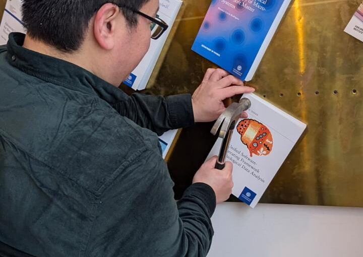
Yu-Wei Chang nailed his PhD thesis on January 7th, 2026. Congrats!
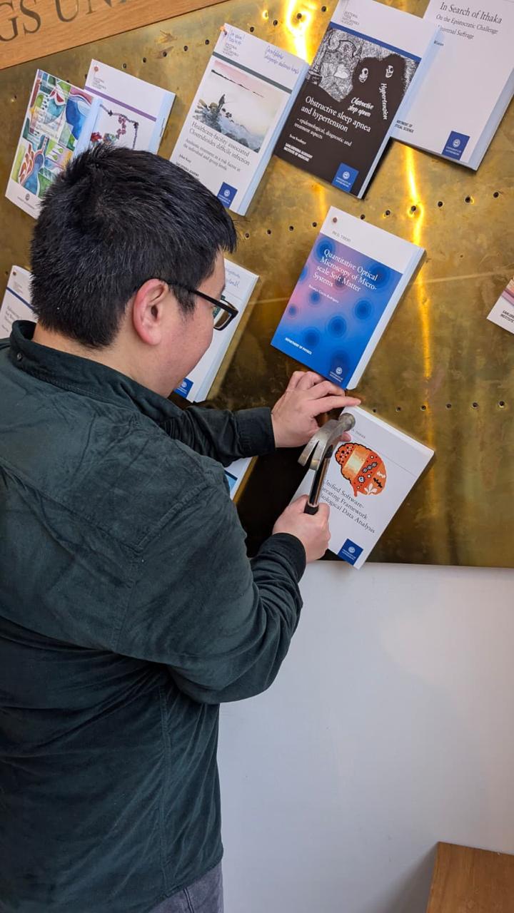
The nailing took place in Universitetsbyggnaden i Vasaparken, Universitetsplatsen 1, Göteborg, at 13:30.
In Swedish academia, “nailing” (spikning) is the formal public announcement and publication of a doctoral thesis. It happens weeks before the defence so that the public has time to read the thesis in advance and prepare questions for the defence. In addition to the physical nailing, the thesis is also published electronically (e-spikning) via GUPEA.
Yu-Wei will defend his thesis on 23 January at 13:00 in SB-H7 lecture hall, SB-Building, Institutionen för fysik, Johanneberg Campus, Göteborg.
Thesis (GUPEA handle): http://hdl.handle.net/2077/90289
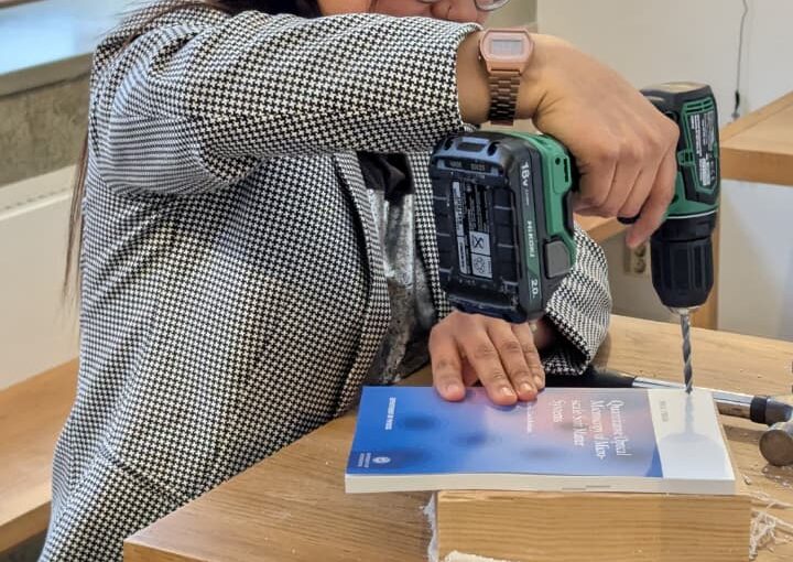
Thesis Nailing by B. García Rodríguez, 7 January 2026. Congrats!
Family and friends were present to mark this milestone.
Berenice will defend her thesis on 28 January at 09:00 in PJ-salen, Institutionen för fysik, Origovägen 6, Göteborg.
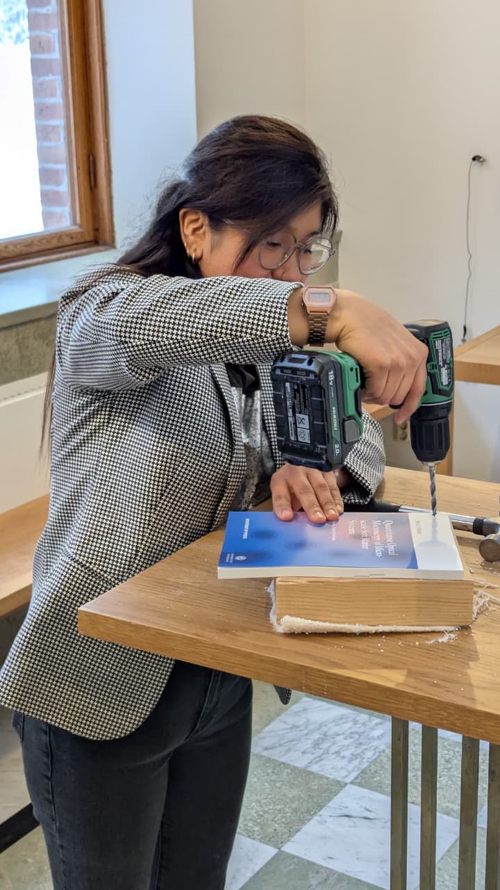
Thesis Nailing by F. Skärberg, 7 January 2026. Congrats!
Fredrik will defend his thesis on 29 January at 09:00 in FB-salen, Institutionen för fysik, Origovägen 6, Göteborg.
Inchworm-Inspired Soft Robot with Groove-Guided Locomotion on ArXiv

flexible PET sheet. (Image by H. P. Thanabalan.)
Hari Prakash Thanabalan, Lars Bengtsson, Ugo Lafont, Giovanni Volpe
arXiv: 2512.07813
Soft robots require directional control to navigate complex terrains. However, achieving such control often requires multiple actuators, which increases mechanical complexity, complicates control systems, and raises energy consumption. Here, we introduce an inchworm-inspired soft robot whose locomotion direction is controlled passively by patterned substrates. The robot employs a single rolled dielectric elastomer actuator, while groove patterns on a 3D-printed substrate guide its alignment and trajectory. Through systematic experiments, we demonstrate that varying groove angles enables precise control of locomotion direction without the need for complex actuation strategies. This groove-guided approach reduces energy consumption, simplifies robot design, and expands the applicability of bio-inspired soft robots in fields such as search and rescue, pipe inspection, and planetary exploration.
MSCA-DN GREENS training event in Göteborg, 10-14 November 2025
![]()
Both Andrea Schiano di Colella and Eduard Andrei Duta Costache, the two doctoral candidates based at the University of Gothenburg, participated to the event along with the other doctoral candidates of the network.
The training event consisted of a series of lectures on different topics such as small scale robots and their actuation mechanisms, theoretical aspects of the simulation of the dynamics of bodies immersed in fluids at low Reynolds numbers, working principles and applications of swarm robotics, and finally artificial intelligence and its applications to data analysis in experiments.
The event included a visit to the Swedish Algae Factory (SAFAB) and Smogenlax on the topic of circular economy, and concluded with a series of meetings between the representatives of the participating institutions, from both accademia and industry, to exchange research questions and plan secondments.
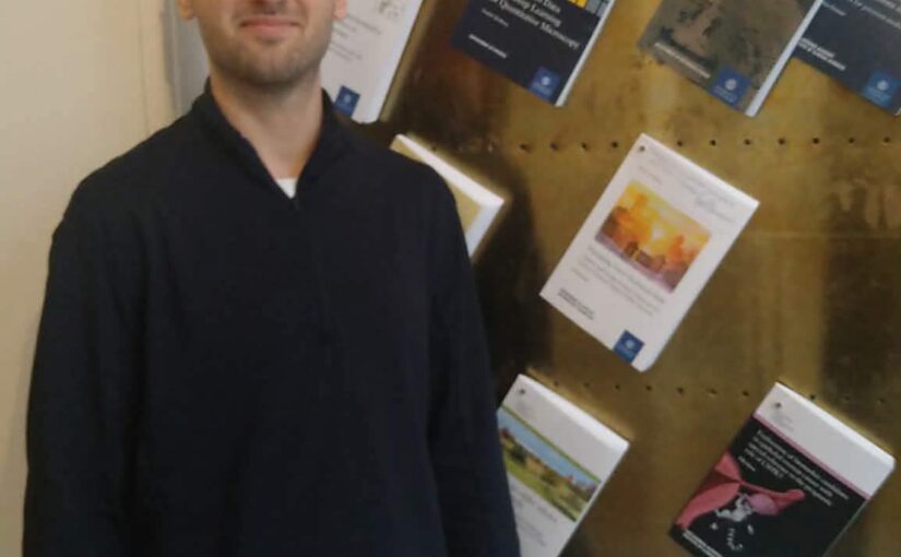
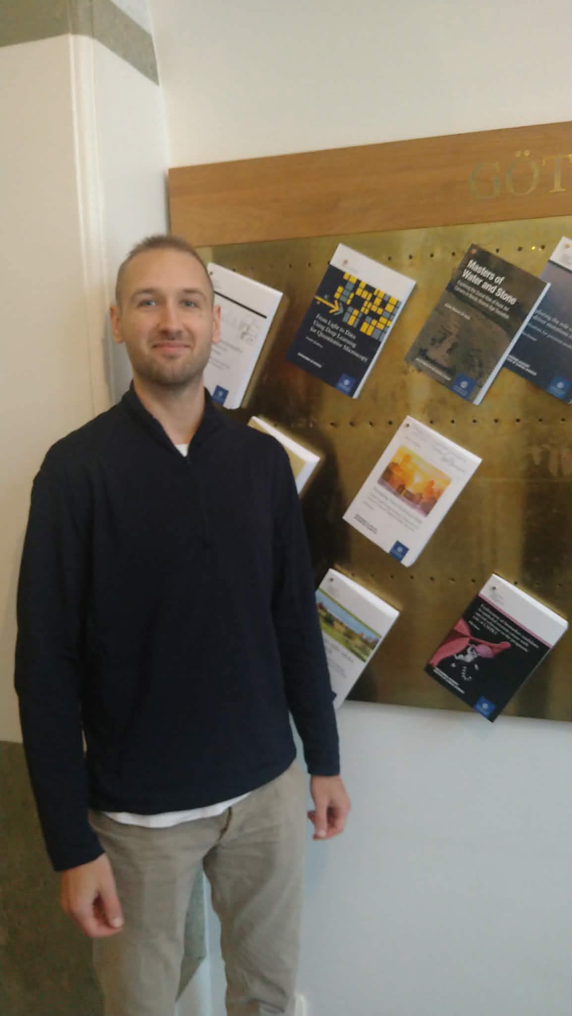
Jesus Pineda defended his PhD thesis on November 11th, 2025. Congrats!
Jesus Pineda defended his PhD thesis on November 11th, 2025. Congrats!
The defense took place in SB-H7 lecture hall, Institutionen för fysik, Johanneberg Campus, Göteborg, at 9:00.
Title: Inductive Biases for Efficient Deep Learning in Microscopy
Abstract: Deep learning has become an indispensable tool for the analysis of microscopy data, yet its integration into routine research remains uneven. Several factors contribute to this gap, including the limited availability of well-annotated datasets and the high computational demands of modern architectures. Microscopy introduces further challenges, as it spans diverse modalities and scales, from proteins to tissues, producing heterogeneous data that defy standardization. Generating reliable annotations also requires expertise and time, while unequal access to high-performance computing further widens the divide between well-resourced institutions and smaller laboratories.
This dissertation argues that the prevailing paradigm of scaling models with ever-larger datasets and computational resources yields diminishing returns for microscopy. Instead, it explores the role of inductive biases as a foundation for building models that are more data-efficient, computationally accessible, and scientifically meaningful. Inductive biases are structural assumptions embedded in model design that guide learning toward patterns aligned with the underlying problem. The first part of this work examines their central role in the advancement of modern deep learning and the diverse ways they shape model behavior.
This potential is demonstrated through three case studies. First, MAGIK employs graph neural networks to analyze biological dynamics in time-lapse microscopy, uncovering local and global properties with high precision, even when trained on limited data. Next, MIRO leverages recurrent graph neural networks to process single-molecule localization datasets, improving the efficiency and reliability of clustering for variable biological structures and scales while retaining strong generalization with minimal supervision. Finally, GAUDI introduces a representation learning framework for characterizing biological systems, providing a physically meaningful representation space for interpretable and transferable analysis.
The findings presented here demonstrate that the integration of inductive biases provides a cohesive strategy to extend the reach of deep learning in the life sciences, enhancing accessibility and ensuring scientific utility under resource constraints.
Thesis: https://gupea.ub.gu.se/items/672c7946-51d6-4773-ad8c-35a3eed41499
Supervisor: Giovanni Volpe
Co-Supervisor: Carlo Manzo
Examiner: Raimund Feifel
Opponent: Anna Kreshuk
Committee: Juliette Griffié, Daniel sage, Daniel Persson
Alternate board member: Jonas Enger