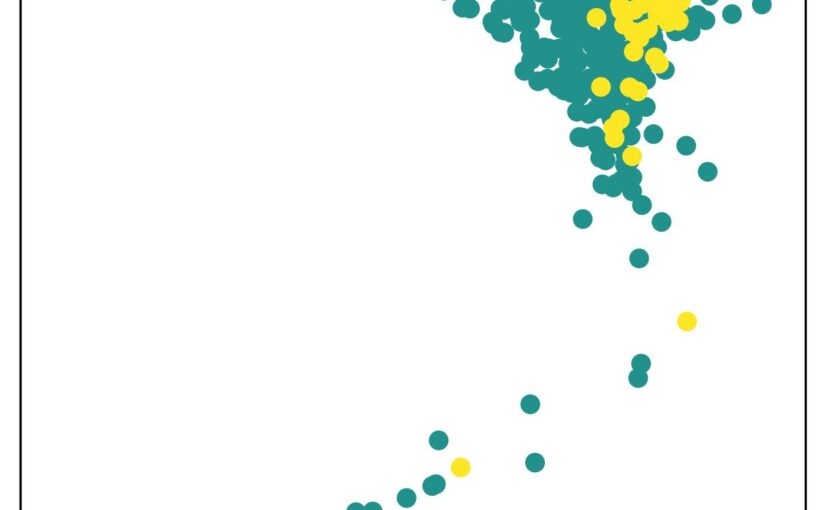
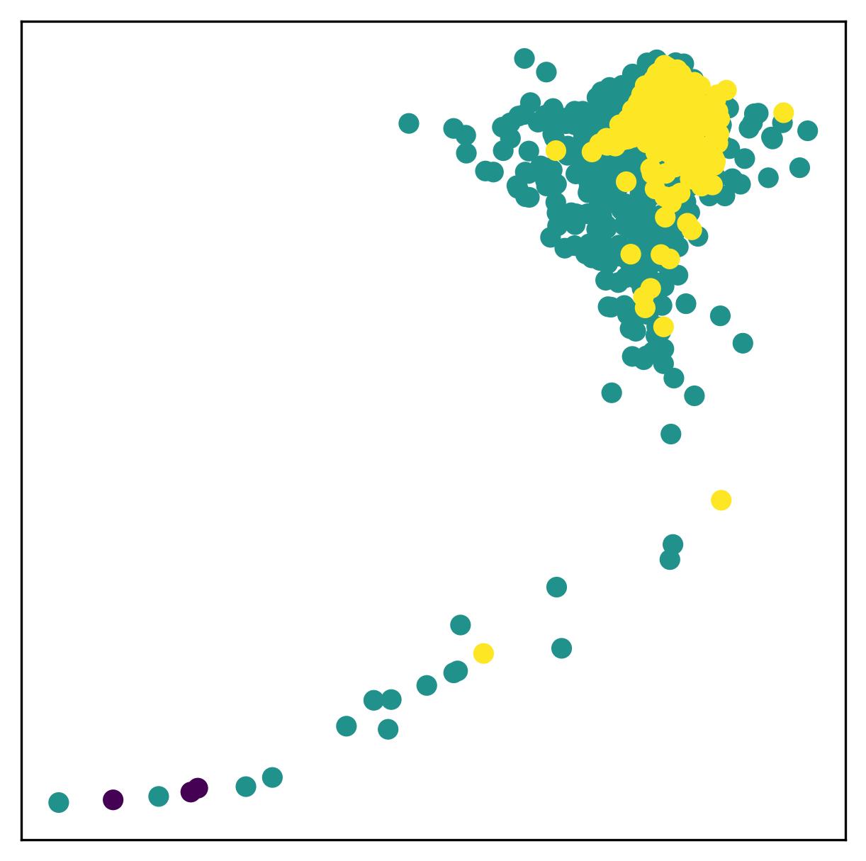
The embedding space generated byvariational autoencoder (VAE), with samples colored by data quality. (Image by J. Huang)Machine Learning-based Data Quality Control for AFM Force Spectroscopy
Jiacheng Huang, Nazli Demirpehlivan, Prakhar Dutta, Rahul Nagshi, Thomas Catley, Sylvia Whittle, Carlo Manzo, Rachel Owen, Giovanni Volpe Date: 11th March 2026 Time: 18:00 – 20:00 Place: Aula Medica, Karolinska Institute, Solna
Conference Protein Folding in Real Time, 11-13 March 2026, Stockholm, Sweden
Atomic Force Microscopy (AFM) force spectroscopy is widely used to probe the mechanical properties and interactions of biological samples at the nanoscale, including living cells. However, large datasets generated during AFM measurements often contain curves affected by experimental artifacts such as poor tip–sample contact, noise, or instrumental instability. These low-quality force curves can significantly affect downstream analysis and typically require time-consuming manual inspection. In this work, we propose a machine learning–based data quality control framework for AFM force spectroscopy using a self-supervised approach.
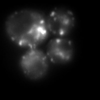
Heat-induced aggregates in Saccharomyces cerevisiae on Slimfield SMLM. Hsp104-mGFP binds to misfolded regions enabling aggregate visualisation for tracking. (Image by L. Viaene.)A single-molecule approach to study the spatial protein quality control system
Linde Viaene Date: 11 March 2026 Time: 18.00-20.00 Place: Aula Medica, Stockholm Sweden
Conference Protein Folding in Real Time, 11-13 March 2026, Stockholm, Sweden
Cell populations are inherently diverse, and averaging measurements across them can mask subtle or rare cellular behaviours. In this work, we use live Slimfield single-molecule microscopy to study the role of Hsp104 in clearing misfolded and aggregated proteins after stress. By analysing endogenously tagged Hsp104, we quantify molecular diffusion and stoichiometry before and after heat stress. Our results show a transition from faster, more mobile molecules to larger, more static assemblies following stress, consistent with Hsp104 functionally engaging with protein aggregates. These measurements provide molecular-level insight into how cells respond to proteotoxic stress.
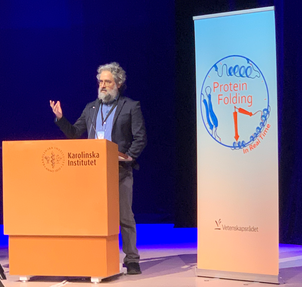
Giovanni Volpe opens the Protein Folding in Real Time conference. (Photo by A. Ciarlo)The international conference Protein Folding in Real Time: From Molecules to Disease opened today, 11 March 2026, at Aula Medica, KI, Stockholm.
The conference brings together researchers from multiple disciplines, including biophysics, molecular biology, computational science, and medicine, to discuss recent advances in the study of protein folding. Proteins must fold into precise three-dimensional structures to perform their biological functions, and failures in this process are associated with several diseases, including neurodegenerative disorders and cancer.
During the three-day meeting, participants attend a series of lectures and discussions covering topics such as single-molecule biophysics, high-resolution experimental techniques for observing folding dynamics, advanced molecular simulations, and artificial intelligence approaches for predicting folding pathways. Particular attention is given to the challenge of observing protein folding in real time, capturing transient intermediate states that determine whether proteins reach their functional structure or misfold.
The event also highlights the interdisciplinary and international nature of the initiative. Representatives from the Embassies of Italy, Japan, and Spain, together with UNESCO, take part in the meeting, emphasizing the global interest in advancing research on protein folding and its biomedical implications. The initiative aims to integrate experimental measurements, computational modeling, and data-driven approaches to build a predictive framework for protein folding dynamics. By combining advanced imaging, force spectroscopy, and machine learning methods, the initiative seeks to better understand how folding processes occur inside living systems and how their disruption can lead to disease.
Overall, the conference provides an opportunity for scientists from different institutions to exchange ideas, establish collaborations, and shape future research directions in the field of protein folding and misfolding. The launch of this initiative represents an important step toward bridging molecular-level observations with biomedical applications, ultimately contributing to improved strategies for diagnosing and treating diseases related to protein misfolding.
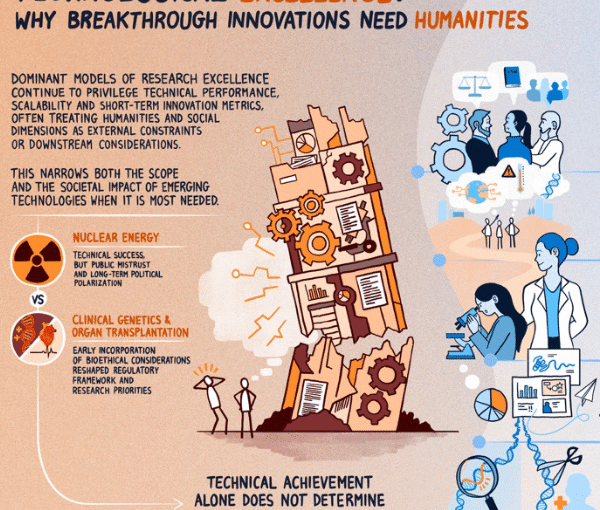
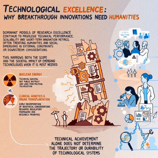
Why breakthrough research needs humanities and social sciences. (From an artwork by Jacopo Sacquegno.)Technological Excellence Requires Human and Social Context
Karl Palmås, Mats Benner, Monica Billger, Ben Clarke, Raimund Feifel, Julia Fernandez-Rodriguez, Anna Foka, Juliette Griffié, Claes Gustafsson, Kerstin Hamilton, Johan Holmén, Kristina Lindström, Tobias Olofsson, Joana B. Pereira, Marisa Ponti, Julia Ravanis, Sviatlana Shashkova, Emma Sparr, Pontus Strimling, Fredrik Höök, Giovanni Volpe
arXiv: 2603.10653
Breakthrough technologies increasingly shape social institutions, economic systems, and political futures. Yet models of research excellence associated with such technologies often prioritize technical performance, scalability, and short-term innovation metrics while treating ethical, social, and cultural dimensions as secondary considerations. This perspective article argues that such separation is no longer tenable. We propose a broader understanding of excellence that combines technical rigor with ethical robustness, social intelligibility, and long-term relevance. The rapid emergence of generative and agentic artificial intelligence further underscores this argument. As technological systems increasingly operate through language, interpretation, and normative alignment, expertise traditionally cultivated in the humanities and social sciences becomes integral to the design, governance, and responsible deployment of such systems. Drawing on historical examples and contemporary research practices, this article examines five interconnected domains where the humanities and social sciences, treated as integrated dimensions of research practice, can strengthen technological development: (1) ethical, legal, and social integration in agenda-setting and research design; (2) plural and reflexive foresight practices that shape technological futures; (3) graduate education as a leverage point for cross-disciplinary literacy; (4) visualization and communication as epistemic and civic practices; and (5) institutional frameworks that move beyond rigid distinctions between basic and applied research. Across these dimensions, we propose practical strategies for embedding interdisciplinary collaboration structurally rather than symbolically.
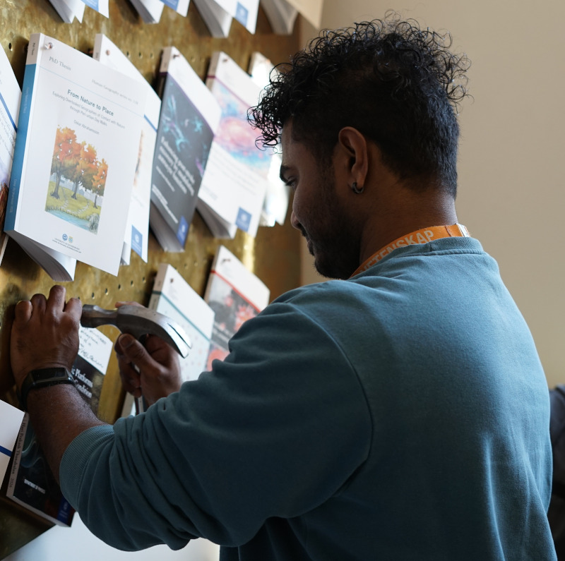
Hari nails his thesis. (Image by A. Ciarlo.)Hari Prakash Thanabalan nailed his PhD thesis, Soft Robotic Platforms for Dynamic Conditions: From Adaptive Locomotion to Space Exploration, on March 5th, 2026.
The nailing took place in Universitetsbyggnaden i Vasaparken, Universitetsplatsen 1, Göteborg, at 14:00.
In Swedish academia, “nailing” (spikning) is the formal public announcement and publication of a doctoral thesis. It happens weeks before the defence so that the public has time to read the thesis in advance and prepare questions for the defence. In addition to the physical nailing, the thesis is also published electronically (e-spikning) via GUPEA.
Hari Prakash will defend his thesis on the 23rd of March 2026 at 13:00 in PJ Salen lecture hall, Institutionen för fysik, Johanneberg Campus, Göteborg.
Per Hillertz. (Photo courtesy of P. Hillertz.)
We are pleased to announce that Per Hillertz (AstraZeneca) joined the Soft Matter Lab on 26 February 2026 as Adjunct Professor.
Per brings a strong scientific background in structural biology and biophysics, with broad experience across multiple therapy areas, including Oncology, CNS-pain, and respiratory diseases. In his current role as IT Site Lead in Gothenburg and Director of M&A IT at AstraZeneca, he focuses on the development and application of AI technologies within the pharmaceutical value chain.
He also plays an active role in shaping academia–industry collaboration through his involvement in several program boards at Chalmers University of Technology and the University of Gothenburg.
We warmly welcome Per to the lab and look forward to strengthening our collaboration at the interface of soft matter, life sciences, and digital innovation.
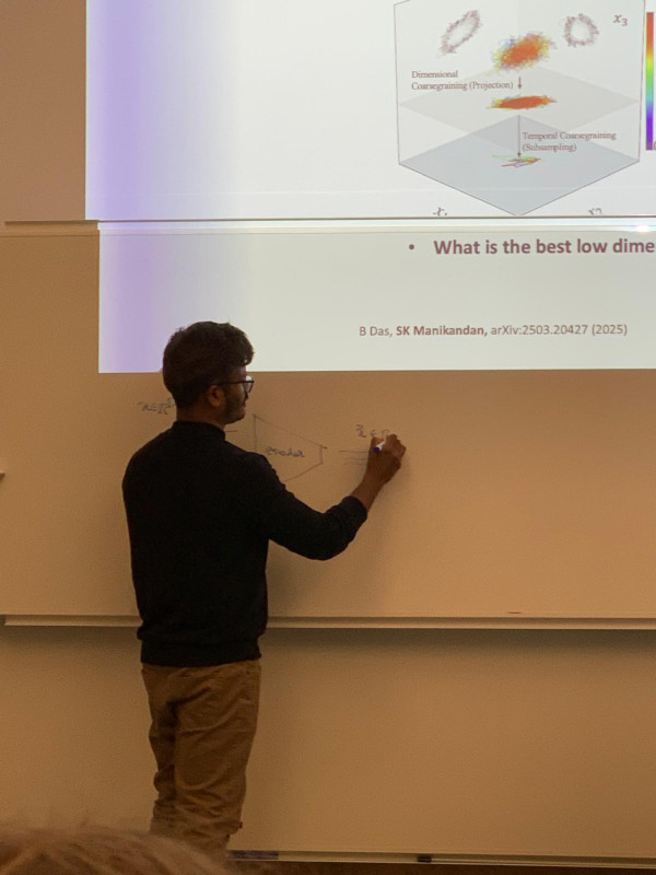
Identifying whether a process is in equilibrium, quantifying how far it lies from equilibrium, and determining optimal reduced descriptions of non-equilibrium processes remain challenging open problems. Here, we discuss how novel data-driven techniques grounded in stochastic thermodynamics can be used to efficiently learn these features directly from experimental data. In particular, we show how entropy production can be localized in space and time, and how maximally dissipative coordinates can be consistently inferred as effective low-dimensional descriptions of non-equilibrium processes. We further discuss applications to experimental biophysical systems and outline key challenges and limitations.
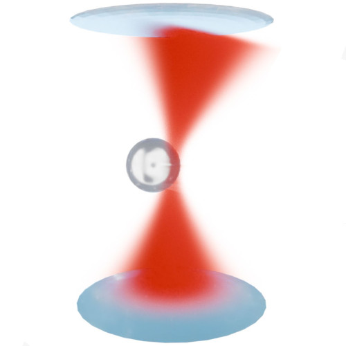
Schematic illustration of the light-momentum detection principle underlying SmartTrap. The momentum change of the trapping laser, induced by its interaction with the trapped particle, is measured to directly quantify optical forces with high precision, enabling real-time feedback and autonomous control in non-equilibrium experiments. (Figure by A. Ciarlo.)SmartTrap: Autonomous Optical Tweezers for Statistical Physics of Non-Equilibrium Systems
Antonio Ciarlo Date: 26th February 2026 Time: 13.30 Place: NORDITA, Stockholm, Sweden The 15th Nordic Workshop on Statistical Physics: Biological, Complex and Non-equilibrium Systems
Optical tweezers are a key tool in non-equilibrium statistical physics, allowing direct measurements of forces, work, and fluctuations in single-molecule and soft matter systems. However, manual operation limits throughput and the systematic study of rare events.
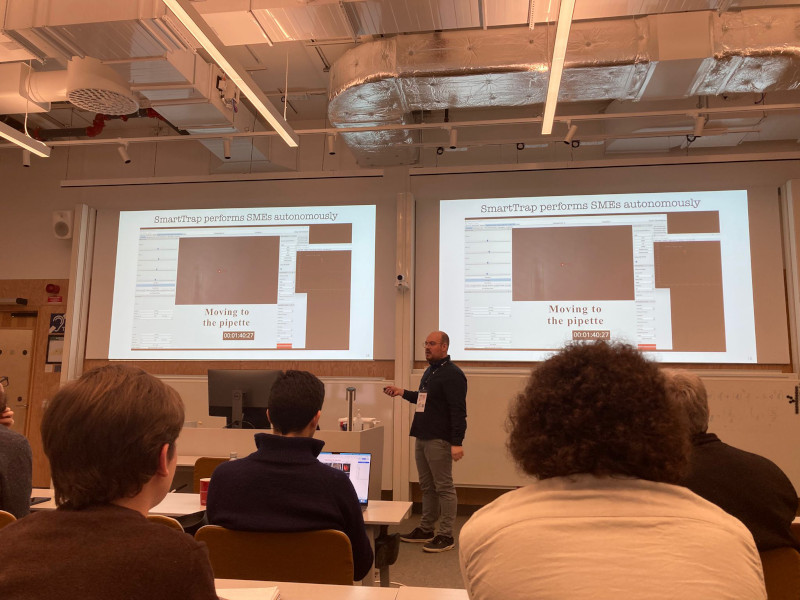
In this talk, Antonio Ciarlo will present SmartTrap, a fully autonomous optical tweezers platform integrating deep learning–based 3D tracking, adaptive feedback control, and automated microfluidics. The system operates without human intervention, executing complete force spectroscopy protocols.
Demonstrated with high-throughput DNA pulling experiments on λ-DNA, SmartTrap enables precise measurements of force–extension curves and folding kinetics. The platform also opens new possibilities for studies of colloids, single cells, and quantitative tests of non-equilibrium statistical physics.
Active Matter: Model Systems and Experimental Tests
Agnese Callegari, Antonio Ciarlo, Sreekanth Manikandan Dates and times:
23 Feb 14:00-15:00 (Agnese)
24 Feb 11:30-12:30 (Antonio)
24 Feb 14:00-15:00 (Sreekanth) Place: PJ Winter school on Geometry of nonequilibrium critical phenomena
Active matter is a broad class of systems that operate intrinsically out of equilibrium. It spans multiple length scales—from macroscopic to micro- and nanoscopic—and includes both biological and artificial realizations, often displaying rich and emerging collective behaviors. The study of active matter aims to explain and interpret these phenomena using concepts and tools from physics. As such, understanding active and non-equilibrium systems requires a combination of theoretical, computational, and experimental approaches.
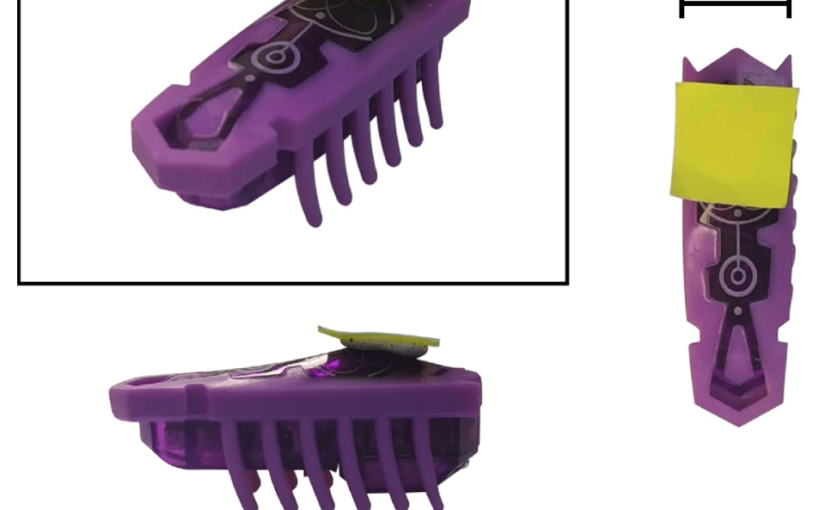
In the first part of the lecture, we introduce the concept of an active particle and demonstrate how it can be embodied in a macroscopic, self-propelled toy robot (a Hexbug). Despite their simplicity, such systems reproduce characteristic—and sometimes counterintuitive—features of microscopic active matter. These experiments have a strong pedagogical value and are designed to help bridge a gap in traditional physics curricula at the primary and secondary education levels.
The second part of the lecture focuses on active matter and non-equilibrium phenomena at the microscopic scale, where advanced experimental tools are essential. Optical tweezers provide precise control over microscopic systems and access to key physical observables. We introduce their operating principles and illustrate how they can be used to construct a minimal, well-controlled experimental model for studying non-equilibrium dynamics at the single-particle level.
In the final part of the lecture, we turn to the theoretical and computational tools required to analyze active matter systems. We discuss how non-equilibrium dynamics can be quantitatively characterized directly from experimental data in a model-independent framework. This naturally leads to an introduction to machine-learning–based inference techniques, which extract dynamical and thermodynamic information from data without relying on a priori assumptions about the underlying physical model.
References:
[1] A. Barona Balda, A. Argun, A. Callegari, G. Volpe. Playing with Active Matter, Am. J. Phys. 92, 847–858 (2024). https://doi.org/10.1119/5.0125111
[2] Martins, T.T., Malavazi, A.H.A., Kamizaki, L.P. et al. Fluctuation theorems with optical tweezers: theory and practice. Eur. Phys. J. Plus 141, 71 (2026). https://doi.org/10.1140/epjp/s13360-025-07181-4
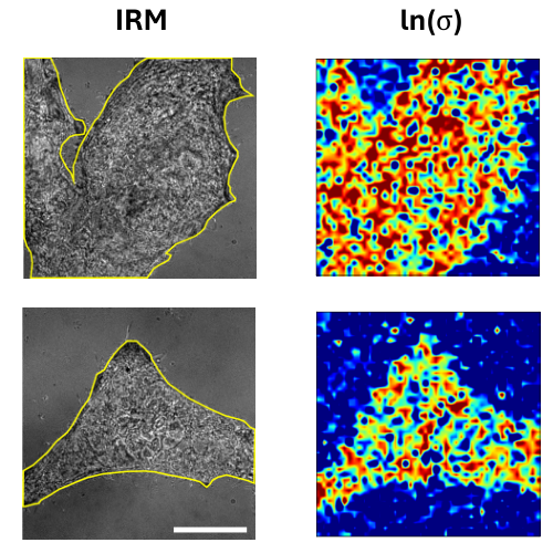
[3] Manikandan, Sreekanth K. and Ghosh, T. and Mandal, T. and Biswas, A. and Sinha, B. and Mitra, D. Estimate of entropy production rate can spatiotemporally resolve the active nature of cell flickering. Phys. Rev. Res. 6, 023310 (2024). https://doi.org/10.1103/PhysRevResearch.6.023310
Photos
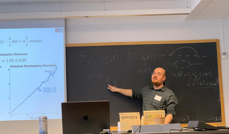
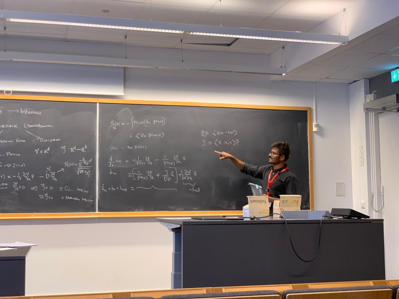
Antonio, presenting. (Photo by M. Orsino)Sreekanth, presenting. (Photo by A. Ciarlo)
Anton Widengård joined the Soft Matter Lab on 9 February 2026.
Anton is a master student in Biomedical Engineering at Chalmers University of Technology.
Alongside with his classmate Max Haraldsson, Anton will be working on his Master’s Thesis at Soft Matter Lab, in collaboration with IFLAI, supervised by Jesús Pineda.
Anton’s and Max’s research focuses on evaluating and efficiently adapting pre-trained deep-learning vision models for cell segmentation and tracking.