The Madrid Institute of Materials Sciences (CSIC-ICMM) hosted the second training workshop for the SPM 4.0 network. Both Prakhar Dutta and Jiacheng Huang, the two doctoral candidates based at the University of Gothenburg, participated to the event along with the other doctoral candidates of the network.
The second training workshop started with presentations from the doctoral candidates on their progress so far. The training event also consisted of a series of lectures on different topics such as a deep dive into atomic force microscopy and the different modes for the same, basics of deep learning, and an overview of data management plans.
ICMM also hosted some practical sessions where hands-on lectures were given on the use of atomic force microscopy machines and their applications.
News

Invited Talk by G. Volpe at DINAMO 2026, Franschhoek, South Africa, 7 April 2026

Giovanni Volpe
DINAMO 2026
Franschhoek, South Africa
7 April 2026
Latent space-driven quantification of biofilm formation using time-resolved droplet microfluidics published in Microchemical Journal

Daniela Pérez Guerrero, Jesús Manuel Antúnez Domínguez, Aurélie Vigne, Daniel Midtvedt, Wylie Ahmed, Lisa D. Muiznieks, Giovanni Volpe, Caroline Beck Adiels
Microchemical Journal 225, 117685 (2026)
arXiv: 2507.07632
DOI: 10.1016/j.microc.2026.117685
Bacterial biofilms play crucial roles across diverse contexts, from public health risks to beneficial applications in bioremediation, biodegradation, and wastewater treatment. However, tools that enable high-resolution, dynamic analysis of their responses to environmental cues and collective cellular behaviors remain limited. Here, we present a droplet-based microfluidic platform that combines continuous in situ microscopy with subsequent unsupervised deep learning for quantitative analysis of biofilm development. In our setup, Bacillus subtilis cells are encapsulated in monodisperse aqueous microdroplets containing Lysogeny Broth, suspended in an oil phase and immobilized within microfabricated traps, providing continuous optical access throughout biofilm formation at the water–oil interface. The platform supports both fluorescence and bright-field imaging, enabling high-throughput, time-resolved monitoring of thousands of droplets under controlled conditions. To extract quantitative information from these large datasets, we developed an automated analysis pipeline based on a Variational Autoencoder (VAE) trained directly on microscopy images from our experiments. This unsupervised model enables segmentation and latent-space representation of bacterial structures without manual annotation or synthetic training data. Post-segmentation size thresholding enables classification of bacterial aggregates and larger biofilm-like clusters, including quantification of biofilm porosity, thereby supporting detailed morphological and temporal analyses across droplets and conditions. By integrating droplet microfluidics with unsupervised deep learning, our platform provides a scalable, robust, and rapid approach for high-throughput quantitative studies of biofilm behavior. It resolves complex structural biofilm patterns, bypasses the need for manual annotation, and opens new opportunities to probe environmental determinants of biofilm formation. Departing from earlier methods, our framework fuses biological training data with unsupervised models to quantify microbial community dynamics across scales, offering a generalizable platform for future high-resolution microbiology.
Nazli Demirpehlivan visits the Soft Matter Lab
Nazli Demirpehlivan will visit the Soft Matter Lab from 30th of March to 30th of May 2026.
Nazli is a doctoral candidate at Bruker Nano GmbH in Berlin and also a part of the MSCA-DN SPM4.0 network.
She will be carrying out her secondment as part of the SPM 4.0 network with the University of Gothenburg.
The focus of her secondment will be the development of ASAP, a deep learning based pipeline for atomic force microscopy applications alongside the doctoral candidates Prakhar Dutta and Jiacheng Huang who are also part of the network. She will also take the Deep learning course offered by the department to facilitate her PhD studies and projects.
Mapping individual molecular connectomes in Alzheimer’s disease published in Alzheimer’s & Dementia
Zhilei Xu, Mite Mijalkov, Jiawei Sun, Yu-Wei Chang, Arianna Sala, Giovanni Volpe, Mario Severino, Mattia Veronese, Sara Garcia-Ptacek, Joana B. Pereira, for the Alzheimer’s Disease Neuroimaging Initiative
Alzheimer’s & Dementia 22, e71310 (2026)
DOI: 10.1002/alz.71310
INTRODUCTION
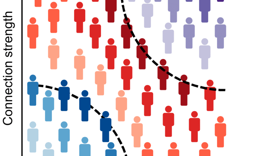
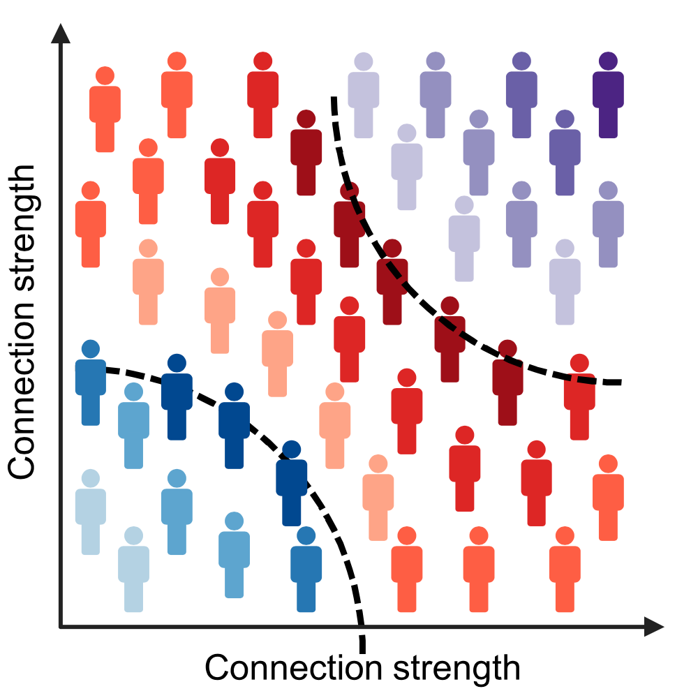
Mapping individual differences is crucial to improve personalized medicine approaches in Alzheimer’s disease (AD), which is characterized by strong inter-individual variability in the accumulation patterns of tau and amyloid beta pathology.
METHODS
We assess the progression of AD across the disease continuum by building individual molecular connectomes using longitudinal positron emission tomography (PET) data.
RESULTS
We demonstrate that these connectomes constitute a unique fingerprint, capable of identifying a single individual from a large group of subjects. Alterations in the connectomes discriminate different diagnostic groups and predict cognitive decline to a higher extent than conventional PET measures. We introduce a novel gene-specific transcription network analysis that linked individual tau and amyloid connectomes to a common transcriptomic profile of apoptosis, with the tau connectome being specifically related to pyrimidine metabolism, and the amyloid connectome to histone acetylation.
DISCUSSION
Individual molecular connectome mapping provides a novel and sensitive framework to monitor AD progression.
Highlights
- Individual molecular connectomes constitute a unique fingerprint, capable of identifying a single individual from a large group of subjects.
- Alterations in individual molecular connectomes significantly increase both across the Alzheimer’s disease (AD) continuum and over time.
- Alterations in individual molecular connectomes discriminate different diagnostic groups and predict cognitive decline to a higher extent than conventional positron emission tomography measures.
- Susceptibilities of individual tau and amyloid connectomes to AD are linked to a common transcriptomic profile of apoptosis, with the tau connectome being specifically related to pyrimidine metabolism, and the amyloid connectome to histone acetylation.
Presentation by S. K. Mondal, online, 22 April, 2026
Optical Fiber Micro/Nano Axicon Tip: An Optical Imaging Platform
Samir K. Mondal
CSIR-CSIO, Chandigarh, India
Date: 22 April 2026
Time: 12:30
Place: Online on zoom
Optical fiber tip under structural modifications enhances light-matter interaction by focusing, collecting or modulating light in microscopic scale and combined with waveguide property, it emerges as a potential optical tool, especially for spectroscopic, endoscopic and imaging application. A chemical etching technique has been introduced to permanently modify the tip as Micro/Nano axicon, capable in generating structured beams. The optics of the axicons have been studied in detail and further used in optical imaging experiments, namely phase microscopy, photonic nanojet and nanoscopy. The seminar will highlight first-hand information about the probe and experiments addressing the above-mentioned application.
Short Bio
Dr. Mondal is Chief Scientist at CSIR-CSIO, Chandigarh. He earned his Ph.D. in Electronic Science and M.Sc. in Physics from the University of Calcutta. After postdoctoral research at the University of California, Irvine and the University of Minnesota, he joined Tyndall Research Institute, Ireland.
With over 25 years in optics and photonics, his work spans optical interconnects, photonic crystals, lasers, and fiber instrumentation. He leads research in optical fiber antennas, near-field optics, imaging, and plasmonics, aiming for sustainable photonics platforms.
He collaborates internationally and is known for pioneering micro/nano axicons on fiber tips. He has over 50 publications and serves as an editor and reviewer.
Hari Prakash Thanabalan defended his PhD thesis on March 23rd, 2026. Congrats!

flexible PET sheet. (Image by H. P. Thanabalan.)
The defense took place in PJ Salen lecture hall, Institutionen för fysik, Johanneberg Campus, Göteborg, at 13:00.
Title: Soft Robotic Platforms for Dynamic Conditions: From Adaptive Locomotion to Space Exploration
Abstract:
Inspired by living organisms, soft robots represent a significant advancement in robotics, offering exceptional flexibility and nearly infinite degrees of freedom. These properties make them ideal for unstructured and remote environments such as planetary surfaces. However, challenges remain in developing efficient and durable soft actuators capable of withstanding complex operational conditions. This work presents two interconnected parts.
In the first part, an inchworm-inspired soft robot was developed that is capable of controlled directionality through a passive alignment mechanism integrated with a 3D-printed grooved substrate. This design enables precise locomotion control using only a single rolled dielectric elastomer actuator (RDEA), eliminating the need for multiple actuators or complex control systems. Experimental validation confirms that manipulating groove angles on the substrate reliably guides locomotion, improving energy efficiency and mechanical simplicity.
In the second part, the fabrication and resilience of fault-tolerant RDEAs were tested. RDEAs utilising Single-Walled Carbon Nanotubes (SWCNTs) as compliant electrodes were developed to withstand multiple damages where they were tested for punctures and cuts. Additionally, the radiation tolerance of these actuators was evaluated under space-like conditions, including Galactic Cosmic Rays and Solar Particle Events, which expose materials to high-energy protons and alpha particles. A computational dual-simulation framework was applied, combining the Stopping and Range of Ions in Matter (SRIM) software for alpha particle interactions and ESA’s SPENVIS Multi-Layered Shielding Simulation Software (MULASSIS) for proton radiation effects.
This framework concerns material selection for robust RDEA fabrication aimed at extraterrestrial applications. Together, these projects advance the development of bioinspired soft robots with improved directional control and environmental resilience, supporting future applications in search and rescue, pipe inspection, and planetary exploration.
Thesis: https://hdl.handle.net/2077/90552
Supervisor: Giovanni Volpe
Examiner: Bernhard Mehlig
Opponent: Maria Guix Noguera
Committee: Juliane Simmchen, Hamid Kellay, Paolo Vinai
Alternate board member: Måns Henningson
Tracking early cognitive decline in preclinical AD with brain MRI similarity published in Alzheimer’s & Dementia
Jiawei Sun, Blanca Zufiria-Gerbolés, Massimiliano Passaretti, Giovanni Volpe, Mite Mijalkov, Joana B. Pereira, for the Alzheimer’s Disease Neuroimaging Initiative
Alzheimer’s & Dementia 22, e71170 (2026)
DOI: 10.1002/alz.71170
INTRODUCTION
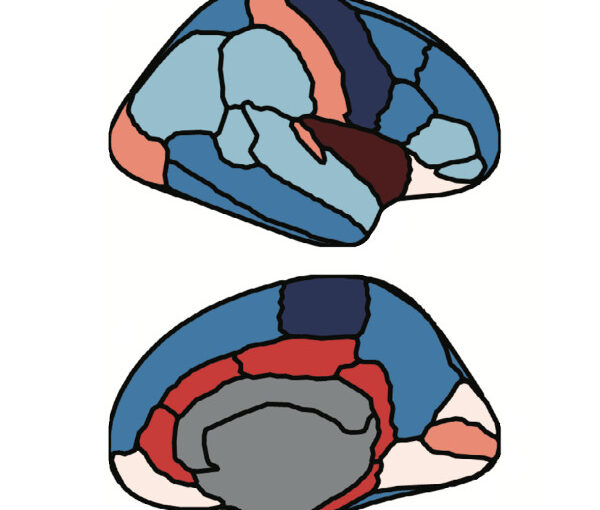
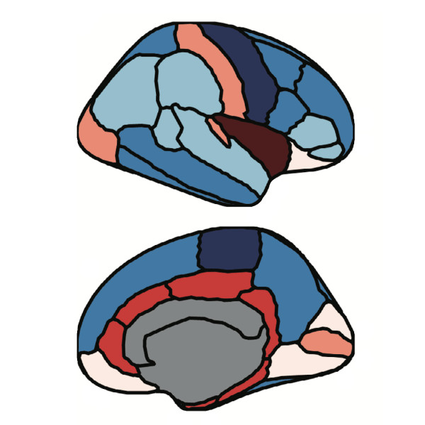
Early detection of neuroanatomical changes in preclinical Alzheimer’s disease (AD) is critical for timely intervention. However, conventional magnetic resonance imaging (MRI) and fluid biomarkers often lack sensitivity to subtle structural alterations in early disease stages.
METHODS
To identify early brain alterations, we applied a perturbation-based brain similarity approach to cognitively normal participants from Alzheimer’s Disease Neuroimaging Initiative (ADNI) and Open Access Series of Imaging Studies (OASIS), stratified by amyloid status. We evaluated its predictive performance for cognition and diagnostic conversion against cortical thickness, volumetric MRI, and fluid biomarkers.
RESULTS
In both cohorts, brain similarity consistently outperformed other biomarkers across cognitive domains and amyloid groups. It also achieved superior accuracy in predicting clinical conversion and exhibited associations with cytoarchitectural organization.
DISCUSSION
These findings highlight brain similarity as a sensitive marker of early neuroanatomical disruption in AD. Its ability to detect subtle structural changes before overt atrophy underscores its potential for early disease monitoring and treatment assessment in preclinical AD trials.
Highlights
- Brain similarity captures early brain changes in preclinical Alzheimer’s disease (AD).
- Brain similarity outperforms conventional biomarkers such as cortical thickness, volume measures, and fluid biomarkers in predicting cognitive decline.
- Brain similarity predicts conversion to mild cognitive impairment and AD more accurately than traditional imaging markers, and its predictive performance is further improved when combined with fluid biomarkers.
- Brain similarity captures structural disruptions associated with cortical layer II of the cytoarchitectonic lamina of human neocortex.
Label-free mass and size characterization of few-kDa biomolecules by hierarchical vision transformer augmented nanofluidic scattering microscopy published in Nature Communications
Henrik K. Moberg, Bohdan Yeroshenko, Joachim Fritzsche, David Albinsson, Barbora Spackova, Daniel Midtvedt, Giovanni Volpe, Christoph Langhammer
Nature Communications 17, 2533 (2026)
DOI: 10.1038/s41467-026-70514-z
Nanofluidic scattering microscopy characterizes single molecules in subwavelength nanofluidic channels label-free, using the interference of visible light scattered by the molecule and nanochannel. It determines a molecule’s hydrodynamic radius by tracking its diffusion trajectory and its molecular weight by analyzing its scattering intensity along that trajectory. However, using standard analysis algorithms, it is limited to characterization of proteins larger than ≈ 60 kDa. Here, we push this limit by one order of magnitude to below ≈ 6 kDa molecular weight and ≈ 1.5 nm hydrodynamic radius — as we exemplify on the peptide hormone insulin — by using ultrasmall nanofluidic channels and by analyzing the data with a hierarchical vision transformer. When we benchmark this approach against the theoretical limit set by the Cramér–Rao Lower Bound, we find that it can be approached with sufficiently long molecular trajectories. This enables quantitative label-free single-molecule microscopy for biologically relevant families of sub-10-kDa molecules, such as cytokines, chemokines and peptide hormones.
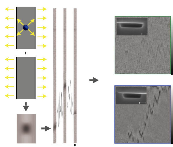
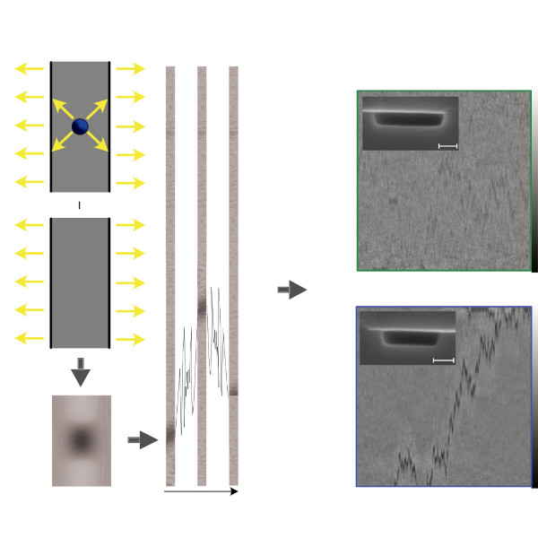
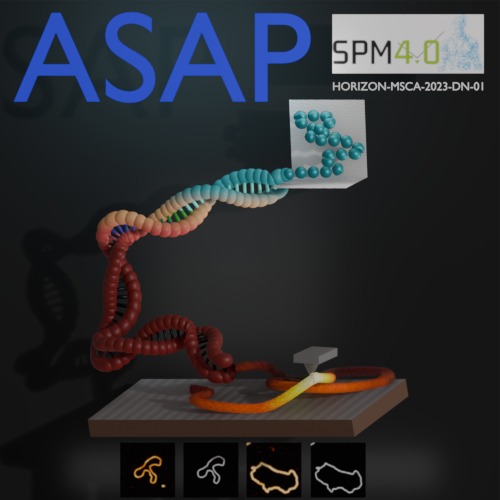
Poster by P. Dutta at the Protein Folding in Real Time Conference, Stockholm, 11th March 2026
Prakhar Dutta, Jiacheng Huang, Nazli Demirpehlivan, Thomas Catley, Sylvia Whittle, Carlo Manzo, Rahul Nagshi, Rachel Owen, Giovanni Volpe
Date: 11th March 2026
Time: 18:00 – 20:00
Place: Aula Medica, Karolinska Institute, Solna
Conference Protein Folding in Real Time, 11-13 March 2026, Stockholm, Sweden
Abstract: Atomic force microscopy (AFM) resolves biological structure and mechanics at high resolution, but produces vast, heterogeneous datasets that are often noisy and very time-consuming to analyse. Although deep learning could automate quality control, segmentation and feature extraction, adoption is limited by scarce ground-truth training data and high technical barriers for experimentalists. Here we present ASAP, an open-source tutorial and pipeline implemented in DeepTrack to provide a reproducible foundation for AI-enabled AFM. At the protein folding conference, a dual-pathway simulation for DNA, offering both molecular dynamics and rapid, non-MD geometries to generate perfect ground truth for segmentation training was presented. By consolidating simulation and learning into a single modular ecosystem, this work enables users to build upon our pipeline to optimize AFM workflows for more efficient data acquisition and robust processing.