Inchworm-inspired soft robot. (Image by H. P. Thanabalan.)Bio-inspired soft robot for multi-directionality
Hari Prakash Thanabalan, Lars Bengtsson, Ugo Lafont, Giovanni Volpe
SPIE Optics+Photonics, San Diego, CA, USA, 3-7 August 2025 Date: 5th August 2025 Time: 8:30 AM – 8:45 AM Place: Conv. Ctr. Room 4
Soft robotics are the forefront of robotics evolution that leverages compliant materials such as silicone elastomer to mimic biological organisms. With infinite degrees of freedom, soft robots surpass rigid robots in adaptability making them ideal for exploration and manipulation tasks. Here we focus on inchworm inspired soft robot achieving multidirectional locomotion through groove-guided movement. By manipulating the groove angles on a substrate, we demonstrate multidirectional locomotion by utilising only a single actuator.
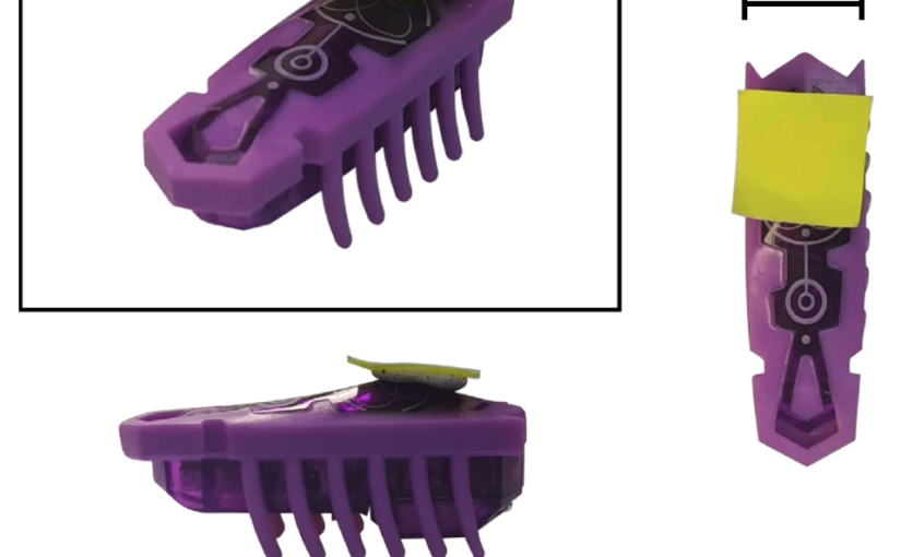
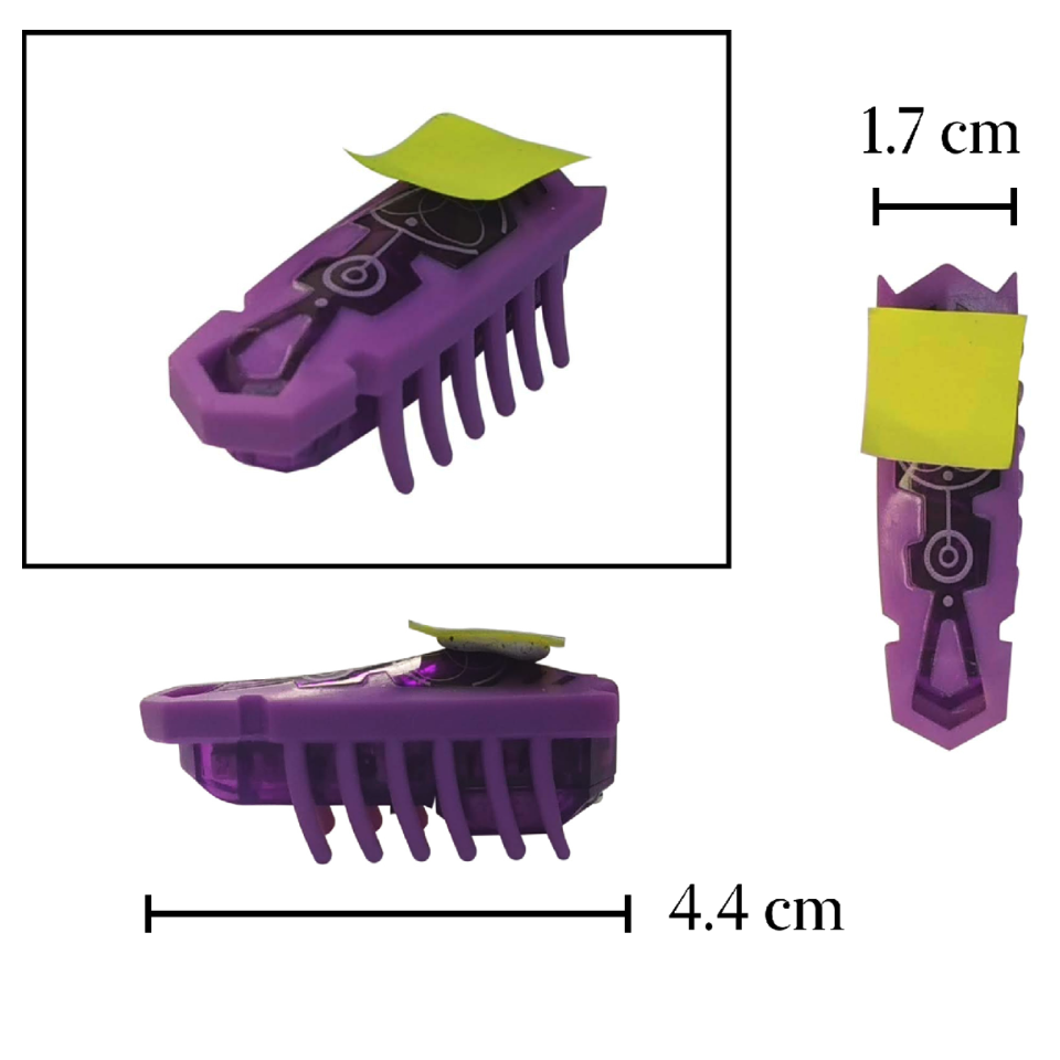
One exemplar of the HEXBUGS used in the experiment. (Image by the Authors of the manuscript.)Experimenting with macroscopic active matter
Angelo Barona Balda, Aykut Argun, Agnese Callegari, Giovanni Volpe
SPIE-OTOM, San Diego, CA, USA, 3 – 7 August 2025 Date: 4 August 2025 Time: 5:30 PM – 7:30 PM PDT Place: Conv. Ctr. Exhibit Hall A
Presenter: Giovanni Volpe
Contribution submitted by Agnese Callegari
Active matter is based on concepts of nonequilibrium thermodynamics applied to the most diverse disciplines. A key concept is the active Brownian particle, which, unlike passive ones, extracts energy from its environment to generate complex motion and emergent behaviors. Despite its significance, active matter remains absent from standard curricula. This work presents macroscopic experiments using commercially available Hexbugs to demonstrate active matter phenomena. We show how Hexbugs can be modified to perform both regular and chiral active Brownian motion and interact with passive objects, inducing movement and rotation. By introducing obstacles, we sort Hexbugs based on motility and chirality. Finally, we demonstrate a Casimir-like attraction effect between planar objects in the presence of active particles.
Reference
Angelo Barona Balda, Aykut Argun, Agnese Callegari, Giovanni Volpe Playing with Active Matter, Americal Journal of Physics 92, 847–858 (2024)
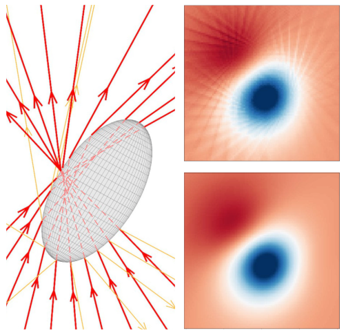
Focused rays scattered by an ellipsoidal particles (left). Optical torque along y calculated in the x-y plane using ray scattering with a grid of 1600 rays (up, right) and using a trained neural network (down, right). (Image by the Authors of the manuscript.)Dense neural networks for geometrical optics
David Bronte Ciriza, Alessandro Magazzù, Agnese Callegari, Gunther Barbosa, Antonio A. R. Neves, Maria Antonia Iatì, Giovanni Volpe, and Onofrio M. Maragò
SPIE-ETAI, San Diego, CA, USA, 3 – 7 August 2025 Date: 4 August 2025 Time: 5:30 PM – 7:30 PM PDT Place: Conv. Ctr. Exhibit Hall A
Presenter: Giovanni Volpe
Contribution submitted by Agnese Callegari
Light can trap and manipulate microscopic objects through optical forces and torques, as seen in optical tweezers. Predicting these forces is crucial for experiments and setup design. This study focuses on the geometrical optics regime, which applies to particles much larger than the light’s wavelength. In this model, a beam is represented by discrete rays that undergo multiple reflections and refractions, transferring momentum and angular momentum. However, the choice of ray discretization affects the balance between computational speed and accuracy. We demonstrate that neural networks overcome this limitation, enabling faster and even more precise simulations. Using an optically trapped spherical particle with an analytical solution as a benchmark, we validate our method and apply it to study complex systems that would otherwise be computationally hard.
Experimental trajectory (blue) of a particle trapped in air when the laser rotates at 1 Hz. The orange line represents the experimental laser trajectory. (Image by A. Ciarlo.)Probing fluid dynamics inertial effects of particles using optical tweezers Antonio Ciarlo, Giuseppe Pesce, Bernhard Mehlig, Antonio Sasso, and Giovanni Volpe Date: 4 August 2025 Time: 11:45 AM – 12:00 PM Place: Conv. Ctr. Room 3
Many natural phenomena involve dense particles suspended in a moving fluid, such as water droplets in clouds or dust grains in circumstellar disks. Studying these systems at the single particle level is challenging and requires precise control of flow and particle motion. Optical tweezers provide a powerful method for studying inertial effects in such environments. Here, we trap micrometer-sized particles in air and induce controlled dynamics by moving the trapping laser. We show that inertia becomes significant when the trap motion frequency is less than the harmonic trapping frequency, while at much higher motion frequencies, inertia has no effect. These results demonstrate the potential of trapping particles in air for studying inertial phenomena in fluids.
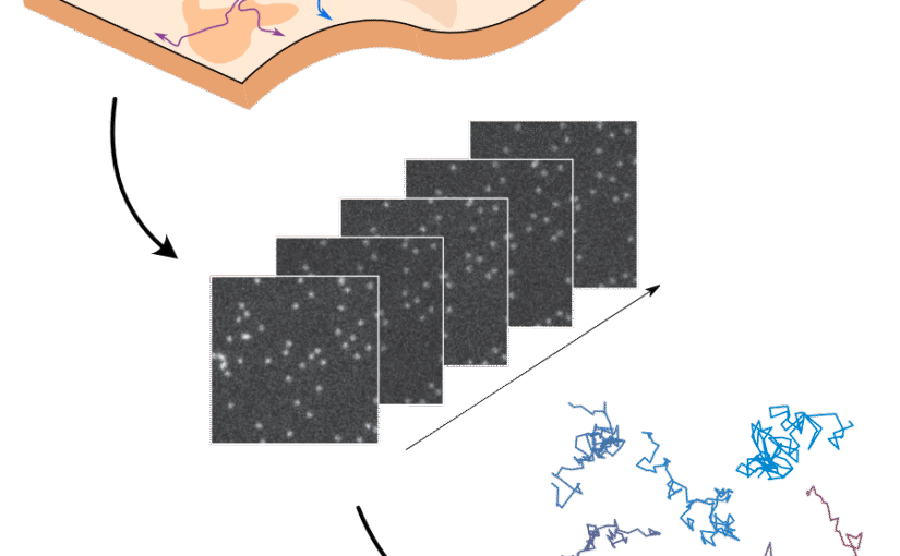
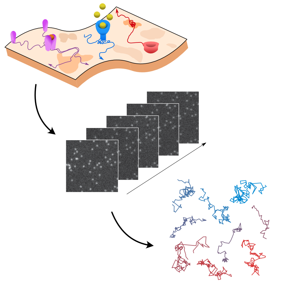
Rationale for the challenge organization. The interactions of biomolecules in complex environments, such as the cell membrane, regulate physiological processes in living systems. These interactions produce changes in molecular motion that can be used as a proxy to measure interaction parameters. Time-lapse single-molecule imaging allows us to visualize these processes with high spatiotemporal resolution and, in combination with single-particle tracking methods, provide trajectories of individual molecules. (Image by the Authors of the manuscript.)Quantitative evaluation of methods to analyze motion changes in single-particle experiments
Gorka Muñoz-Gil, Harshith Bachimanchi, Jesús Pineda, Benjamin Midtvedt, Gabriel Fernández-Fernández, Borja Requena, Yusef Ahsini, Solomon Asghar, Jaeyong Bae, Francisco J. Barrantes, Steen W. B. Bender, Clément Cabriel, J. Alberto Conejero, Marc Escoto, Xiaochen Feng, Rasched Haidari, Nikos S. Hatzakis, Zihan Huang, Ignacio Izeddin, Hawoong Jeong, Yuan Jiang, Jacob Kæstel-Hansen, Judith Miné-Hattab, Ran Ni, Junwoo Park, Xiang Qu, Lucas A. Saavedra, Hao Sha, Nataliya Sokolovska, Yongbing Zhang, Giorgio Volpe, Maciej Lewenstein, Ralf Metzler, Diego Krapf, Giovanni Volpe, Carlo Manzo
Nature Communications 16, 6749 (2025)
arXiv: 2311.18100
doi: https://doi.org/10.1038/s41467-025-61949-x
The analysis of live-cell single-molecule imaging experiments can reveal valuable information about the heterogeneity of transport processes and interactions between cell components. These characteristics are seen as motion changes in the particle trajectories. Despite the existence of multiple approaches to carry out this type of analysis, no objective assessment of these methods has been performed so far. Here, we report the results of a competition to characterize and rank the performance of these methods when analyzing the dynamic behavior of single molecules. To run this competition, we implemented a software library that simulates realistic data corresponding to widespread diffusion and interaction models, both in the form of trajectories and videos obtained in typical experimental conditions. The competition constitutes the first assessment of these methods, providing insights into the current limitations of the field, fostering the development of new approaches, and guiding researchers to identify optimal tools for analyzing their experiments.
In this work, we present an unsupervised deep learning framework using Variational Autoencoders (VAEs) to decode stress-specific biomolecular fingerprints directly from Raman spectral data across multiple plant species and genotypes. (Image by the Authors of the manuscript. A part of the image was designed using Biorender.com.)From Spectra to Stress: Unsupervised Deep Learning for Plant Health Monitoring
Anoop C. Patil, Benny Jian Rong Sng, Yu-Wei Chang, Joana B. Pereira, Chua Nam-Hai, Rajani Sarojam, Gajendra Pratap Singh, In-Cheol Jang, and Giovanni Volpe
ArXiv: 2507.15772
Detecting stress in plants is crucial for both open-farm and controlled-environment agriculture. Biomolecules within plants serve as key stress indicators, offering vital markers for continuous health monitoring and early disease detection. Raman spectroscopy provides a powerful, non-invasive means to quantify these biomolecules through their molecular vibrational signatures. However, traditional Raman analysis relies on customized data-processing workflows that require fluorescence background removal and prior identification of Raman peaks of interest-introducing potential biases and inconsistencies. Here, we introduce DIVA (Deep-learning-based Investigation of Vibrational Raman spectra for plant-stress Analysis), a fully automated workflow based on a variational autoencoder. Unlike conventional approaches, DIVA processes native Raman spectra-including fluorescence backgrounds-without manual preprocessing, identifying and quantifying significant spectral features in an unbiased manner. We applied DIVA to detect a range of plant stresses, including abiotic (shading, high light intensity, high temperature) and biotic stressors (bacterial infections). By integrating deep learning with vibrational spectroscopy, DIVA paves the way for AI-driven plant health assessment, fostering more resilient and sustainable agricultural practices.
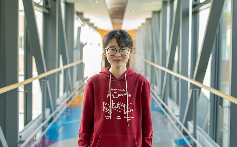
(Photo by A. CiarloXinwen Zhang started her PhD at the Physics Department of Gothenburg University on 7 July 2025.
Xinwen holds a master’s degree in Physics (biophysics) from the University of Science and Technology of China (USTC), Hefei, China.
During her PhD, she will focus on label-free optical microscopy combined with deep learning, aiming to characterize nanoparticles and uncover their physical mechanisms.
GAUDI leverages a hierarchical graph-convolutional variational autoencoder architecture, where an encoder progressively compresses the graph into a low-dimensional latent space, and a decoder reconstructs the graph from the latent embedding. (Image by M. Granfors and J. Pineda.)Cutting Training Data Needs through Inductive Bias & Unsupervised Learning
Giovanni Volpe and Carlo Manzo Computational Intelligence Group (CIG), Weekly Reading Session Date: 3 July 2025 Time: 17:00 Place: Makerere University, Kampala, Uganda (Online)
Graphs provide a powerful framework for modeling complex systems, but their structural variability makes analysis and classification challenging. To address this, we introduce GAUDI (Graph Autoencoder Uncovering Descriptive Information), a novel unsupervised geometric deep learning framework that captures both local details and global structure. GAUDI employs an innovative hourglass architecture with hierarchical pooling and upsampling layers, linked through skip connections to preserve essential connectivity information throughout the encoding–decoding process. By mapping different realizations of a system — generated from the same underlying parameters — into a continuous, structured latent space, GAUDI disentangles invariant process-level features from stochastic noise. We demonstrate its power across multiple applications, including modeling small-world networks, characterizing protein assemblies from super-resolution microscopy, analyzing collective motion in the Vicsek model, and capturing age-related changes in brain connectivity. This approach not only improves the analysis of complex graphs but also provides new insights into emergent phenomena across diverse scientific domains.
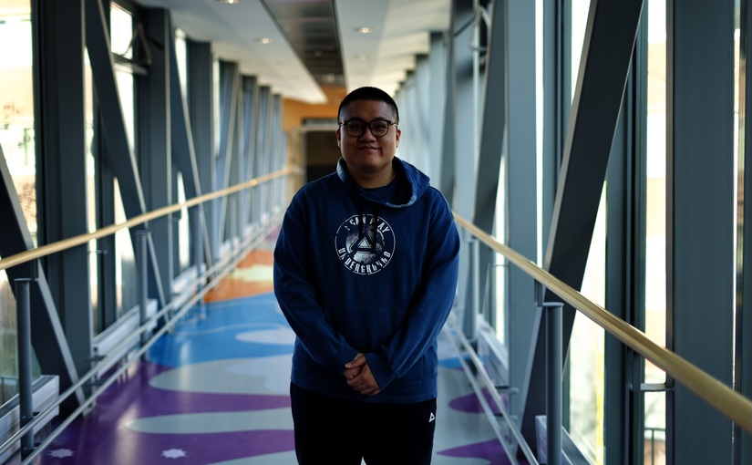
(Photo by A. Ciarlo.)Jiacheng Huang started his PhD at the Physics Department of the University of Gothenburg on 1st July 2025.
Jiacheng has a Master degree in Material and Chemical Engineering from the Department of Chemical and Biochemical Engineering, Xiamen University, China.
In his PhD, which is part of the MSCA-DN SPM4.0, he will focus on machine learning and smart microscopy.
Electromagnetic light scattering underpins a wide range of phenomena in both fundamental and applied research, from characterizing complex materials to tracking particles and cells in microfluidic devices. Video microscopy, in particular, has become a powerful method for studying scattering processes and extracting quantitative information. Yet, conventional algorithmic approaches for analyzing scattering data often prove cumbersome, computationally expensive, and highly specialized.
Recent advances in deep learning offer a compelling alternative. By leveraging data-driven models, we can automate the extraction of scattering characteristics with unprecedented speed and accuracy—uncovering insights that classical techniques might miss or require substantial computation to achieve. Despite these advantages, deep-learning-based tools remain underutilized in light-scattering research, largely because of the steep learning curve required to design and train such models.
To address these challenges, we have developed a user-friendly software platform (DeepTrack, now in version 2.2) that simplifies the entire workflow of deep-learning applications in digital microscopy. DeepTrack enables straightforward creation of custom datasets, network architectures, and training pipelines specifically tailored for quantitative scattering analyses. In this talk, I will discuss how emerging deep-learning methods can be combined with advanced imaging technologies to push the boundaries of electromagnetic light scattering research—reducing computational overhead, improving accuracy, and ultimately broadening access to powerful, data-driven solutions.