Eduard Andrei Duta Costache started his PhD at the Physics Department of the University of Gothenburg on the 19th of January 2026.
Eduard has a double Master’s degree in Artificial Intelligence from the University of Alicante (Spain) and in Machine Learning & Data Mining from Jean Monnet University (France).
During the course of his PhD, as part of the GREENS MSCA Doctoral Network, he will focus on developing AI frameworks to model and optimize the lifecycle of micro-robotic platforms.
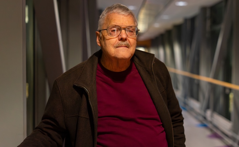
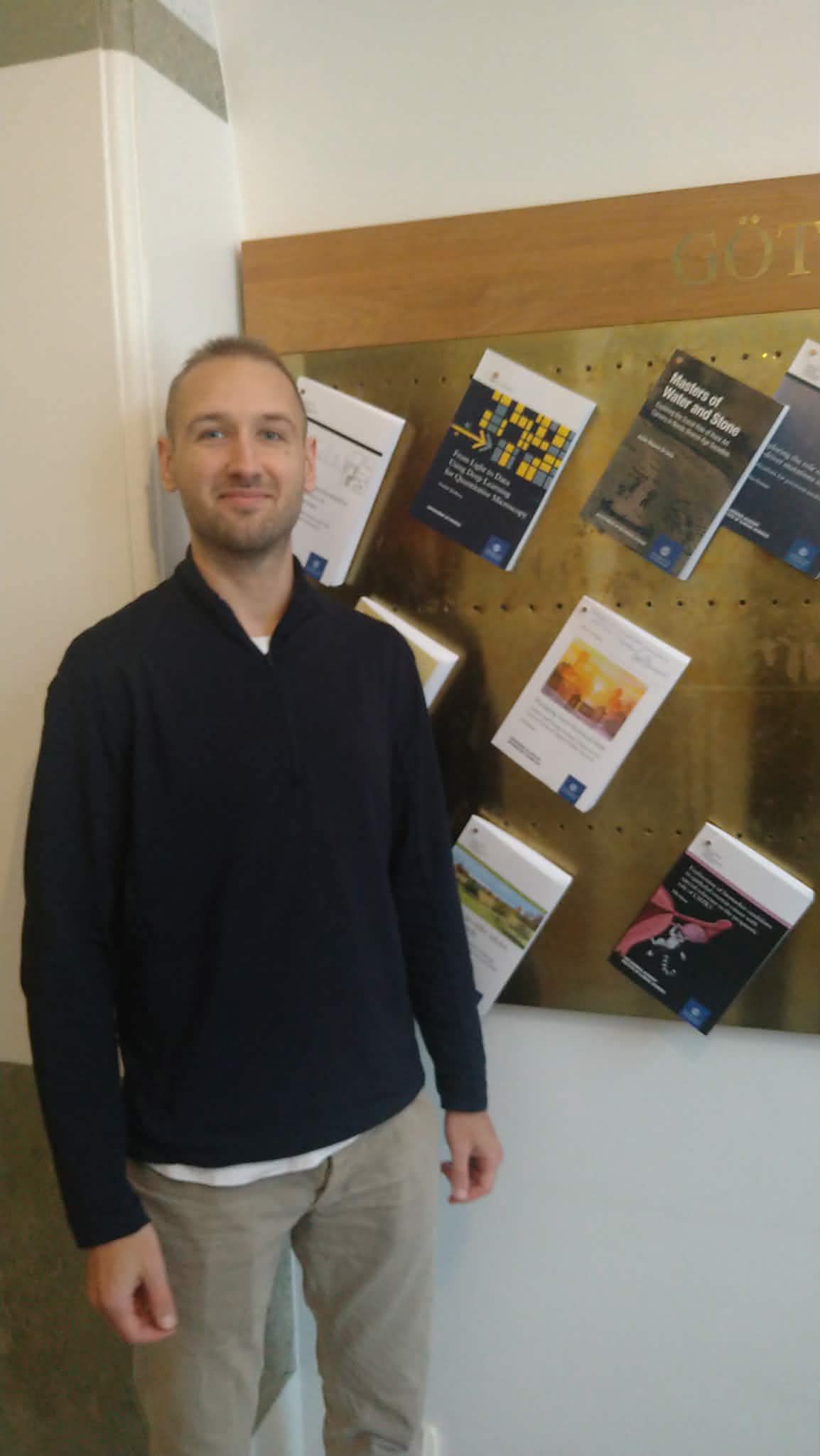
Steven B. Smith. (Photo by A. Ciarlo)Steven Smith will visit the Soft Matter Lab from 17 to 28 January 2026.
Steven brings years of expertise from the laboratory of Professor Carlos Bustamante, where they pioneered the use and development of optical tweezers. As the main developer of the widely used ‘minitweezers’ instrument, which today is used by dozens of research groups, he has helped shape the field of single-molecule biophysics on a global scale. We look forward to his visit, during which he will work on refining cutting-edge single-molecule measurement techniques.
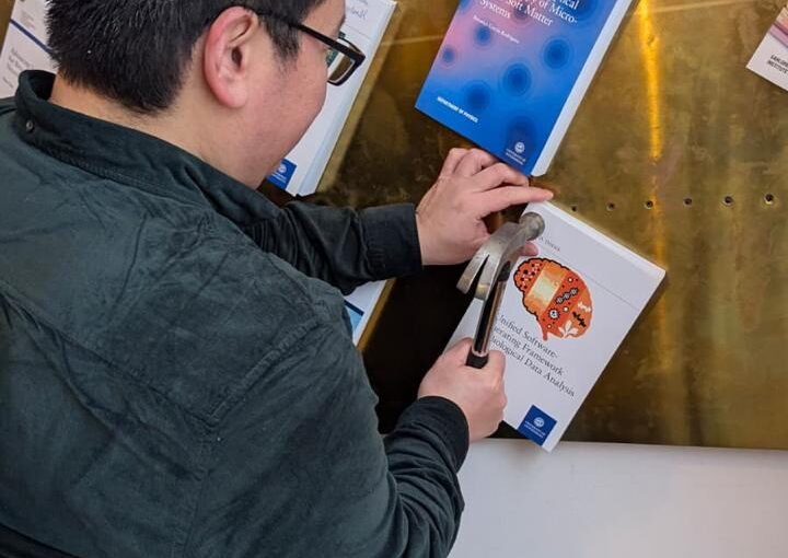
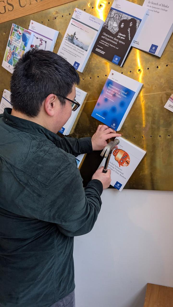
Thesis nailing by Yu-Wei Chang. (Photo by C. Khanolkar.)Yu-Wei Chang nailed his PhD thesis, A Unified Software-Generating Framework for Biological Data Analysis, on January 7th, 2026. Congrats!
The nailing took place in Universitetsbyggnaden i Vasaparken, Universitetsplatsen 1, Göteborg, at 13:30.
In Swedish academia, “nailing” (spikning) is the formal public announcement and publication of a doctoral thesis. It happens weeks before the defence so that the public has time to read the thesis in advance and prepare questions for the defence. In addition to the physical nailing, the thesis is also published electronically (e-spikning) via GUPEA.
Yu-Wei will defend his thesis on 23 January at 13:00 in SB-H7 lecture hall, SB-Building, Institutionen för fysik, Johanneberg Campus, Göteborg.
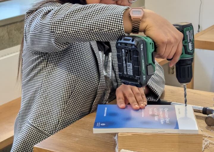
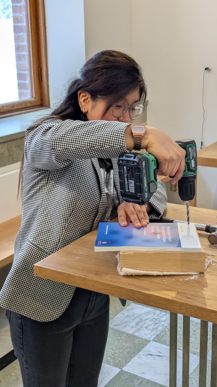
Thesis nailing by Berenice García Rodríguez. (Photo by C. Khanolkar.)On 7 January at 13:00, Berenice García Rodríguez nailed her PhD thesis, Quantitative Optical Microscopy of Microscale Soft Matter Systems, at the University of Gothenburg in Vasaparken.
Family and friends were present to mark this milestone.
Berenice will defend her thesis on 28 January at 09:00 in PJ-salen, Institutionen för fysik, Origovägen 6, Göteborg.
Thesis nailing by Fredrik Skärberg. (Photo by E. Skärberg.)On 7 January at 12:30, Fredrik Skärberg nailed his PhD thesis, From Light to Data: Using Deep Learning for Quantitative Microscopy, at the University of Gothenburg in Vasaparken. Family and friends were present to mark this milestone.
Fredrik will defend his thesis on 29 January at 09:00 in FB-salen, Institutionen för fysik, Origovägen 6, Göteborg.
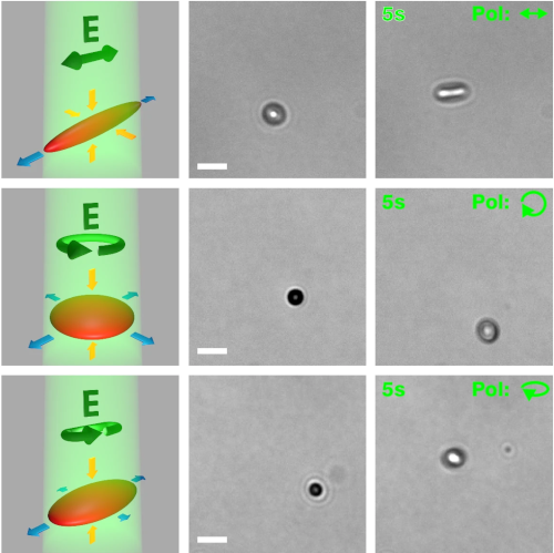
(Image from the manuscript.)Directional flows using capillary assembly of photo-deformable colloidal particles at water-air interfaces
David Urban, Marcel Rey, Antonio Ciarlo, Marie Friederike Schulte, Emiliano Descrovi & Giovanni Volpe
Nature Communications 17, 1004 (2026)
doi: 10.1038/s41467-025-67739-9
Colloidal particles at liquid interfaces experience long-ranged capillary interactions, whose magnitude and directionality depend on the particle shapes. When particle shapes are determined by fabrication or synthesis, the resulting shape-mediated interactions are predefined and often lead to the formation of persistent interfacial structures. Here, we introduce polymer particles at water-air interfaces whose shape and, therefore, interactions can be altered by illumination with polarized light. Specifically, we selectively trigger capillary self-assembly by anisotropically deforming the particles at the interface. Intriguingly, further deformation of already assembled particles induces sustained interfacial flows with velocities of up to 90 μm/s. Benefitting from polarization-defined deformation directions, we create flow-patterns that do not simply follow the illumination intensity pattern, such as shear flows along a single rectangular illumination stripe. We anticipate that this interplay between photo-deformation and capillary interactions of particles will enable various forms of mixing, manipulation, and assembly of soft matter at liquid interfaces.
Photograph of the soft robot, consisting of a multilayer rolled dielectric elastomer actuator integrated with a
flexible PET sheet. (Image by H. P. Thanabalan.)Inchworm-Inspired Soft Robot with Groove-Guided Locomotion
Hari Prakash Thanabalan, Lars Bengtsson, Ugo Lafont, Giovanni Volpe
arXiv: 2512.07813
Soft robots require directional control to navigate complex terrains. However, achieving such control often requires multiple actuators, which increases mechanical complexity, complicates control systems, and raises energy consumption. Here, we introduce an inchworm-inspired soft robot whose locomotion direction is controlled passively by patterned substrates. The robot employs a single rolled dielectric elastomer actuator, while groove patterns on a 3D-printed substrate guide its alignment and trajectory. Through systematic experiments, we demonstrate that varying groove angles enables precise control of locomotion direction without the need for complex actuation strategies. This groove-guided approach reduces energy consumption, simplifies robot design, and expands the applicability of bio-inspired soft robots in fields such as search and rescue, pipe inspection, and planetary exploration.
Charite University and Bruker Nano GmbH hosted the first training workshop for the SPM 4.0 network. Both Prakhar Dutta and Jiacheng Huang, the two doctoral candidates based at the University of Gothenburg, participated to the event along with the other doctoral candidates of the network.
The training event consisted of a series of lectures on different topics such as basics of scanning probe microscopy, medical radiation based elastography, and the basics of machine learning.
Two of the days of the training event were held at Bruker Nano GmbH in Berlin, where hands-on lectures and practicals were given on the use of atomic force microscopy machines and their applications.
The training event consisted of a series of lectures on different topics such as small scale robots and their actuation mechanisms, theoretical aspects of the simulation of the dynamics of bodies immersed in fluids at low Reynolds numbers, working principles and applications of swarm robotics, and finally artificial intelligence and its applications to data analysis in experiments.
The event included a visit to the Swedish Algae Factory (SAFAB) and Smogenlax on the topic of circular economy, and concluded with a series of meetings between the representatives of the participating institutions, from both accademia and industry, to exchange research questions and plan secondments.