Individual and collective motion of nematic, polar, and chiral actively driven objects
Andreas Menzel
19 January 2023
15:30, Nexus
Abstract:
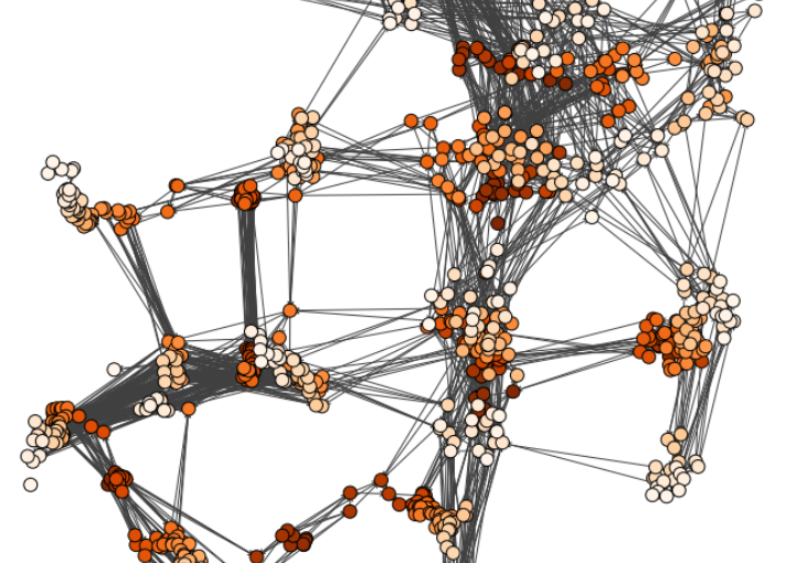
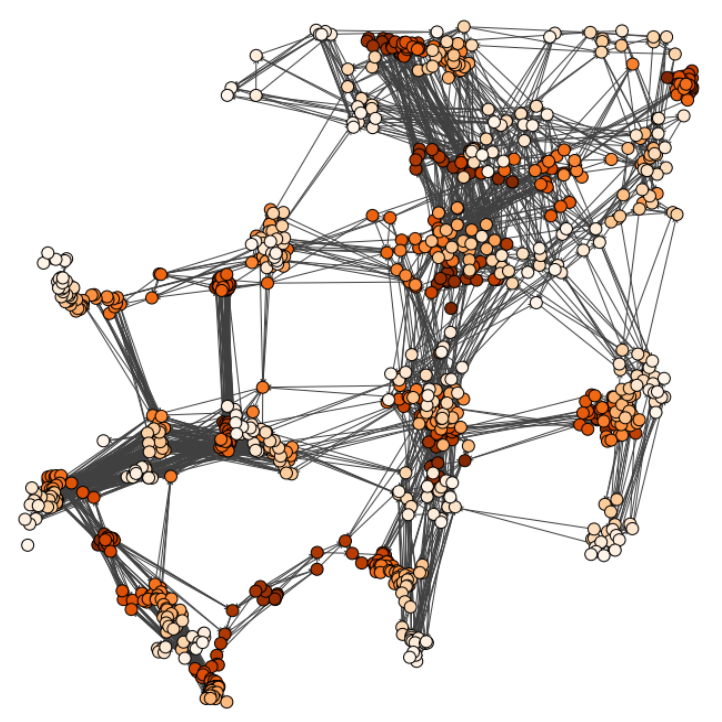
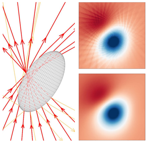
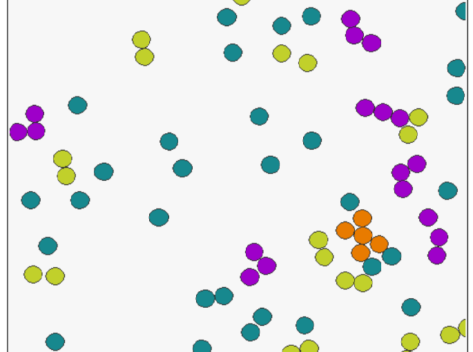
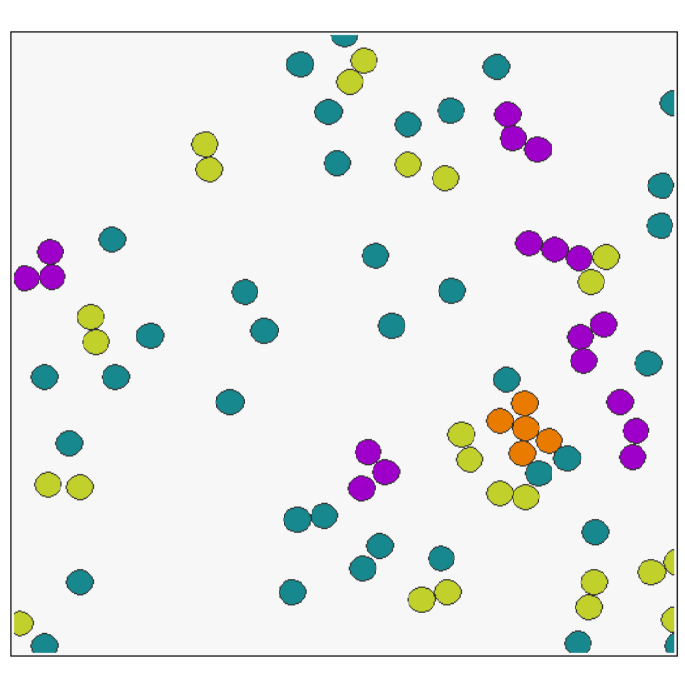
Actively driven objects comprise a manifold of possible different realizations: from self-propelling bacteria and artificial phoretically driven colloidal particles via vibrated hoppers to walking pedestrians. We analyze basic theoretical models to identify generic features of subclasses of such agents. Within this framework, we first address nematic objects [1]. They predominantly propel along one specific axis of their body, but do not feature an explicit head or tail. That is, they can move either way by spontaneous symmetry breaking. This leads to characteristic kinks along their trajectories. Second, we study chiral objects that show persistent bending of their trajectories and migrate in discrete steps [2]. When, additionally, they tend to migrate towards a fixed remote target, rich nonlinear dynamics emerges. It comprises period doubling and chaotic behavior as a function of the tendency of alignment, which is reflected by the trajectories. Third, we consider the collective motion of continuously moving chiral objects in crystal-like arrangements [3]. We here identify a localization transition with increasing chirality or self-shearing phenomena within the crystal-like structures. Overall, we hope by our work to stimulate experimental realization and observation of the various investigated systems and phenomena.
References
[1] A. M. Menzel, J. Chem. Phys. 157, 011102 (2022).
[2] A. M. Menzel, resubmitted.
[3] Z.-F. Huang, A. M. Menzel, H. Löwen, Phys. Rev. Lett. 125, 218002 (2020).
Short Bio:
Andreas Menzel studied physics at the University of Bayreuth (Germany), where he also completed his PhD on the continuum theory of soft elastic liquid-crystalline composite materials. After postdoctoral stays at the University of Illinois at Urbana-Champaign with Prof. Nigel Goldenfeld and at the Max Planck Institute for Polymer Research in Mainz in the department headed by Prof. Kurt Kremer, as well as research stays at Kyoto University with Prof. Takao Ohta, he completed his Habilitation at Heinrich Heine University Düsseldorf at the Theory Institute for Soft Matter headed by Prof. Hartmut Löwen. Amongst others, Andreas is interested in developing and applying explicit Green’s functions methods, statistical descriptions, and continuum theories on soft matter, addressing, for example, functionalized elastic composite materials and active matter. In 2020 he moved as a Heisenberg Fellow of the German Research Foundation to Otto von Guericke University Magdeburg (Germany), where he now heads the department on Theory of Soft Matter / Biophysics.