Giovanni Volpe
AI Photonics, Donostia, Spain
Date: 12 September 2023
Giovanni Volpe
AI Photonics, Donostia, Spain
Date: 12 September 2023
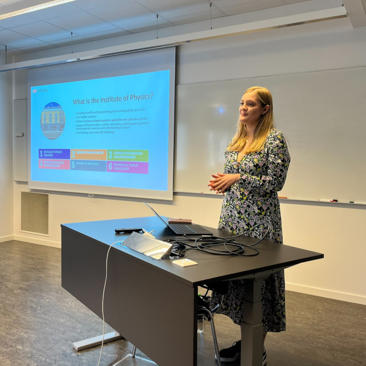
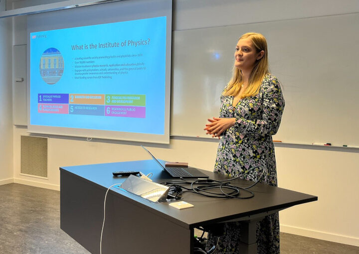
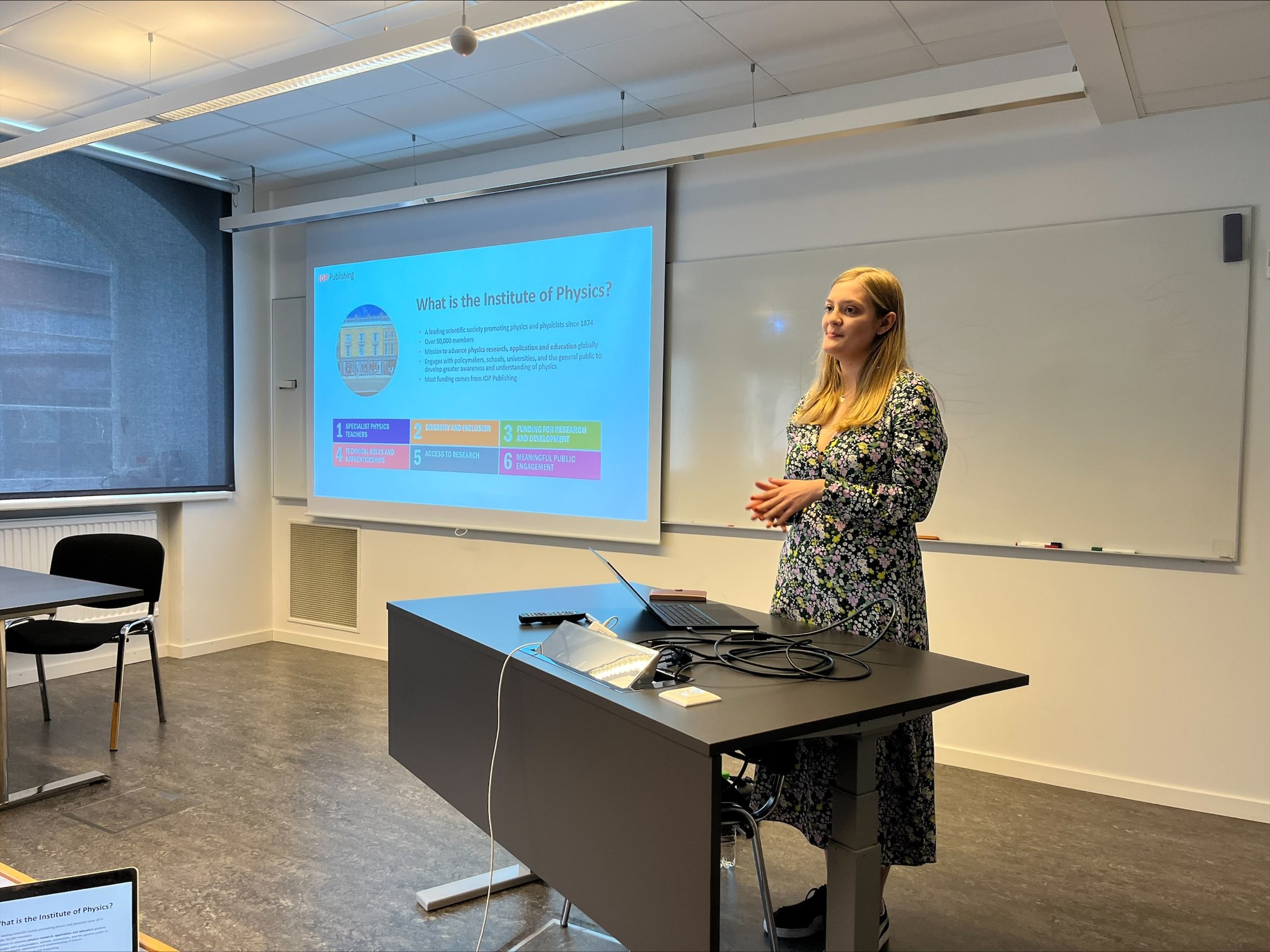
Kate Porter
IOP Publishing
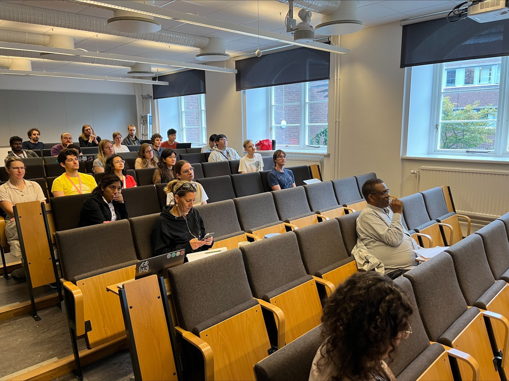
Do you want your article to stand out from the crowd, improving your chances of publication in this highly competitive industry? If so, you won’t want to miss this talk from Kate Porter, Senior Publisher from IOP Publishing! During this talk, Kate will provide you with a toolkit to help you navigate the world of academic publishing and share some top tips to help you get published.
Topics covered in this talk include:
Date: 6 Sep 2023
Time: 12:30 PM
Location: PJ
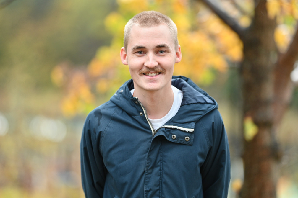
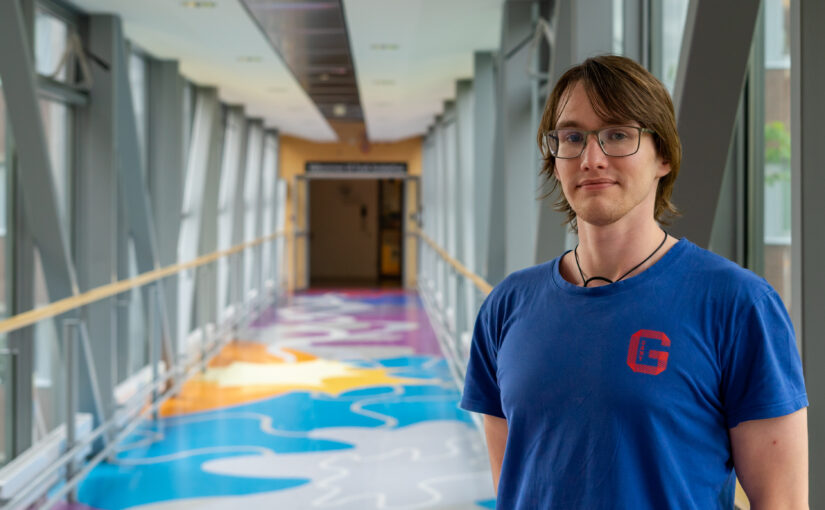
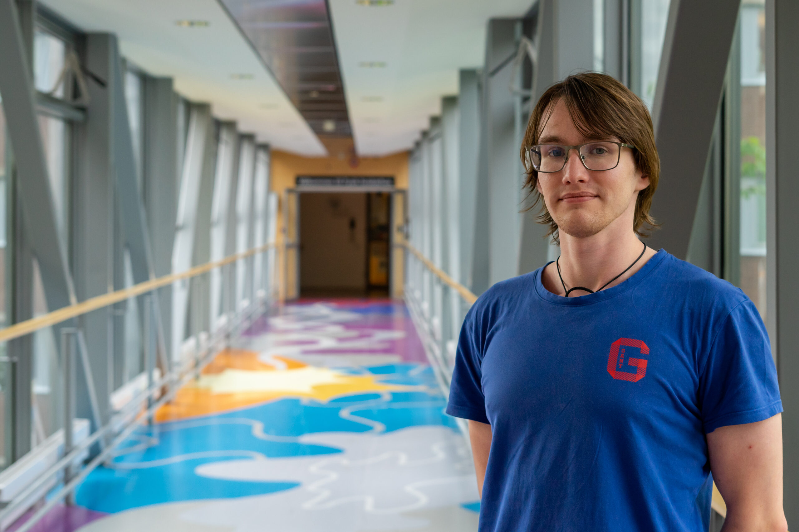
Gideon has a master degree in Complex Adaptive Systems from Chalmers University of Technology.
In his PhD, Gideon will focus his research on deep learning.
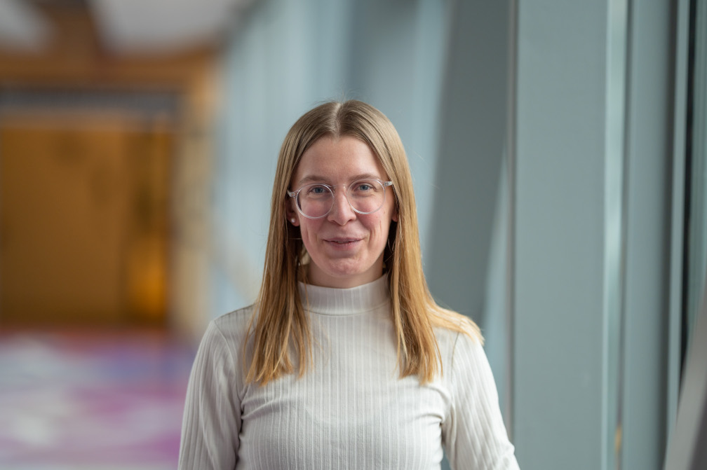
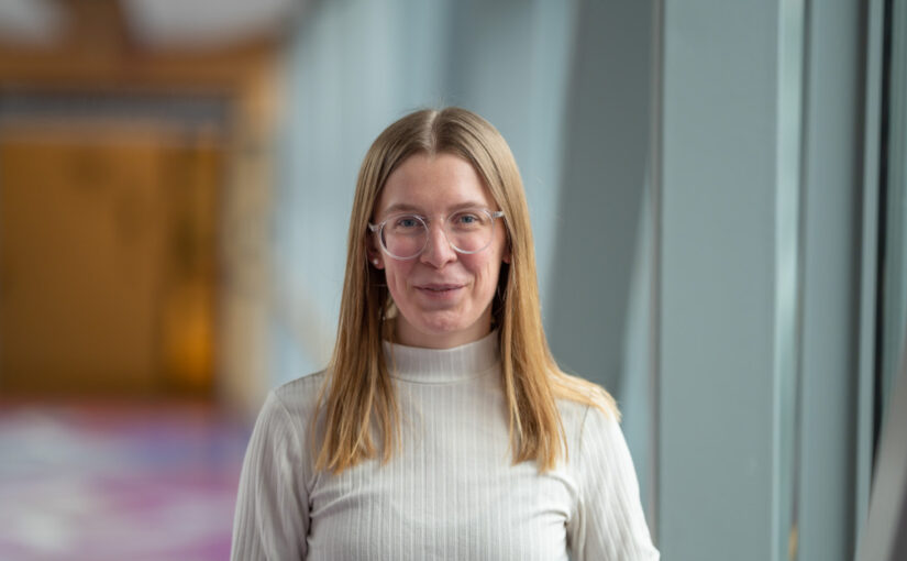
Mirja has a Master degree in Physics from the University of Gothenburg.
In her PhD, Mirja will focus on graph neural networks and deep learning.
Dániel Veréb, Mite Mijalkov, Anna Canal-Garcia, Yu-Wei Chang, Emiliano Gomez-Ruiz, Blanca Zufiria Gerboles, Miia Kivipelto, Per Svenningsson, Henrik Zetterberg, Giovanni Volpe, Matthew Betts, Heidi IL Jacobs, Joana B Pereira
eLife 12, RP87188 (2023)
doi: 10.7554/eLife.87188.3
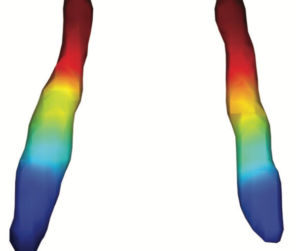
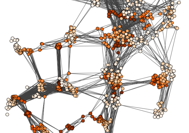
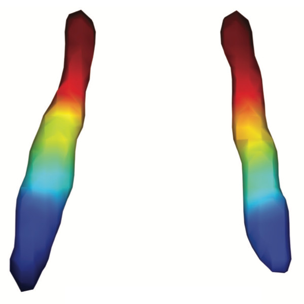
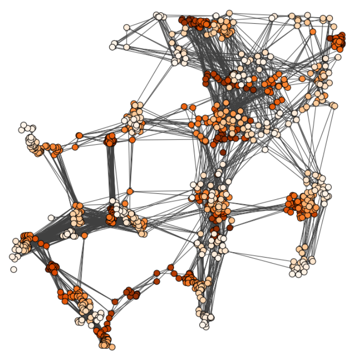
The locus coeruleus (LC) is an important noradrenergic nucleus that has recently attracted a lot of attention because of its emerging role in cognitive and psychiatric disorders. Although previous histological studies have shown that the LC has heterogeneous connections and cellular features, no studies have yet assessed its functional topography in vivo, how this heterogeneity changes over aging, and whether it is associated with cognition and mood. Here, we employ a gradient-based approach to characterize the functional heterogeneity in the organization of the LC over aging using 3T resting-state fMRI in a population-based cohort aged from 18 to 88 years of age (Cambridge Centre for Ageing and Neuroscience cohort, n=618). We show that the LC exhibits a rostro-caudal functional gradient along its longitudinal axis, which was replicated in an independent dataset (Human Connectome Project [HCP] 7T dataset, n=184). Although the main rostro-caudal direction of this gradient was consistent across age groups, its spatial features varied with increasing age, emotional memory, and emotion regulation. More specifically, a loss of rostral-like connectivity, more clustered functional topography, and greater asymmetry between right and left LC gradients was associated with higher age and worse behavioral performance. Furthermore, participants with higher-than-normal Hospital Anxiety and Depression Scale (HADS) ratings exhibited alterations in the gradient as well, which manifested in greater asymmetry. These results provide an in vivo account of how the functional topography of the LC changes over aging, and imply that spatial features of this organization are relevant markers of LC-related behavioral measures and psychopathology.
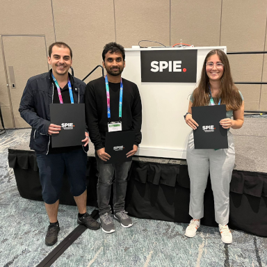
Alex is a master student in Physics at the University of Gothenburg.
During His time at the Soft Matter Lab, he will work on simulating colloidal systems with phoretic interactions.
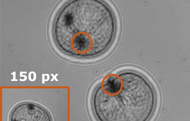
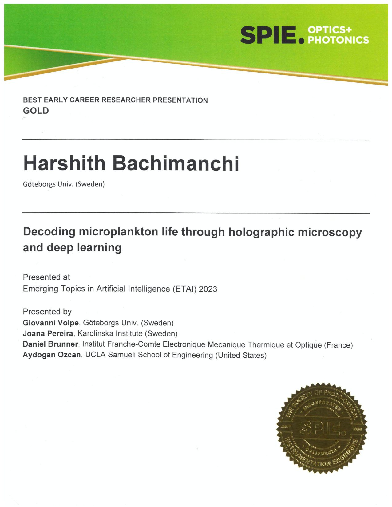
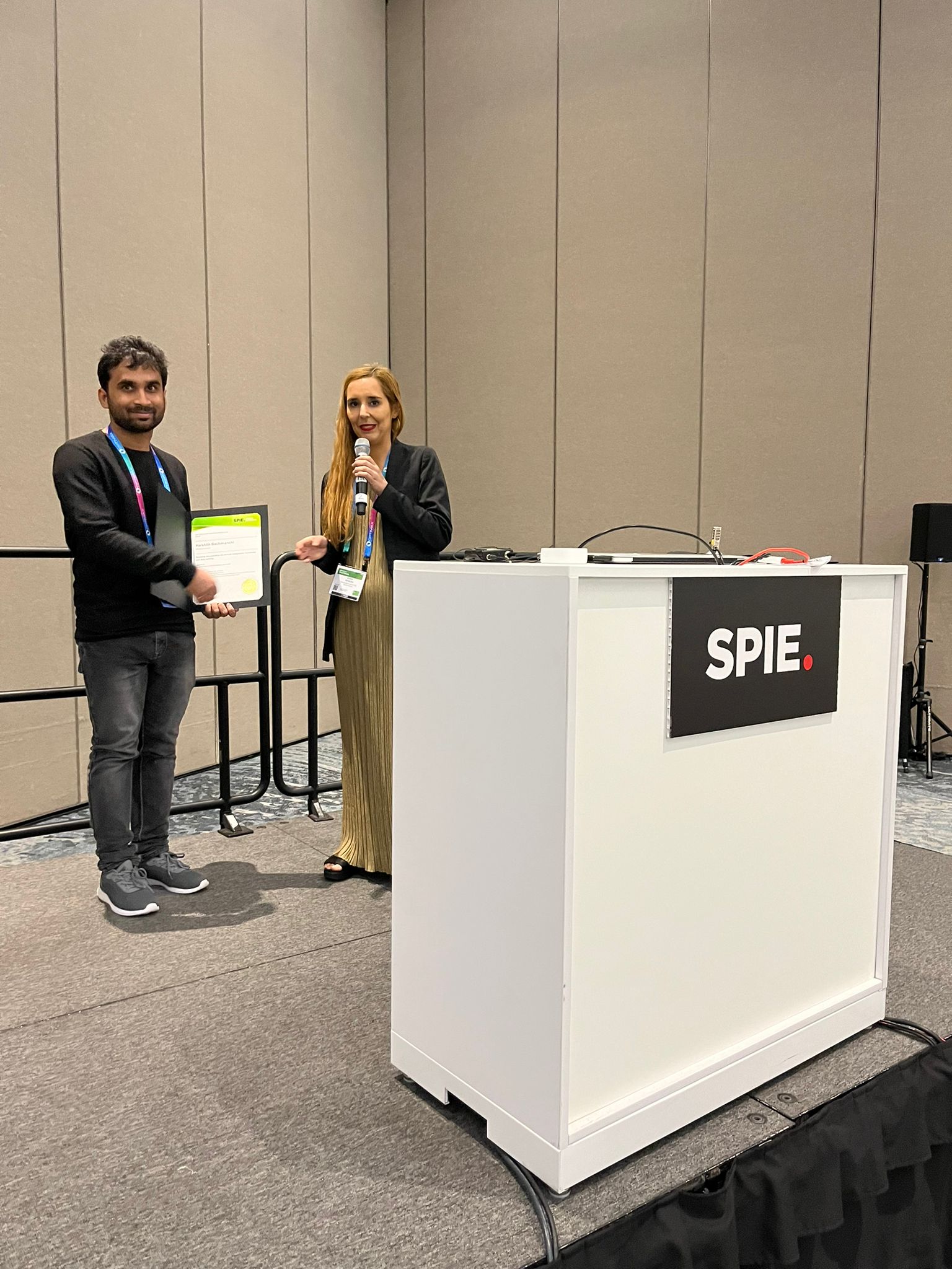
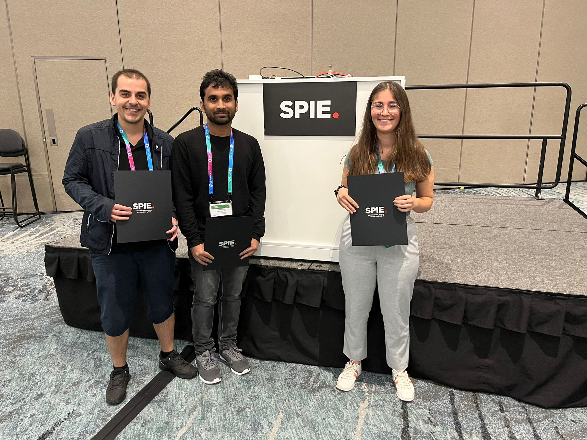
In this work, Harshith presented his recent work on combining holographic microscopy and deep learning to study the marine microplankton. He demonstrated that the combination of holographic microscopy and deep learning can be used to follow the marine microorganisms throughout their lifespan, continuously measuring their three-dimensional positions and dry mass. The deep-learning algorithms circumvent the computationally intensive processing of holographic data and allow rapid measurements over extended periods of time. This enables to reliably estimate growth rates, both in terms of dry mass increase and cell divisions, as well as to measure trophic interactions between species such as predation events. Studying individual interactions in idealized small systems provides insights that help us understand microbial food webs and ultimately larger-scale processes. He exemplified this by showing detailed descriptions of micro-zooplankton feeding events, cell divisions, and long-term monitoring of single cells from division to division.
The article related to this presentation can be found at the following link: Microplankton life histories revealed by holographic microscopy and deep learning.
Jesús Pineda
Date: 23 August 2023
Time: 2:30 PM PDT
Characterizing dynamic processes in living systems provides essential information for advancing our understanding of life processes in health and diseases and for developing new technologies and treatments. In the past two decades, optical microscopy has undergone significant developments, enabling us to study the motion of cells, organelles, and individual molecules with unprecedented detail at various scales in space and time. However, analyzing the dynamic processes that occur in complex and crowded environments remains a challenge. This work introduces MAGIK, a deep-learning framework for the analysis of biological system dynamics from time-lapse microscopy. MAGIK models the movement and interactions of particles through a directed graph where nodes represent detections and edges connect spatiotemporally close nodes. The framework utilizes an attention-based graph neural network (GNN) to process the graph and modulate the strength of associations between its elements, enabling MAGIK to derive insights into the dynamics of the systems. MAGIK provides a key enabling technology to estimate any dynamic aspect of the particles, from reconstructing their trajectories to inferring local and global dynamics. We demonstrate the flexibility and reliability of the framework by applying it to real and simulated data corresponding to a broad range of biological experiments.
Reference
Pineda, J., Midtvedt, B., Bachimanchi, H. et al. Geometric deep learning reveals the spatiotemporal features of microscopic motion. Nat Mach Intell 5, 71–82 (2023)
Benjamin Midtvedt, Jesus Pineda, Fredrik Skärberg, Erik Olsén, Harshith Bachimanchi, Emelie Wesén, Elin Esbjörner, Erik Selander, Fredrik Höök, Daniel Midtvedt, Giovanni Volpe
Date: 23 August 2023
Time: 10:30 AM (PDT)
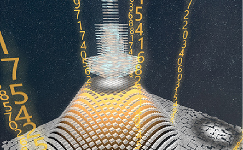
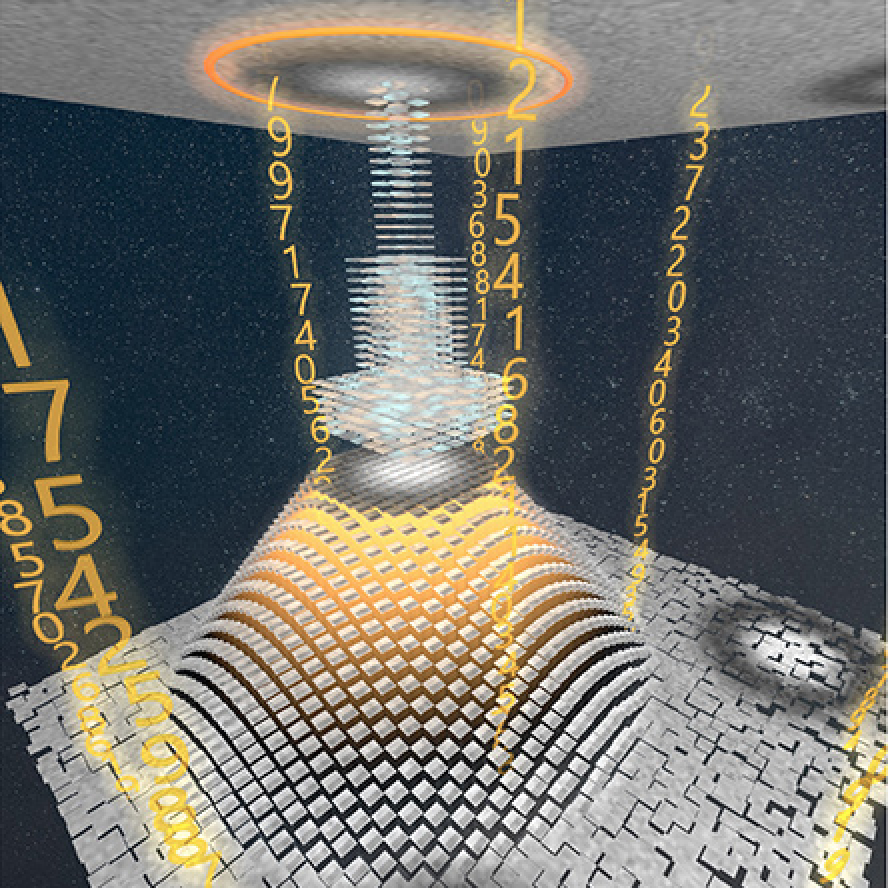
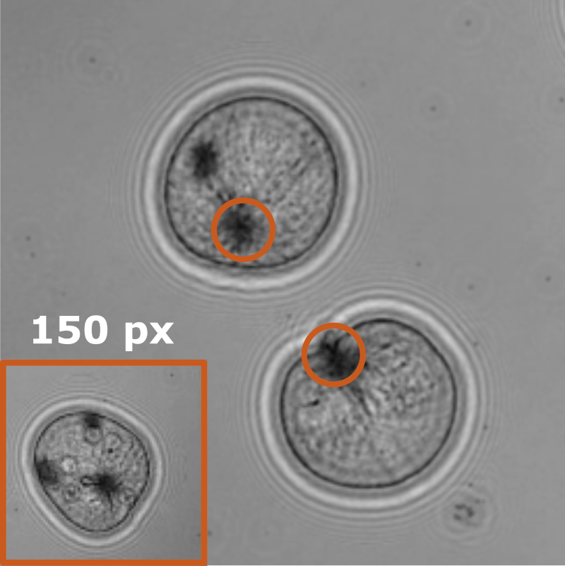
Object detection is a fundamental task in digital microscopy. Recently, machine-learning approaches have made great strides in overcoming the limitations of more classical approaches. The training of state-of-the-art machine-learning methods almost universally relies on either vast amounts of labeled experimental data or the ability to numerically simulate realistic datasets. However, the data produced by experiments are often challenging to label and cannot be easily reproduced numerically. Here, we propose a novel deep-learning method, named LodeSTAR (Low-shot deep Symmetric Tracking And Regression), that learns to detect small, spatially confined, and largely homogeneous objects that have sufficient contrast to the background with sub-pixel accuracy from a single unlabeled experimental image. This is made possible by exploiting the inherent roto-translational symmetries of the data. We demonstrate that LodeSTAR outperforms traditional methods in terms of accuracy. Furthermore, we analyze challenging experimental data containing densely packed cells or noisy backgrounds. We also exploit additional symmetries to extend the measurable particle properties to the particle’s vertical position by propagating the signal in Fourier space and its polarizability by scaling the signal strength. Thanks to the ability to train deep-learning models with a single unlabeled image, LodeSTAR can accelerate the development of high-quality microscopic analysis pipelines for engineering, biology, and medicine.
Henrik Klein Moberg
Date: 23 August 2023
Time: 8:15 AM PDT
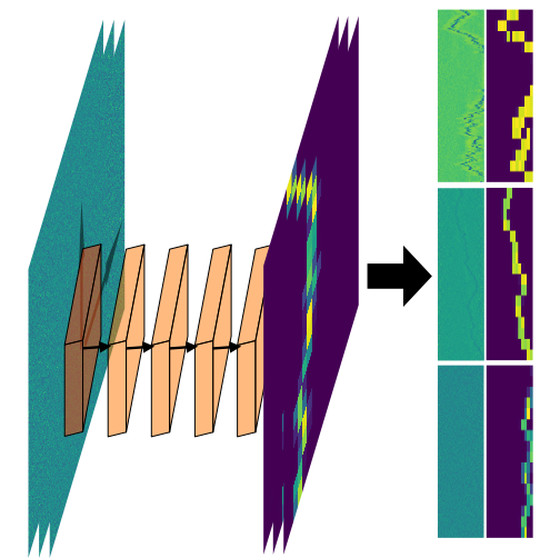
We show that a custom ResNet-inspired CNN architecture trained on simulated biomolecule trajectories surpasses the performance of standard algorithms in terms of tracking and determining the molecular weight and hydrodynamic radius of biomolecules in the low-kDa regime in optical microscopy. We show that high accuracy and precision is retained even below the 10-kDa regime, constituting approximately an order of magnitude improvement in limit of detection compared to current state-of-the-art, enabling analysis of hitherto elusive species of biomolecules such as cytokines (~5-25 kDa) important for cancer research and the protein hormone insulin (~5.6 kDa), potentially opening up entirely new avenues of biological research.