Invited Talk by G. Volpe at 12th Nordic Workshop on Statistical Physics, Nordita, Stockholm, 15 March 2023

Logo of the AnDi challenge.
An Anomalous Competition: Assessment of methods for anomalous diffusion through a community effort
Giovanni Volpe
Nordita, Stockholm, 15 March 2023, 14:00

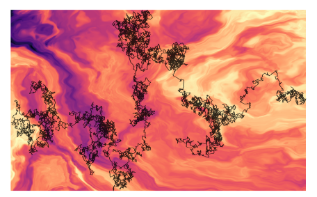
Deviations from the law of Brownian motion, typically referred to as anomalous diffusion, are ubiquitous in science and associated with non-equilibrium phenomena, flows of energy and information, and transport in living systems. In the last years, the booming of machine learning has boosted the development of new methods to detect and characterize anomalous diffusion from individual trajectories, going beyond classical calculations based on the mean squared displacement. We thus designed the AnDi challenge, an open community effort to objectively assess the performance of conventional and novel methods. We developed a python library for generating simulated datasets according to the most popular theoretical models of diffusion. We evaluated 16 methods over 3 different tasks and 3 different dimensions, involving anomalous exponent inference, model classification, and trajectory segmentation. Our analysis provides the first assessment of methods for anomalous diffusion in a variety of realistic conditions of trajectory length and noise. Furthermore, we compared the prediction provided by these methods for several experimental datasets. The results of this study further highlight the role that anomalous diffusion has in defining the biological function while revealing insight into the current state of the field and providing a benchmark for future developers.

Invited Talk by G. Volpe at BIST Symposium on Microscopy, Nanoscopy and Imaging Sciences, Castelldefels, 10 March 2023

DeepTrack 2.1 Logo. (Image from DeepTrack 2.1 Project)
AI and deep learning for microscopy
Giovanni Volpe
BIST Symposium on Microscopy, Nanoscopy and Imaging Sciences
Castedefells, 10 March 2023

Video microscopy has a long history of providing insights and breakthroughs for a broad range of disciplines, from physics to biology. Image analysis to extract quantitative information from video microscopy data has traditionally relied on algorithmic approaches, which are often difficult to implement, time consuming, and computationally expensive. Recently, alternative data-driven approaches using deep learning have greatly improved quantitative digital microscopy, potentially offering automatized, accurate, and fast image analysis. However, the combination of deep learning and video microscopy remains underutilized primarily due to the steep learning curve involved in developing custom deep-learning solutions.

To overcome this issue, we have introduced a software, currently at version DeepTrack 2.1, to design, train and validate deep-learning solutions for digital microscopy. We use it to exemplify how deep learning can be employed for a broad range of applications, from particle localization, tracking and characterization to cell counting and classification. Thanks to its user-friendly graphical interface, DeepTrack 2.1 can be easily customized for user-specific applications, and, thanks to its open-source object-oriented programming, it can be easily expanded to add features and functionalities, potentially introducing deep-learning-enhanced video microscopy to a far wider audience.

Geometric deep learning reveals the spatiotemporal fingerprint of microscopic motion published in Nature Machine Intelligence

Input graph structure including a redundant number of edges. (Image by J. Pineda.)
Geometric deep learning reveals the spatiotemporal fingerprint of microscopic motion
Jesús Pineda, Benjamin Midtvedt, Harshith Bachimanchi, Sergio Noé, Daniel Midtvedt, Giovanni Volpe, Carlo Manzo
Nature Machine Intelligence 5, 71–82 (2023)
arXiv: 2202.06355
doi: 10.1038/s42256-022-00595-0

The characterization of dynamical processes in living systems provides important clues for their mechanistic interpretation and link to biological functions. Thanks to recent advances in microscopy techniques, it is now possible to routinely record the motion of cells, organelles, and individual molecules at multiple spatiotemporal scales in physiological conditions. However, the automated analysis of dynamics occurring in crowded and complex environments still lags behind the acquisition of microscopic image sequences. Here, we present a framework based on geometric deep learning that achieves the accurate estimation of dynamical properties in various biologically-relevant scenarios. This deep-learning approach relies on a graph neural network enhanced by attention-based components. By processing object features with geometric priors, the network is capable of performing multiple tasks, from linking coordinates into trajectories to inferring local and global dynamic properties. We demonstrate the flexibility and reliability of this approach by applying it to real and simulated data corresponding to a broad range of biological experiments.

Faster and more accurate geometrical-optics optical force calculation using neural networks published in ACS Photonics

Focused rays scattered by an ellipsoidal particles (left). Optical torque along y calculated in the x-y plane using ray scattering with a grid of 1600 rays (up, right) and using a trained neural network (down, right). (Image by the Authors of the manuscript.)
Faster and more accurate geometrical-optics optical force calculation using neural networks
David Bronte Ciriza, Alessandro Magazzù, Agnese Callegari, Gunther Barbosa, Antonio A. R. Neves, Maria A. Iatì, Giovanni Volpe, Onofrio M. Maragò
ACS Photonics 10, 234–241 (2023)
doi: 10.1021/acsphotonics.2c01565
arXiv: 2209.04032

Optical forces are often calculated by discretizing the trapping light beam into a set of rays and using geometrical optics to compute the exchange of momentum. However, the number of rays sets a trade-off between calculation speed and accuracy. Here, we show that using neural networks permits one to overcome this limitation, obtaining not only faster but also more accurate simulations. We demonstrate this using an optically trapped spherical particle for which we obtain an analytical solution to use as ground truth. Then, we take advantage of the acceleration provided by neural networks to study the dynamics of an ellipsoidal particle in a double trap, which would be computationally impossible otherwise.

Corneal endothelium assessment in specular microscopy images with Fuchs’ dystrophy via deep regression of signed distance maps published in Biomedical Optics Express

Example of final segmentation with the UNet-dm of the specular microscopy image of a severe case of cornea guttata. (Image by the Authors of the manuscript.)
Corneal endothelium assessment in specular microscopy images with Fuchs’ dystrophy via deep regression of signed distance maps
Juan S. Sierra, Jesus Pineda, Daniela Rueda, Alejandro Tello, Angelica M. Prada, Virgilio Galvis, Giovanni Volpe, Maria S. Millan, Lenny A. Romero, Andres G. Marrugo
Biomedical Optics Express 14, 335-351 (2023)
doi: 10.1364/BOE.477495
arXiv: 2210.07102

Specular microscopy assessment of the human corneal endothelium (CE) in Fuchs’ dystrophy is challenging due to the presence of dark image regions called guttae. This paper proposes a UNet-based segmentation approach that requires minimal post-processing and achieves reliable CE morphometric assessment and guttae identification across all degrees of Fuchs’ dystrophy. We cast the segmentation problem as a regression task of the cell and gutta signed distance maps instead of a pixel-level classification task as typically done with UNets. Compared to the conventional UNet classification approach, the distance-map regression approach converges faster in clinically relevant parameters. It also produces morphometric parameters that agree with the manually-segmented ground-truth data, namely the average cell density difference of -41.9 cells/mm2 (95% confidence interval (CI) [-306.2, 222.5]) and the average difference of mean cell area of 14.8 um2 (95% CI [-41.9, 71.5]). These results suggest a promising alternative for CE assessment.

Single-shot self-supervised object detection in microscopy published in Nature Communications

LodeSTAR tracks the plankton Noctiluca scintillans. (Image by the Authors of the manuscript.)
Single-shot self-supervised particle tracking
Benjamin Midtvedt, Jesús Pineda, Fredrik Skärberg, Erik Olsén, Harshith Bachimanchi, Emelie Wesén, Elin K. Esbjörner, Erik Selander, Fredrik Höök, Daniel Midtvedt, Giovanni Volpe
Nature Communications 13, 7492 (2022)
arXiv: 2202.13546
doi: 10.1038/s41467-022-35004-y

Object detection is a fundamental task in digital microscopy, where machine learning has made great strides in overcoming the limitations of classical approaches. The training of state-of-the-art machine-learning methods almost universally relies on vast amounts of labeled experimental data or the ability to numerically simulate realistic datasets. However, experimental data are often challenging to label and cannot be easily reproduced numerically. Here, we propose a deep-learning method, named LodeSTAR (Localization and detection from Symmetries, Translations And Rotations), that learns to detect microscopic objects with sub-pixel accuracy from a single unlabeled experimental image by exploiting the inherent roto-translational symmetries of this task. We demonstrate that LodeSTAR outperforms traditional methods in terms of accuracy, also when analyzing challenging experimental data containing densely packed cells or noisy backgrounds. Furthermore, by exploiting additional symmetries we show that LodeSTAR can measure other properties, e.g., vertical position and polarizability in holographic microscopy.

Active matter in space published in npj Microgravity

Effect of gravity on matter: Sedimentation and creaming. Fv and Fg represent the viscous force and gravitational force, respectively. (Image by Authors.)
Active matter in space
Giorgio Volpe, Clemens Bechinger, Frank Cichos, Ramin Golestanian, Hartmut Löwen, Matthias Sperl and Giovanni Volpe
npj Microgravity, 8, 54 (2022)
doi: 10.1038/s41526-022-00230-7

In the last 20 years, active matter has been a highly dynamic field of research, bridging fundamental aspects of non-equilibrium thermodynamics with applications to biology, robotics, and nano-medicine. Active matter systems are composed of units that can harvest and harness energy and information from their environment to generate complex collective behaviours and forms of self-organisation. On Earth, gravity-driven phenomena (such as sedimentation and convection) often dominate or conceal the emergence of these dynamics, especially for soft active matter systems where typical interactions are of the order of the thermal energy. In this review, we explore the ongoing and future efforts to study active matter in space, where low-gravity and microgravity conditions can lift some of these limitations. We envision that these studies will help unify our understanding of active matter systems and, more generally, of far-from-equilibrium physics both on Earth and in space. Furthermore, they will also provide guidance on how to use, process and manufacture active materials for space exploration and colonisation.

Recent eLife article on plankton tracking gets featured on Swedish national radio

Planktons imaged under a holographic microscope. (Illustration by J. Heuschele.)
The article Microplankton life histories revealed by holographic microscopy and deep learning gets featured on Vetenskapradion Nyheter (Science radio) operated by Sveriges Radio (Swedish national radio) on November 7, 2022.

The short audio feature (Hologram hjälper forskare att förstå plankton) which highlights the important results of the paper (in Swedish) is now available for public listening.

Vetenskapradion Nyheter airs daily news, reports and in-depth discussions about latest research.

Press release on Tunable critical Casimir forces counteract Casimir-Lifshitz attraction

An illustration of microscopic gold flakes on surface. (Image by F. Schmidt.)
The article Tunable critical Casimir forces counteract Casimir-Lifshitz attraction has been featured in the News of the University of Gothenburg (in English and in Swedish), SISSA-International School of Advanced Studies in Trieste, Italy, Heinrich-Heine-Universität Düsseldorf, and Friedrich-Schiller-Universität Jena.

The study, published in Nature Physics and co-written by researchers at the Soft Matter Lab of the Department of Physics at the University of Gothenburg, demonstrate that tunable repulsive critical Casimir forces can be used to counteract stiction, i.e., the tendency of tiny parts of micro- and nanoelectromechanical devices to stick together, which is caused by the Casimir-Lifshitz interaction.

The study is featured also in Phys.org, NanoWerk.

Here the links to the press releases:
Casimir vs Casimir – using opposing forces to improve nanotechnology (GU, English)
https://www.gu.se/nyheter/casimir-vs-casimir-klaschande-krafter-kan-forbattra-nanotekniken (GU, Swedish)
Casimir vs Casimir – usare forze opposte per migliorare le nanotecnologie (SISSA, Italian)
Casimir vs Casimir – using opposing forces to improve nanotechnology (SISSA, English)
Nano-Bauteile clever voneinander lösen (Heinrich-Heine-Universität Düsseldorf)
Clever method for separating nano-components (Friedrich-Schiller-Universität Jena)
Clever method for separating nano-components (Phys.org)
Clever method for separating nano-components (NanoWerk)

Tunable critical Casimir forces counteract Casimir-Lifshitz attraction published in Nature Physics

Gold flake suspended over a functionalized gold-coated substrate. (Image by F. Schmidt.)
Tunable critical Casimir forces counteract Casimir-Lifshitz attraction
Falko Schmidt, Agnese Callegari, Abdallah Daddi-Moussa-Ider, Battulga Munkhbat, Ruggero Verre, Timur Shegai, Mikael Käll, Hartmut Löwen, Andrea Gambassi and Giovanni Volpe
Nature Physics 19, 271-278 (2023)
arXiv: 2202.10926
doi: 10.1038/s41567-022-01795-6

Casimir forces in quantum electrodynamics emerge between microscopic metallic objects because of the confinement of the vacuum electromagnetic fluctuations occurring even at zero temperature. Their generalization at finite temperature and in material media are referred to as Casimir-Lifshitz forces. These forces are typically attractive, leading to the widespread problem of stiction between the metallic parts of micro- and nanodevices. Recently, repulsive Casimir forces have been experimentally realized but their reliance on specialized materials prevents their dynamic control and thus limits their further applicability. Here, we experimentally demonstrate that repulsive critical Casimir forces, which emerge in a critical binary liquid mixture upon approaching the critical temperature, can be used to actively control microscopic and nanoscopic objects with nanometer precision. We demonstrate this by using critical Casimir forces to prevent the stiction caused by the Casimir-Lifshitz forces. We study a microscopic gold flake above a flat gold-coated substrate immersed in a critical mixture. Far from the critical temperature, stiction occurs because of dominant Casimir-Lifshitz forces. Upon approaching the critical temperature, however, we observe the emergence of repulsive critical Casimir forces that are sufficiently strong to counteract stiction. This experimental demonstration can accelerate the development of micro- and nanodevices by preventing stiction as well as providing active control and precise tunability of the forces acting between their constituent parts.