Artificial Intelligence for particle detection and tracking
Giovanni Volpe
Virtual talk at AMR-Educate Workshop, 13-14 July 2023
Tag: Giovanni Volpe
Invited Lecture by G. Volpe at PSL Soft and Active Matter Days 2023, Paris, 3 July 2023
Giovanni Volpe
PSL Soft and Active Matter Days 2023, Paris, France
Date: 3 July 2023
Invited Talk by G. Volpe at XVII Congress of the Spanish Biophysical Society, Castelldefels, Spain, 30 June 2023
Giovanni Volpe
XVII Congress of the Spanish Biophysical Society, Castelldefels, Spain, 30 June 2023
Date: 30 June 2023
Time: 11:00
Invited Talk by G. Volpe at Active Matter at Surfaces and in Complex Environments, Dresden, 20 June 2023
Giovanni Volpe
Active Matter at Surfaces and in Complex Environments, Dresden, Germany
Date: 20 June 2023
Time: 15:30
Invited Talk by G. Volpe at DINAMO 2023, Svolvaer, Lofoten Islands, Norway, 15 June 2023
Giovanni Volpe
DINAMO 2023, Svolvaer, Lofoten Islands, Norway
Date: 15 June 2023
Time: 08:30
Invited Seminar by G. Volpe at BPPB Seminar, 19 May 2023
Giovanni Volpe
BPPB Seminar 2023
Date: 19 May 2023
Time: 17:00
Invited Seminar by G. Volpe at LOMA, Bordeaux, 2 May 2023
Giovanni Volpe
Seminar at LOMA, Bordeaux
2 May 2023, 14:00
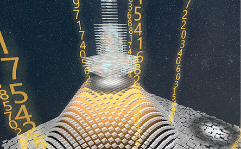
Video microscopy has a long history of providing insights and breakthroughs for a broad range of disciplines, from physics to biology. Image analysis to extract quantitative information from video microscopy data has traditionally relied on algorithmic approaches, which are often difficult to implement, time consuming, and computationally expensive. Recently, alternative data-driven approaches using deep learning have greatly improved quantitative digital microscopy, potentially offering automatized, accurate, and fast image analysis. However, the combination of deep learning and video microscopy remains underutilized primarily due to the steep learning curve involved in developing custom deep-learning solutions. To overcome this issue, we have introduced a software, currently at version DeepTrack 2.1, to design, train and validate deep-learning solutions for digital microscopy.
Plenary Lecture by G. Volpe at SPIE Optics + Optoelectronics, Prague, 25 April 2023
Giovanni Volpe
SPIE Optics + Optoelectronics, Prague, 25 April 2023
Time: 09:45
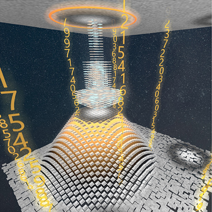
Video microscopy has a long history of providing insights and breakthroughs for a broad range of disciplines, from physics to biology. Image analysis to extract quantitative information from video microscopy data has traditionally relied on algorithmic approaches, which are often difficult to implement, time consuming, and computationally expensive. Recently, alternative data-driven approaches using deep learning have greatly improved quantitative digital microscopy, potentially offering automatized, accurate, and fast image analysis. However, the combination of deep learning and video microscopy remains underutilized primarily due to the steep learning curve involved in developing custom deep-learning solutions.
To overcome this issue, we have introduced a software, currently at version DeepTrack 2.1, to design, train and validate deep-learning solutions for digital microscopy. We use it to exemplify how deep learning can be employed for a broad range of applications, from particle localization, tracking and characterization to cell counting and classification. Thanks to its user-friendly graphical interface, DeepTrack 2.1 can be easily customized for user-specific applications, and, thanks to its open-source object-oriented programming, it can be easily expanded to add features and functionalities, potentially introducing deep-learning-enhanced video microscopy to a far wider audience.
Functional gradients of the medial parietal cortex in a healthy cohort with family history of sporadic Alzheimer’s disease published in Alzheimer’s Research & Therapy
Dániel Veréb, Mite Mijalkov, Yu-Wei Chang, Anna Canal-Garcia, Emiliano Gomez-Ruis, Anne Maass, Sylvia Villeneuve, Giovanni Volpe Joana B. Pereira
Alzheimer’s Research & Therapy 15, 82 (2023)
doi: 10.1186/s13195-023-01228-3
Background
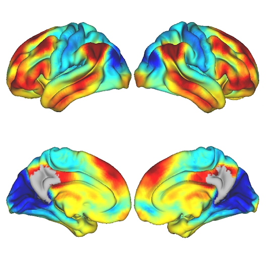
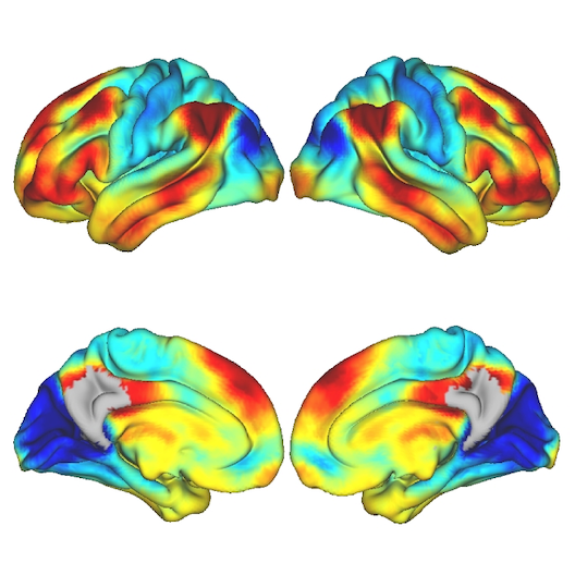
The medial parietal cortex is an early site of pathological protein deposition in Alzheimer’s disease (AD). Previous studies have identified different subregions within this area; however, these subregions are often heterogeneous and disregard individual differences or subtle pathological alterations in the underlying functional architecture. To address this limitation, here we measured the continuous connectivity gradients of the medial parietal cortex and assessed their relationship with cerebrospinal fluid (CSF) biomarkers, ApoE ε4 carriership and memory in asymptomatic individuals at risk to develop AD.
Methods
Two hundred sixty-three cognitively normal participants with a family history of sporadic AD who underwent resting-state and task-based functional MRI using encoding and retrieval tasks were included from the PREVENT-AD cohort. A novel method for characterizing spatially continuous patterns of functional connectivity was applied to estimate functional gradients in the medial parietal cortex during the resting-state and task-based conditions. This resulted in a set of nine parameters that described the appearance of the gradient across different spatial directions. We performed correlation analyses to assess whether these parameters were associated with CSF biomarkers of phosphorylated tau181 (p-tau), total tau (t-tau), and amyloid-ß1-42 (Aß). Then, we compared the spatial parameters between ApoE ε4 carriers and noncarriers, and evaluated the relationship between these parameters and memory.
Results
Alterations involving the superior part of the medial parietal cortex, which was connected to regions of the default mode network, were associated with higher p-tau, t-tau levels as well as lower Aß/p-tau levels during the resting-state condition (p < 0.01). Similar alterations were found in ApoE ε4 carriers compared to non-carriers (p < 0.003). In contrast, lower immediate memory scores were associated with changes in the middle part of the medial parietal cortex, which was connected to inferior temporal and posterior parietal regions, during the encoding task (p = 0.001). No results were found when using conventional connectivity measures.
Conclusions
Functional alterations in the medial parietal gradients are associated with CSF AD biomarkers, ApoE e4 carriership, and lower memory in an asymptomatic cohort with a family history of sporadic AD, suggesting that functional gradients are sensitive to subtle changes associated with early AD stages.
Light, Matter, Action: Shining light on active matter published in ACS Photonics
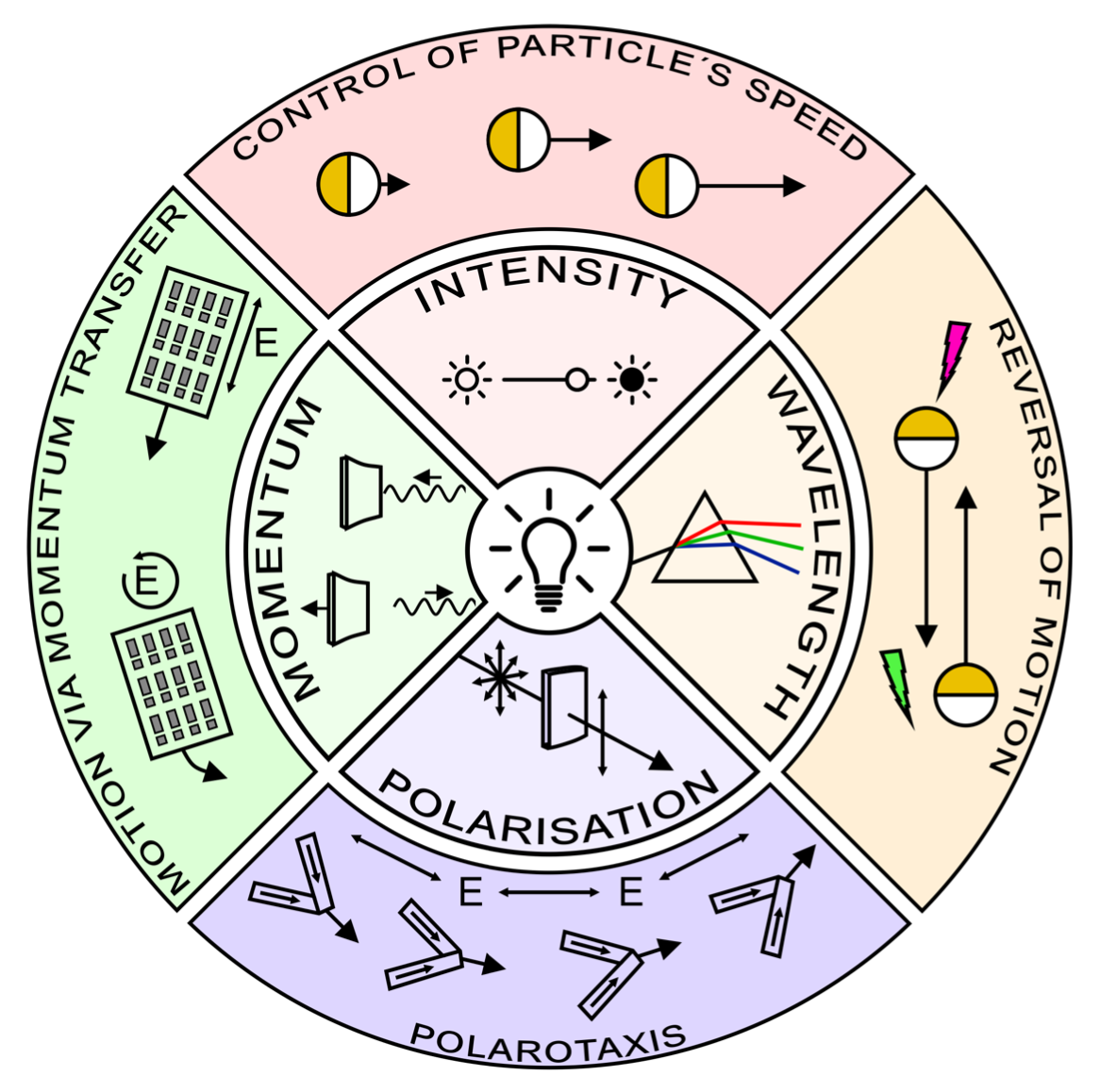
Marcel Rey, Giovanni Volpe, Giorgio Volpe
ACS Photonics, 10, 1188–1201 (2023)
arXiv: 2301.13034
doi: 10.1021/acsphotonics.3c00140
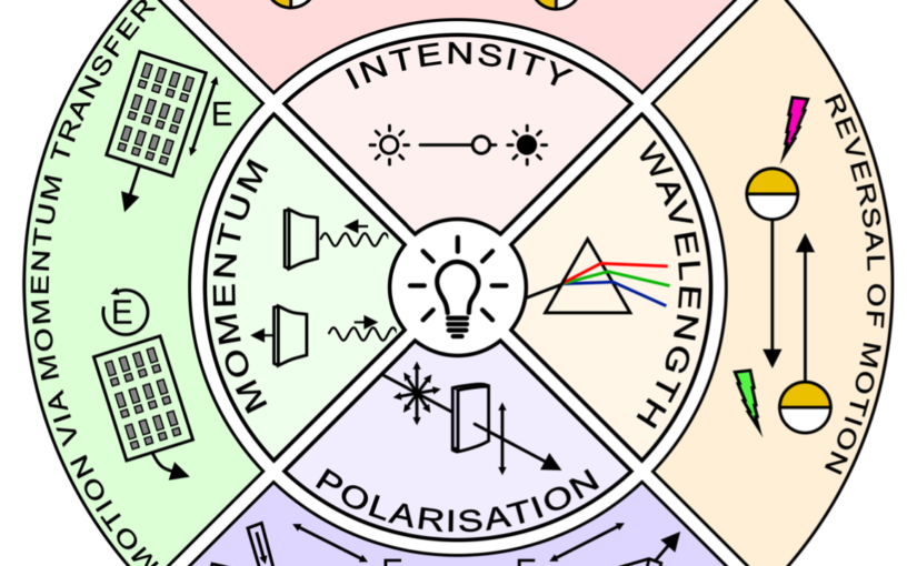
Light carries energy and momentum. It can therefore alter the motion of objects from atomic to astronomical scales. Being widely available, readily controllable and broadly biocompatible, light is also an ideal tool to propel microscopic particles, drive them out of thermodynamic equilibrium and make them active. Thus, light-driven particles have become a recent focus of research in the field of soft active matter. In this perspective, we discuss recent advances in the control of soft active matter with light, which has mainly been achieved using light intensity. We also highlight some first attempts to utilize light’s additional degrees of freedom, such as its wavelength, polarization, and momentum. We then argue that fully exploiting light with all of its properties will play a critical role to increase the level of control over the actuation of active matter as well as the flow of light itself through it. This enabling step will advance the design of soft active matter systems, their functionalities and their transfer towards technological applications.