Angelo Barona Balda, Aykut Argun, Agnese Callegari, Giovanni Volpe
Americal Journal of Physics 92, 847–858 (2024)
doi: 10.1119/5.0125111
arXiv: 2209.04168
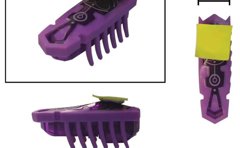
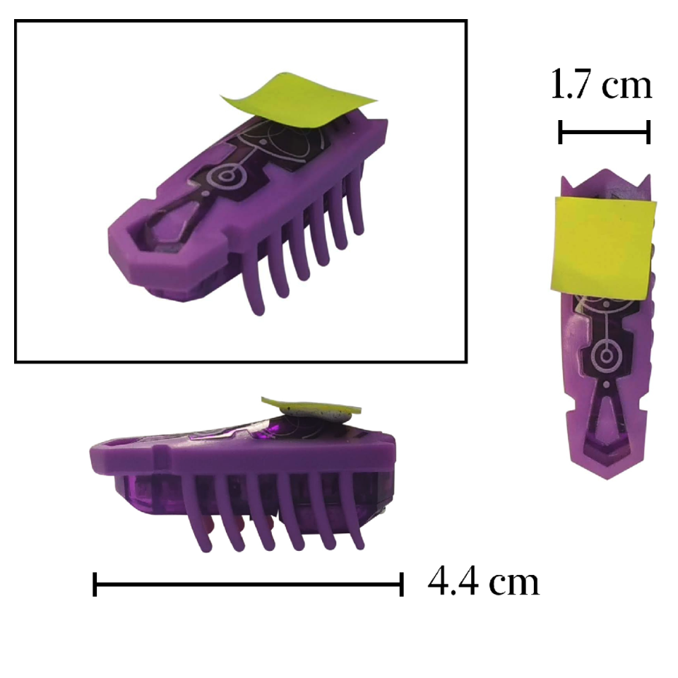
In the past 20 years, active matter has been a very successful research field, bridging the fundamental physics of nonequilibrium thermodynamics with applications in robotics, biology, and medicine. Active particles, contrary to Brownian particles, can harness energy to generate complex motions and emerging behaviors. Most active-matter experiments are performed with microscopic particles and require advanced microfabrication and microscopy techniques. Here, we propose some macroscopic experiments with active matter employing commercially available toy robots (the Hexbugs). We show how they can be easily modified to perform regular and chiral active Brownian motion and demonstrate through experiments fundamental signatures of active systems such as how energy and momentum are harvested from an active bath, how obstacles can sort active particles by chirality, and how active fluctuations induce attraction between planar objects (a Casimir-like effect). These demonstrations enable hands-on experimentation with active matter and showcase widely used analysis methods.