Recent eLife article on plankton tracking gets featured on Swedish national radio

Planktons imaged under a holographic microscope. (Illustration by J. Heuschele.)
The article Microplankton life histories revealed by holographic microscopy and deep learning gets featured on Vetenskapradion Nyheter (Science radio) operated by Sveriges Radio (Swedish national radio) on November 7, 2022.

The short audio feature (Hologram hjälper forskare att förstå plankton) which highlights the important results of the paper (in Swedish) is now available for public listening.

Vetenskapradion Nyheter airs daily news, reports and in-depth discussions about latest research.

Press release on Microplankton life histories revealed by holographic microscopy and deep learning

Planktons imaged under a holographic microscope. (Illustration by J. Heuschele.)
The article Microplankton life histories revealed by holographic microscopy and deep learning has been featured in the news of University of Gothenburg (in English & Swedish) and in the press release of eLife (in English).

The study, now published in eLife, and co-written by researchers at the Soft Matter Lab of the Department of Physics at the University of Gothenburg, demonstrates how the combination of holographic microscopy and deep learning provides a strong complimentary tool in marine microbial ecology. The research allows quantitative assessments of microplankton feeding behaviours, and biomass increase throughout the cell cycle from generation to generation.

The study is featured also in eLife digest.

Here are the links to the press releases:
Researchers combine microscopy with AI to characterise marine microbial food web (eLife, English)
Holographic microscopy provides insights into the life of microplankton (GU, English)
Hologram ger insyn i planktonens liv (GU, Swedish)
The secret lives of microbes (eLife digest)

Microplankton life histories revealed by holographic microscopy and deep learning published in eLife

Tracking of microplankton by holographic optical microscopy and deep learning. (Image by H. Bachimanchi.)
Microplankton life histories revealed by holographic microscopy and deep learning
Harshith Bachimanchi, Benjamin Midtvedt, Daniel Midtvedt, Erik Selander, and Giovanni Volpe
eLife 11, e79760 (2022)
arXiv: 2202.09046
doi: 10.7554/eLife.79760

The marine microbial food web plays a central role in the global carbon cycle. Our mechanistic understanding of the ocean, however, is biased towards its larger constituents, while rates and biomass fluxes in the microbial food web are mainly inferred from indirect measurements and ensemble averages. Yet, resolution at the level of the individual microplankton is required to advance our understanding of the oceanic food web. Here, we demonstrate that, by combining holographic microscopy with deep learning, we can follow microplanktons throughout their lifespan, continuously measuring their three dimensional position and dry mass. The deep learning algorithms circumvent the computationally intensive processing of holographic data and allow rapid measurements over extended time periods. This permits us to reliably estimate growth rates, both in terms of dry mass increase and cell divisions, as well as to measure trophic interactions between species such as predation events. The individual resolution provides information about selectivity, individual feeding rates and handling times for individual microplanktons. This method is particularly useful to explore the flux of carbon through micro-zooplankton, the most important and least known group of primary consumers in the global oceans. We exemplify this by detailed descriptions of micro-zooplankton feeding events, cell divisions, and long term monitoring of single cells from division to division.

Presentation by D. Midtvedt at SPIE-ETAI, San Diego, 23 August 2022

LodeSTAR tracks the plankton Noctiluca scintillans. (Image by the Authors of the manuscript.)
Single-shot self-supervised object detection
Benjamin Midtvedt, Jesus Pineda, Fredrik Skärberg, Erik Olsén, Harshith Bachimanchi, Emelie Wesén, Elin Esbjörner, Erik Selander, Fredrik Höök, Daniel Midtvedt, Giovanni Volpe
Submitted to SPIE-ETAI
Date: 23 August 2022
Time: 2:20 PM (PDT)

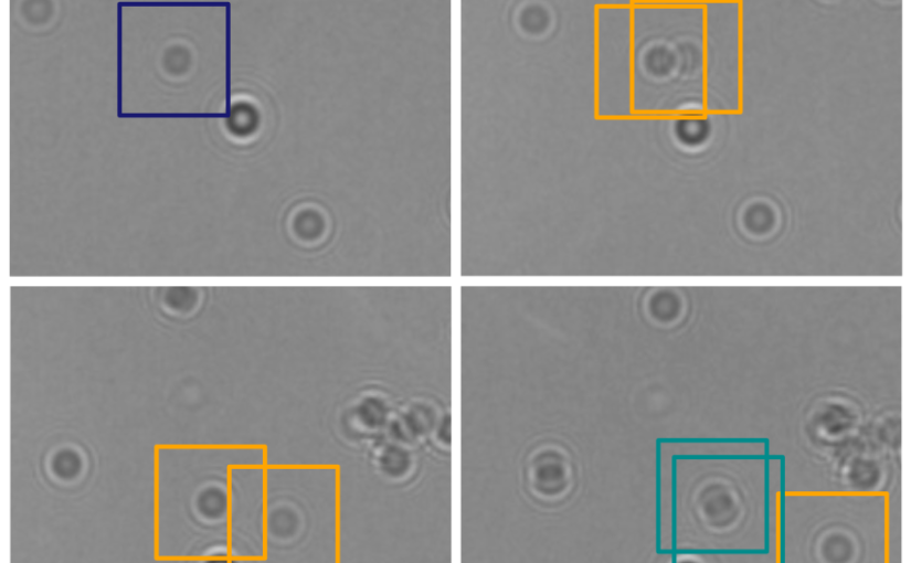
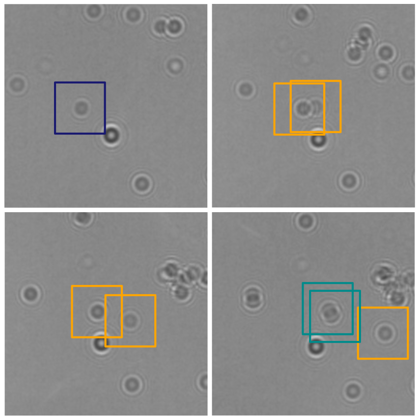
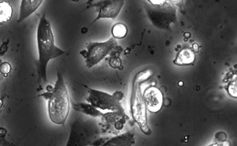
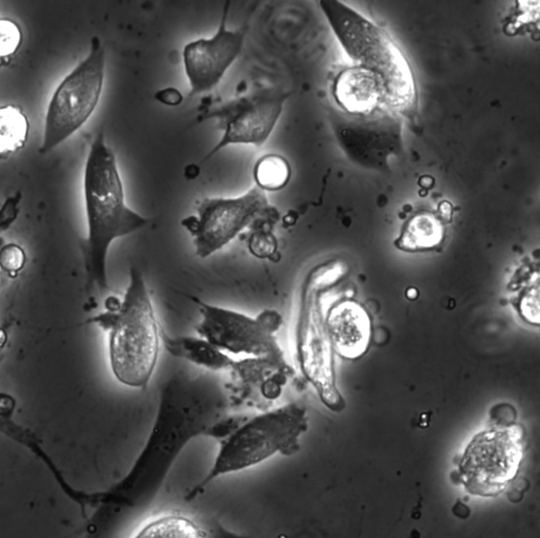
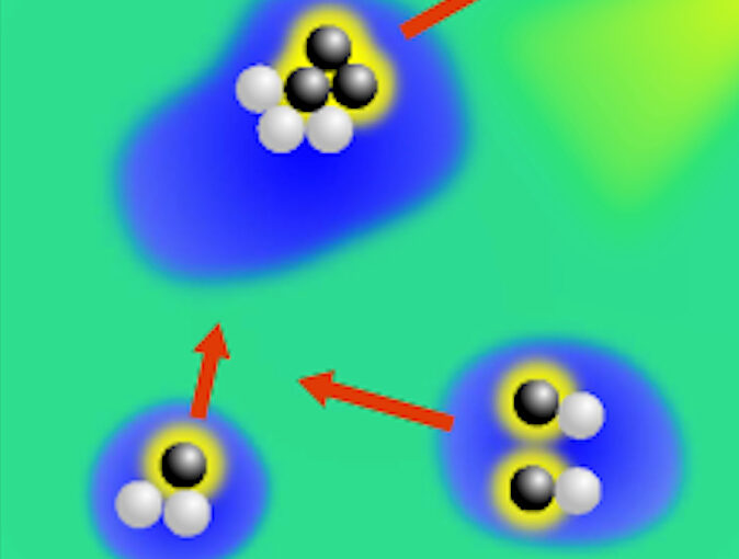
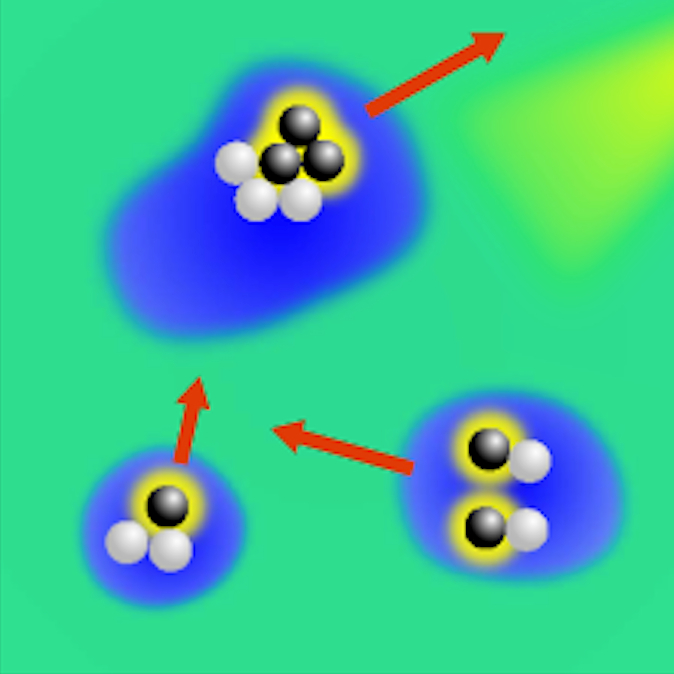
Object detection is a fundamental task in digital microscopy. Recently, machine-learning approaches have made great strides in overcoming the limitations of more classical approaches. The training of state-of-the-art machine-learning methods almost universally relies on either vast amounts of labeled experimental data or the ability to numerically simulate realistic datasets. However, the data produced by experiments are often challenging to label and cannot be easily reproduced numerically. Here, we propose a novel deep-learning method, named LodeSTAR (Low-shot deep Symmetric Tracking And Regression), that learns to detect small, spatially confined, and largely homogeneous objects that have sufficient contrast to the background with sub-pixel accuracy from a single unlabeled experimental image. This is made possible by exploiting the inherent roto-translational symmetries of the data. We demonstrate that LodeSTAR outperforms traditional methods in terms of accuracy. Furthermore, we analyze challenging experimental data containing densely packed cells or noisy backgrounds. We also exploit additional symmetries to extend the measurable particle properties to the particle’s vertical position by propagating the signal in Fourier space and its polarizability by scaling the signal strength. Thanks to the ability to train deep-learning models with a single unlabeled image, LodeSTAR can accelerate the development of high-quality microscopic analysis pipelines for engineering, biology, and medicine.

Soft Matter Lab members present at SPIE Optics+Photonics conference in San Diego, 21-25 August 2022

The Soft Matter Lab participates to the SPIE Optics+Photonics conference in San Diego, CA, USA, 21-25 August 2022, with the presentations listed below.

Giovanni Volpe is also co-author of the presentations:

Dynamic live/apoptotic cell assay using phase-contrast imaging and deep learning on bioRxiv

Phase-contrast image before virtual staining. (Image by the Authors.)
Dynamic live/apoptotic cell assay using phase-contrast imaging and deep learning
Zofia Korczak, Jesús Pineda, Saga Helgadottir, Benjamin Midtvedt, Mattias Goksör, Giovanni Volpe, Caroline B. Adiels
bioRxiv: 10.1101/2022.07.18.500422

Chemical live/dead assay has a long history of providing information about the viability of cells cultured in vitro. The standard methods rely on imaging chemically-stained cells using fluorescence microscopy and further analysis of the obtained images to retrieve the proportion of living cells in the sample. However, such a technique is not only time-consuming but also invasive. Due to the toxicity of chemical dyes, once a sample is stained, it is discarded, meaning that longitudinal studies are impossible using this approach. Further, information about when cells start programmed cell death (apoptosis) is more relevant for dynamic studies. Here, we present an alternative method where cell images from phase-contrast time-lapse microscopy are virtually-stained using deep learning. In this study, human endothelial cells are stained live or apoptotic and subsequently counted using the self-supervised single-shot deep-learning technique (LodeSTAR). Our approach is less labour-intensive than traditional chemical staining procedures and provides dynamic live/apoptotic cell ratios from a continuous cell population with minimal impact. Further, it can be used to extract data from dense cell samples, where manual counting is unfeasible.

DeepTrack won the pitching competition at the Startup Camp 2022. Congrats!

DeepTrack team members (left to right) Henrik, Giovanni and Jesus. (Picture by Jonas Sandwall, Chalmers Ventures.)
The DeepTrack team, composed by Henrik Klein Moberg, Jesus Pineda, Benjamin Midtvedt and Giovanni Volpe, won the pitching competition at the Startup Camp 2022 organised by Chalmers Ventures.

In the event, held on Tuesday, 15 March 2022, 16:00-19:00, the ten teams that had gone through the training at the Startup Camp and developed their company ideas, pitched their companies on stage to a panel of entrepreneur experts, the other nine teams, and all business coaches at Chalmers Ventures. DeepTrack obtained the first place among the ten participants. Congrats!

Here a few pictures from the final pitching event of the Startup Camp.

Henrik. (Picture by Jonas Sandwall, Chalmers Ventures.)
DeepTrack team members (left to right) Henrik, Giovanni and Jesus. (Picture by Jonas Sandwall, Chalmers Ventures.)
Panelists. (Picture by Jonas Sandwall, Chalmers Ventures.)

Featured in:
University of Gothenburg – News and Events: AI tool that analyses microscope images won startup competition and AI-verktyg som analyserar mikroskopbilder vann startup-tävling
(Swedish)

Press release on Active Droploids

The article Active Droploids has been featured in a press release of the University of Gothenburg.

The study, published in Nature Communications, examines a special system of colloidal particles and demonstrates a new kind of active matter, which interacts with and modifies its environment. In the long run, the result of the study can be used for drug delivery inside the human body or to perform sensing of environmental pollutants and their clean-up.

Here the links to the press releases:
English: Feedback creates a new class of active biomimetic materials.
Swedish: Feedback möjliggör en ny form av aktiva biomimetiska material.

The article has been features also in Mirage News, Science Daily, Phys.org, Innovations Report, Informationsdienst Wissenschaft (idw) online, Nanowerk.

Active droploids published in Nature Communications

Active droploids. (Image taken from the article.)
Active droploids
Jens Grauer, Falko Schmidt, Jesús Pineda, Benjamin Midtvedt, Hartmut Löwen, Giovanni Volpe & Benno Liebchen
Nat. Commun. 12, 6005 (2021)
doi: 10.1038/s41467-021-26319-3
arXiv: 2109.10677

Active matter comprises self-driven units, such as bacteria and synthetic microswimmers, that can spontaneously form complex patterns and assemble into functional microdevices. These processes are possible thanks to the out-of-equilibrium nature of active-matter systems, fueled by a one-way free-energy flow from the environment into the system. Here, we take the next step in the evolution of active matter by realizing a two-way coupling between active particles and their environment, where active particles act back on the environment giving rise to the formation of superstructures. In experiments and simulations we observe that, under light-illumination, colloidal particles and their near-critical environment create mutually-coupled co-evolving structures. These structures unify in the form of active superstructures featuring a droplet shape and a colloidal engine inducing self-propulsion. We call them active droploids—a portmanteau of droplet and colloids. Our results provide a pathway to create active superstructures through environmental feedback.

Press release on Extracting quantitative biological information from bright-field cell images using deep learning

Virtually-stained generated image for lipid-droplet.

The article Extracting quantitative biological information from bright-field cell images using deep learning has been featured in a press release of the University of Gothenburg.

The study, recently published in Biophysics Reviews, shows how artificial intelligence can be used to develop faster, cheaper and more reliable information about cells, while also eliminating the disadvantages from using chemicals in the process.

Here the links to the press releases on Cision:
Swedish: Effektivare studier av celler med ny AI-metod
English: More effective cell studies using new AI method

Here the links to the press releases in the News of the University of Gothenburg:
Swedish: Effektivare studier av celler med ny AI-metod
English: More effective cell studies using new AI method