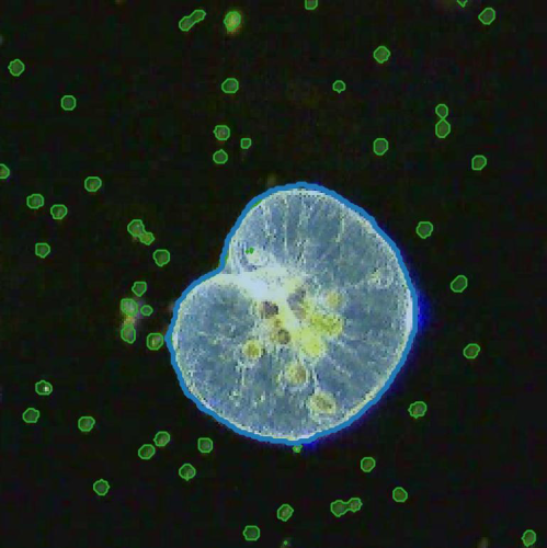
Segmentation of two plankton species using deep learning (N. scintillans in blue, D. tertiolecta in green). (Image by H. Bachimanchi.)Deep-learning-powered data analysis in plankton ecology
Harshith Bachimanchi, Matthew I. M. Pinder, Chloé Robert, Pierre De Wit, Jonathan Havenhand, Alexandra Kinnby, Daniel Midtvedt, Erik Selander, Giovanni Volpe
Limnology and Oceanography Letters (2024)
doi: 10.1002/lol2.10392
arXiv: 2309.08500
The implementation of deep learning algorithms has brought new perspectives to plankton ecology. Emerging as an alternative approach to established methods, deep learning offers objective schemes to investigate plankton organisms in diverse environments. We provide an overview of deep-learning-based methods including detection and classification of phytoplankton and zooplankton images, foraging and swimming behavior analysis, and finally ecological modeling. Deep learning has the potential to speed up the analysis and reduce the human experimental bias, thus enabling data acquisition at relevant temporal and spatial scales with improved reproducibility. We also discuss shortcomings and show how deep learning architectures have evolved to mitigate imprecise readouts. Finally, we suggest opportunities where deep learning is particularly likely to catalyze plankton research. The examples are accompanied by detailed tutorials and code samples that allow readers to apply the methods described in this review to their own data.
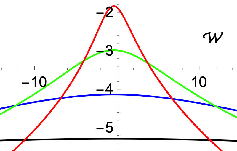
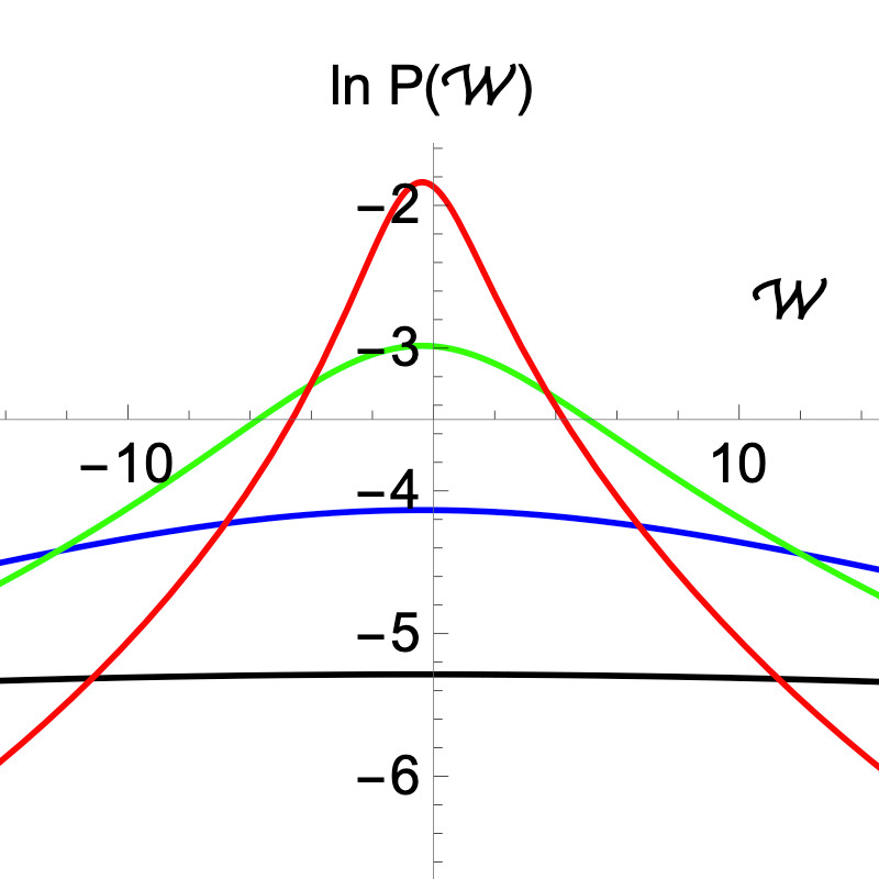
Angular velocity in the steady-state. (Excerpt from Fig. 2 of the manuscript.)Destructive effect of fluctuations on the performance of a Brownian gyrator
Pascal Viot, Aykut Argun, Giovanni Volpe, Alberto Imparato, Lamberto Rondoni, Gleb Oshanin
Soft Matter, 20, 3154-3160 (2024)
arxiv: 2307.05248
doi: 10.1039/D3SM01606D
The Brownian gyrator (BG) is often called a minimal model of a nano-engine performing a rotational motion, judging solely upon the fact that in non-equilibrium conditions its torque, specific angular momentum L and specific angular velocity W have non-zero mean values. For a time-discretised (with time-step δt) model we calculate here the previously unknown probability density functions (PDFs) of L and W. We show that for finite δt, the PDF of L has exponential tails and all moments are therefore well-defined. At the same time, this PDF appears to be effectively broad – the noise-to-signal ratio is generically bigger than unity meaning that L is strongly not self-averaging. Concurrently, the PDF of W exhibits heavy power-law tails and its mean W is the only existing moment. The BG is therefore not an engine in the common sense: it does not exhibit regular rotations on each run and its fluctuations are not only a minor nuisance – on contrary, their effect is completely destructive for the performance. Our theoretical predictions are confirmed by numerical simulations and experimental data. We discuss some plausible improvements of the model which may result in a more systematic rotational motion.
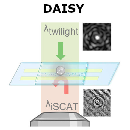
Conceptual schematic of dual-angle interferometric scattering microscopy (DAISY). (Image by the Authors of the manuscript.)Dual-Angle Interferometric Scattering Microscopy for Optical Multiparametric Particle Characterization
Erik Olsén, Berenice García Rodríguez, Fredrik Skärberg, Petteri Parkkila, Giovanni Volpe, Fredrik Höök, and Daniel Sundås Midtvedt
Nano Letters, 24(6), 1874-1881 (2024)
doi: 10.1021/acs.nanolett.3c03539
arXiv: 2309.07572
Traditional single-nanoparticle sizing using optical microscopy techniques assesses size via the diffusion constant, which requires suspended particles to be in a medium of known viscosity. However, these assumptions are typically not fulfilled in complex natural sample environments. Here, we introduce dual-angle interferometric scattering microscopy (DAISY), enabling optical quantification of both size and polarizability of individual nanoparticles (radius <170 nm) without requiring a priori information regarding the surrounding media or super-resolution imaging. DAISY achieves this by combining the information contained in concurrently measured forward and backward scattering images through twilight off-axis holography and interferometric scattering (iSCAT). Going beyond particle size and polarizability, single-particle morphology can be deduced from the fact that the hydrodynamic radius relates to the outer particle radius, while the scattering-based size estimate depends on the internal mass distribution of the particles. We demonstrate this by differentiating biomolecular fractal aggregates from spherical particles in fetal bovine serum at the single-particle level.
Different sampling methods for the trajectory of a particle. (Adapted from the manuscript.)Optimal calibration of optical tweezers with arbitrary integration time and sampling frequencies — A general framework
Laura Pérez-García, Martin Selin, Antonio Ciarlo, Alessandro Magazzù, Giuseppe Pesce, Antonio Sasso, Giovanni Volpe, Isaac Pérez Castillo, Alejandro V. Arzola
Biomedical Optics Express, 14, 6442-6469 (2023)
doi: 10.1364/BOE.495468
arXiv: 2305.07245
Optical tweezers (OT) have become an essential technique in several fields of physics, chemistry, and biology as precise micromanipulation tools and microscopic force transducers. Quantitative measurements require the accurate calibration of the trap stiffness of the optical trap and the diffusion constant of the optically trapped particle. This is typically done by statistical estimators constructed from the position signal of the particle, which is recorded by a digital camera or a quadrant photodiode. The finite integration time and sampling frequency of the detector need to be properly taken into account. Here, we present a general approach based on the joint probability density function of the sampled trajectory that corrects exactly the biases due to the detector’s finite integration time and limited sampling frequency, providing theoretical formulas for the most widely employed calibration methods: equipartition, mean squared displacement, autocorrelation, power spectral density, and force reconstruction via maximum-likelihood-estimator analysis (FORMA). Our results, tested with experiments and Monte Carlo simulations, will permit users of OT to confidently estimate the trap stiffness and diffusion constant, extending their use to a broader set of experimental conditions.
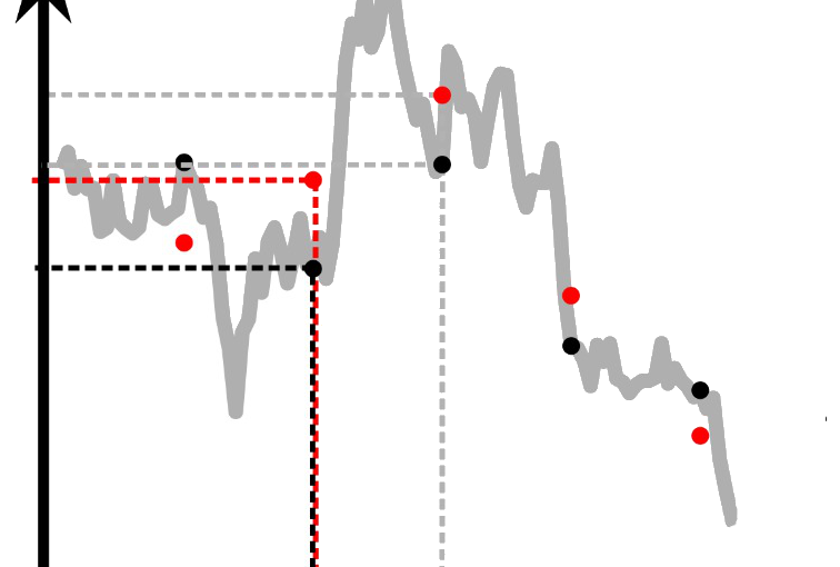
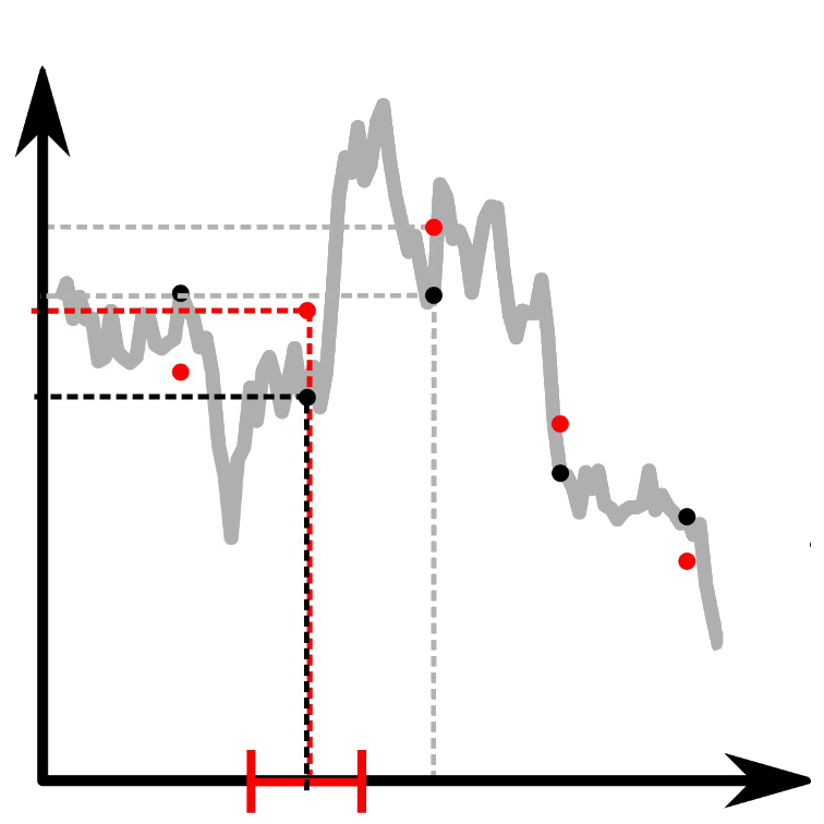
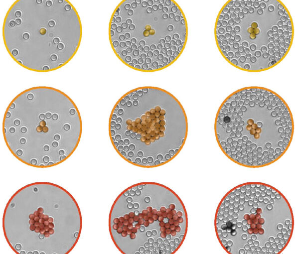
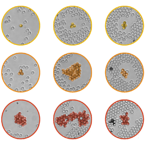
Non-monotonic size dependence of group formation on environmental crowding. (Excerpt from Fig. 2 of the manuscript.)Environmental Memory Boosts Group Formation of Clueless Individuals
Cristóvão S. Dias, Manish Trivedi, Giovanni Volpe, Nuno A. M. Araújo, Giorgio Volpe
Nature Communications, 14, 7324 (2023)
doi: 10.1038/s41467-023-43099-0
arXiv: 2306.00516
The formation of groups of interacting individuals improves performance and fitness in many decentralised systems, from micro-organisms to social insects, from robotic swarms to artificial intelligence algorithms. Often, group formation and high-level coordination in these systems emerge from individuals with limited information-processing capabilities implementing low-level rules of communication to signal to each other. Here, we show that, even in a community of clueless individuals incapable of processing information and communicating, a dynamic environment can coordinate group formation by transiently storing memory of the earlier passage of individuals. Our results identify a new mechanism of indirect coordination via shared memory that is primarily promoted and reinforced by dynamic environmental factors, thus overshadowing the need for any form of explicit signalling between individuals. We expect this pathway to group formation to be relevant for understanding and controlling self-organisation and collective decision making in both living and artificial active matter in real-life environments.
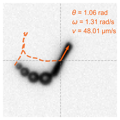
Bubble-propelled micromotors tracked by deep learning. (Image by H. Bachimanchi.)Bubble-propelled micromotors for ammonia generation
Rebeca Ferrer Campos, Harshith Bachimanchi, Giovanni Volpe, Katherine Villa
Nanoscale (2023)
doi: 10.1039/D3NR03804A
Micromotors have emerged as promising tools for environmental remediation, thanks to their ability to autonomously navigate and perform specific tasks at the microscale. In this study, we present the development of MnO2 tubular micromotors modified with laccase for enhanced oxidation of organic pollutants by providing an additional oxidative catalytic pathway for pollutant removal. These modified micromotors exhibit efficient ammonia generation through the catalytic decomposition of urea, suggesting their potential application in the field of green energy generation. Compared to bare micromotors, the MnO2 micromotors modified with laccase exhibit a 20% increase in rhodamine B degradation. Moreover, the generation of ammonia increased from 2 to 31 ppm in only 15 min, evidencing their high catalytic activity. To enable precise tracking of the micromotors and measurement of their speed, a deep-learning-based tracking system was developed. Overall, this work expands the potential applicability of bio-catalytic tubular micromotors in the energy field.
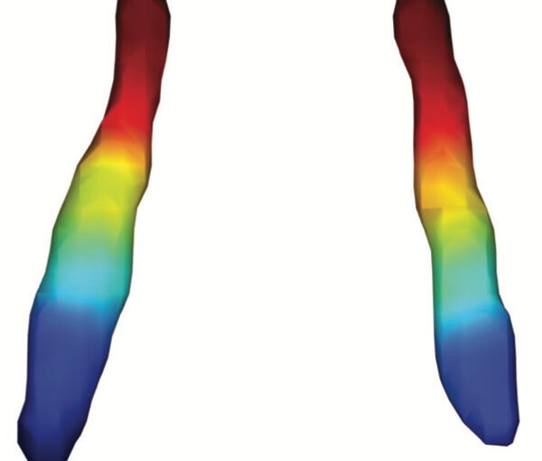
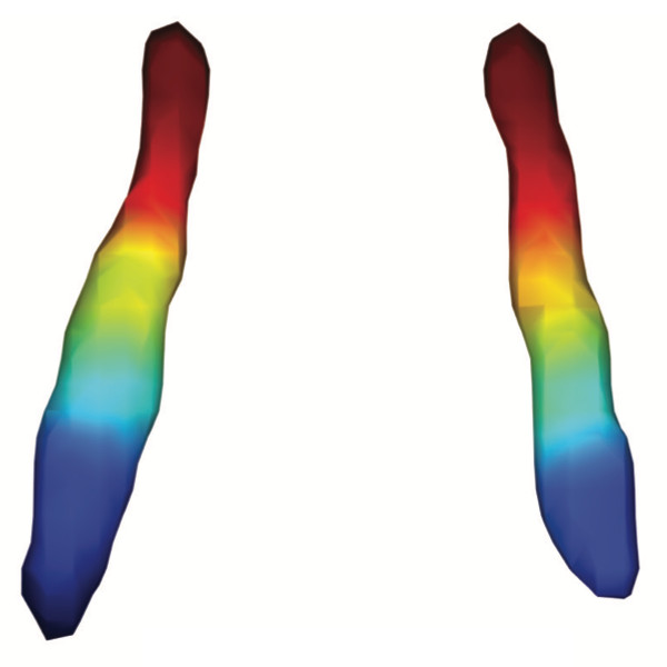
Average functional gradients of the locus coeruleus in the CamCAN 3T dataset. (Image from the publication.)Age-related differences in the functional topography of the locus coeruleus and their implications for cognitive and affective functions
Dániel Veréb, Mite Mijalkov, Anna Canal-Garcia, Yu-Wei Chang, Emiliano Gomez-Ruiz, Blanca Zufiria Gerboles, Miia Kivipelto, Per Svenningsson, Henrik Zetterberg, Giovanni Volpe, Matthew Betts, Heidi IL Jacobs, Joana B Pereira
eLife 12, RP87188 (2023)
doi: 10.7554/eLife.87188.3
The locus coeruleus (LC) is an important noradrenergic nucleus that has recently attracted a lot of attention because of its emerging role in cognitive and psychiatric disorders. Although previous histological studies have shown that the LC has heterogeneous connections and cellular features, no studies have yet assessed its functional topography in vivo, how this heterogeneity changes over aging, and whether it is associated with cognition and mood. Here, we employ a gradient-based approach to characterize the functional heterogeneity in the organization of the LC over aging using 3T resting-state fMRI in a population-based cohort aged from 18 to 88 years of age (Cambridge Centre for Ageing and Neuroscience cohort, n=618). We show that the LC exhibits a rostro-caudal functional gradient along its longitudinal axis, which was replicated in an independent dataset (Human Connectome Project [HCP] 7T dataset, n=184). Although the main rostro-caudal direction of this gradient was consistent across age groups, its spatial features varied with increasing age, emotional memory, and emotion regulation. More specifically, a loss of rostral-like connectivity, more clustered functional topography, and greater asymmetry between right and left LC gradients was associated with higher age and worse behavioral performance. Furthermore, participants with higher-than-normal Hospital Anxiety and Depression Scale (HADS) ratings exhibited alterations in the gradient as well, which manifested in greater asymmetry. These results provide an in vivo account of how the functional topography of the LC changes over aging, and imply that spatial features of this organization are relevant markers of LC-related behavioral measures and psychopathology.
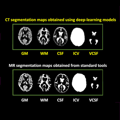
Imaging-based volumetric measures. (Image by the Authors of the manuscript.)CT-based volumetric measures obtained through deep learning: Association with biomarkers of neurodegeneration
Meera Srikrishna, Nicholas J. Ashton, Alexis Moscoso, Joana B. Pereira, Rolf A. Heckemann, Danielle van Westen, Giovanni Volpe, Joel Simrén, Anna Zettergren, Silke Kern, Lars-Olof Wahlund, Bibek Gyanwali, Saima Hilal, Joyce Chong Ruifen, Henrik Zetterberg, Kaj Blennow, Eric Westman, Christopher Chen, Ingmar Skoog, Michael Schöll
Alzheimer’s & Dementia 20, 629–640 (2024)
arXiv: 2401.06260
doi: 10.1002/alz.13445
INTRODUCTION
Cranial computed tomography (CT) is an affordable and widely available imaging modality that is used to assess structural abnormalities, but not to quantify neurodegeneration. Previously we developed a deep-learning–based model that produced accurate and robust cranial CT tissue classification.
MATERIALS AND METHODS
We analyzed 917 CT and 744 magnetic resonance (MR) scans from the Gothenburg H70 Birth Cohort, and 204 CT and 241 MR scans from participants of the Memory Clinic Cohort, Singapore. We tested associations between six CT-based volumetric measures (CTVMs) and existing clinical diagnoses, fluid and imaging biomarkers, and measures of cognition.
RESULTS
CTVMs differentiated cognitively healthy individuals from dementia and prodromal dementia patients with high accuracy levels comparable to MR-based measures. CTVMs were significantly associated with measures of cognition and biochemical markers of neurodegeneration.
DISCUSSION
These findings suggest the potential future use of CT-based volumetric measures as an informative first-line examination tool for neurodegenerative disease diagnostics after further validation.
Illustration of resting state network activity. (Image by the Authors of the manuscript.)Peripheral inflammatory subgroup differences in anterior Default Mode network and multiplex functional network topology are associated with cognition in psychosis
Paulo Lizano, Chelsea Kiely, Mite Mijalkov, Shashwath A. Meda, Sarah K. Keedy, Dung Hoang, Victor Zeng, Olivia Lutz, Joana B. Pereira, Elena I. Ivleva, Giovanni Volpe, Yanxun Xu, Adam M. Lee, Leah H. Rubin, S Kristian Hill, Brett A. Clementz, Carol A. Tamminga, Godfrey D. Pearlson, John A. Sweeney, Elliot S. Gershon, Matcheri S. Keshavan, Jeffrey R. Bishop
Brain Behavior and Immunity, 114, 3-15 (2023)
doi: 10.1016/j.bbi.2023.07.014
Introduction
High-inflammation subgroups of patients with psychosis demonstrate cognitive deficits and neuroanatomical alterations. Systemic inflammation assessed using IL-6 and C-reactive protein may alter functional connectivity within and between resting-state networks, but the cognitive and clinical implications of these alterations remain unknown. We aim to determine the relationships of elevated peripheral inflammation subgroups with resting-state functional networks and cognition in psychosis spectrum disorders.
Methods
Serum and resting-state fMRI were collected from psychosis probands (schizophrenia, schizoaffective, psychotic bipolar disorder) and healthy controls (HC) from the B-SNIP1 (Chicago site) study who were stratified into inflammatory subgroups based on factor and cluster analyses of 13 cytokines (HC Low n = 32, Proband Low n = 65, Proband High n = 29). Nine resting-state networks derived from independent component analysis were used to assess functional and multilayer connectivity. Inter-network connectivity was measured using Fisher z-transformation of correlation coefficients. Network organization was assessed by investigating networks of positive and negative connections separately, as well as investigating multilayer networks using both positive and negative connections. Cognition was assessed using the Brief Assessment of Cognition in Schizophrenia. Linear regressions, Spearman correlations, permutations tests and multiple comparison corrections were used for analyses in R.
Results
Anterior default mode network (DMNa) connectivity was significantly reduced in the Proband High compared to Proband Low (Cohen’s d = -0.74, p = 0.002) and HC Low (d = -0.85, p = 0.0008) groups. Inter-network connectivity between the DMNa and the right-frontoparietal networks was lower in Proband High compared to Proband Low (d = -0.66, p = 0.004) group. Compared to Proband Low, the Proband High group had lower negative (d = 0.54, p = 0.021) and positive network (d = 0.49, p = 0.042) clustering coefficient, and lower multiplex network participation coefficient (d = -0.57, p = 0.014). Network findings in high inflammation subgroups correlate with worse verbal fluency, verbal memory, symbol coding, and overall cognition.
Conclusion
These results expand on our understanding of the potential effects of peripheral inflammatory signatures and/or subgroups on network dysfunction in psychosis and how they relate to worse cognitive performance. Additionally, the novel multiplex approach taken in this study demonstrated how inflammation may disrupt the brain’s ability to maintain healthy co-activation patterns between the resting-state networks while inhibiting certain connections between them.
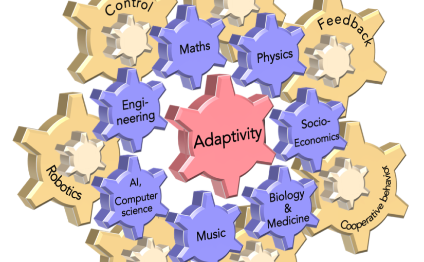
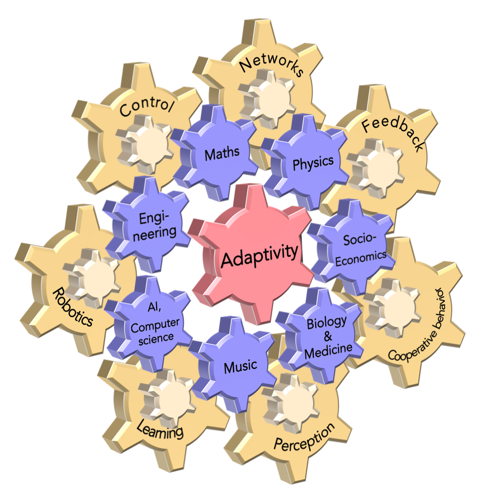
Adaptivity across different scientific disciplines (blue) and applications (yellow) as well as its strong inter- linking and interlocking, similar to a system of gears. (Image by the Authors of the manuscript)Perspectives on adaptive dynamical systems
Jakub Sawicki, Rico Berner, Sarah A. M. Loos, Mehrnaz Anvari, Rolf Bader, Wolfram Barfuss, Nicola Botta, Nuria Brede, Igor Franović, Daniel J. Gauthier, Sebastian Goldt, Aida Hajizadeh, Philipp Hövel, Omer Karin, Philipp Lorenz-Spreen, Christoph Miehl, Jan Mölter, Simona Olmi, Eckehard Schöll, Alireza Seif, Peter A. Tass, Giovanni Volpe, Serhiy Yanchuk, Jürgen Kurths
Chaos 33, 071501 (2023)
doi: 10.1063/5.0147231
arXiv: 2303.01459
Adaptivity is a dynamical feature that is omnipresent in nature, socio-economics, and technology. For example, adaptive couplings appear in various real-world systems like the power grid, social, and neural networks, and they form the backbone of closed-loop control strategies and machine learning algorithms. In this article, we provide an interdisciplinary perspective on adaptive systems. We reflect on the notion and terminology of adaptivity in different disciplines and discuss which role adaptivity plays for various fields. We highlight common open challenges, and give perspectives on future research directions, looking to inspire interdisciplinary approaches.