Gideon is a Master student in the Complex Adaptive Systems at Chalmers University of Technology.
During his time at the Soft Matter Lab, he will work on his Master thesis project on particle representation and graph neural networks.
Gideon is a Master student in the Complex Adaptive Systems at Chalmers University of Technology.
During his time at the Soft Matter Lab, he will work on his Master thesis project on particle representation and graph neural networks.
An anomalous competition: assessment of methods for anomalous diffusion through a community effort
Carlo Manzo, Giovanni Volpe
Submitted to SPIE-ETAI
Date: 25 August 2022
Time: 9:00 (PDT)
Deviations from the law of Brownian motion, typically referred to as anomalous diffusion, are ubiquitous in science and associated with non-equilibrium phenomena, flows of energy and information, and transport in living systems. In the last years, the booming of machine learning has boosted the development of new methods to detect and characterize anomalous diffusion from individual trajectories, going beyond classical calculations based on the mean squared displacement. We thus designed the AnDi challenge, an open community effort to objectively assess the performance of conventional and novel methods. We developed a python library for generating simulated datasets according to the most popular theoretical models of diffusion. We evaluated 16 methods over 3 different tasks and 3 different dimensions, involving anomalous exponent inference, model classification, and trajectory segmentation. Our analysis provides the first assessment of methods for anomalous diffusion in a variety of realistic conditions of trajectory length and noise. Furthermore, we compared the prediction provided by these methods for several experimental datasets. The results of this study further highlight the role that anomalous diffusion has in defining the biological function while revealing insight into the current state of the field and providing a benchmark for future developers.
Presenter: Giovanni Volpe
Periodic feedback effect in counterpropagating intracavity optical tweezers
Antonio Ciarlo, Giuseppe Pesce, Fatemeh Kalantarifard, Parviz Elahi, Agnese Callegari, Giovanni Volpe, Antonio Sasso
Submitted to SPIE-OTOM
Date: 24 August 2022
Time: 14:00 (PDT)
Intracavity optical tweezers are a powerful tool to trap microparticles in water using the nonlinear feedback effect produced by the particle motion when it is trapped inside the laser cavity. In such systems two configurations are possible: a single-beam configuration and counterpropagating one. A removable isolator allows to switch between these configurations by suppressing one of the beams. Trapping a particle in the counterpropagating configuration, the measure of the optical power shows a feedback effect for each beam, that is present also when the two beams are misaligned and the trapped particle periodically jumps between them.
Label-free characterization of biological matter across scales
Daniel Midtvedt, Erik Olsén, Benjamin Midtvedt, Elin K. Esbjörner, Fredrik Skärberg, Berenice Garcia, Caroline B. Adiels, Fredrik Höök, Giovanni Volpe
SPIE-ETAI
Date: 24 August 2022
Time: 09:10 (PDT)
Agnese Callegari, Mathias Samuelsson, Antonio Ciarlo, Giuseppe Pesce, David Bronte Ciriza, Alessandro Magazzù, Onofrio M. Maragò, Antonio Sasso, Giovanni Volpe
Submitted to SPIE-ETAI
Date: 23 August 2022
Time: 13:40 (PDT)
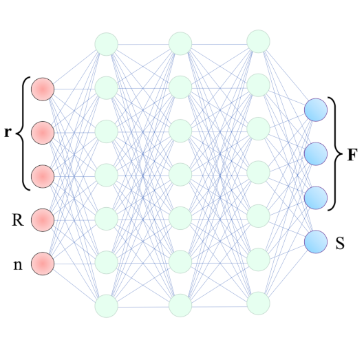
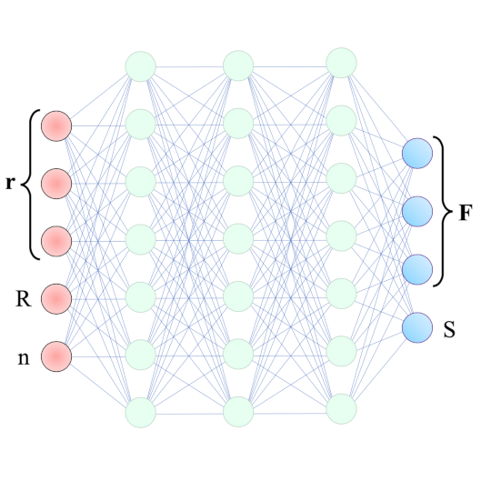
Intracavity optical tweezers have been proven successful for trapping microscopic particles at very low average power intensity – much lower than the one in standard optical tweezers. This feature makes them particularly promising for the study of biological samples. The modeling of such systems, though, requires time-consuming numerical simulations that affect its usability and predictive power. With the help of machine learning, we can overcome the numerical bottleneck – the calculation of optical forces, torques, and losses – reproduce the results in the literature and generalize to the case of counterpropagating-beams intracavity optical trapping.
Henrik Klein Moberg, Christoph Langhammer, Daniel Midtvedt, Barbora Spackova, Bohdan Yeroshenko, David Albinsson, Joachim Fritzsche, Giovanni Volpe
Submitted to SPIE-ETAI
Date: 23 August 2022
Time: 9:15 (PDT)
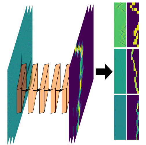
We show that a custom ResNet-inspired CNN architecture trained on simulated biomolecule trajectories surpasses the performance of standard algorithms in terms of tracking and determining the molecular weight and hydrodynamic radius of biomolecules in the low-kDa regime in NSM optical microscopy. We show that high accuracy and precision is retained even below the 10-kDa regime, constituting approximately an order of magnitude improvement in limit of detection compared to current state-of-the-art, enabling analysis of hitherto elusive species of biomolecules such as cytokines (~5-25 kDa) important for cancer research and the protein hormone insulin (~5.6 kDa), potentially opening up entirely new avenues of biological research.
Fredrik Skärberg, Erik Olsén, Benjamin Midtvedt, Emelie V. Wesén, Elin K. Esbjörner, Giovanni Volpe, Fredrik Höök, Daniel Midtvedt
Submitted to SPIE-ETAI
Date: 22 August 2022
Time: 17:30 (PDT)
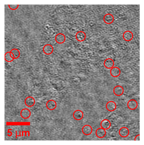
Optical characterization of nanoparticles in/outside cells is a challenging task, as the scattering from the nanoparticles is distorted by the scattering from the cells. In this work, we demonstrate a framework for optical mass quantification of intra- and extracellular nanoparticles by leveraging a novel deep learning method, LodeSTAR, in combination with off-axis twilight holography. The result provides new means for the exploration of nanoparticle/cell interactions.
Zofia Korczak, Jesús D. Pineda, Saga Helgadottir, Benjamin Midtvedt, Mattias Goksör, Giovanni Volpe, Caroline B. Adiels
Submitted to SPIE-ETAI
Date: 22 August 2022
Time: 17:30 (PDT)
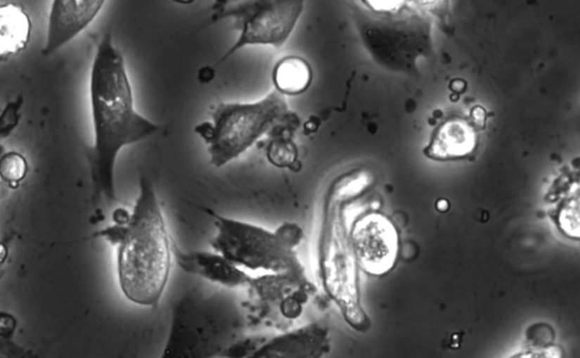
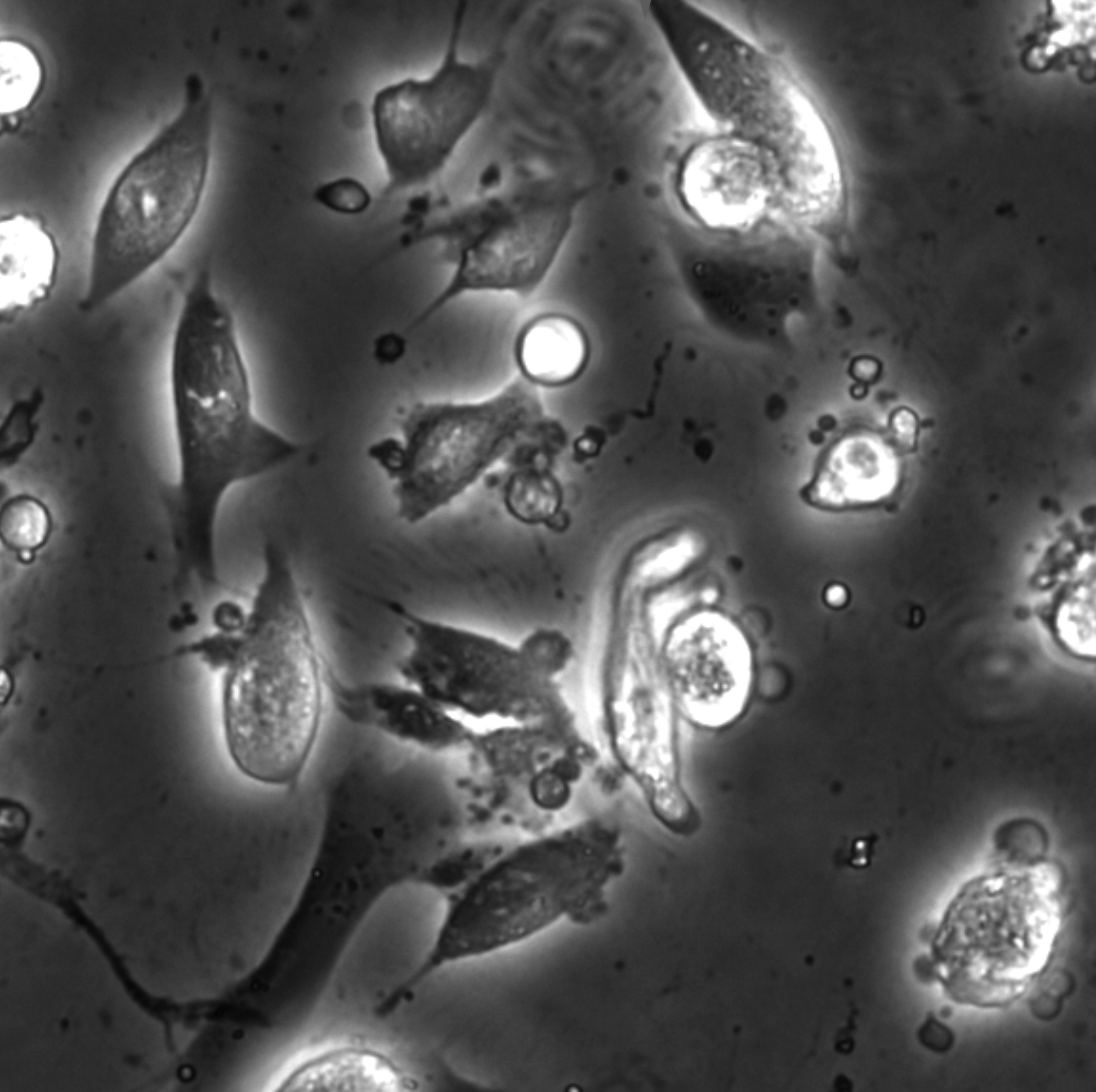
In vitro cell cultures rely on that the cultured cells thrive and behave in a physiologically relevant way. A standard approach to evaluate cells behaviour is to perform chemical staining in which fluorescent probes are added to the cell culture for further imaging and analysis. However, such a technique is invasive and sometimes even toxic to cells, hence, alternative methods are requested. Here, we describe an analysis method for the detection and discrimination of live and apoptotic cells using deep learning. This approach is less labour-intensive than traditional chemical staining procedures and enables cell imaging with minimal impact.
The Soft Matter Lab participates to the SPIE Optics+Photonics conference in San Diego, CA, USA, 21-25 August 2022, with the presentations listed below.
Giovanni Volpe is also co-author of the presentations:
Quantitative Digital Microscopy with Deep Learning
Giovanni Volpe
19 August 2022, 14:40 (PDT)
At the intersection of Photonics, Neuroscience, and AI
Ozcan Lab, UCLA, 19 August 2022
Video microscopy has a long history of providing insights and breakthroughs for a broad range of disciplines, from physics to biology. Image analysis to extract quantitative information from video microscopy data has traditionally relied on algorithmic approaches, which are often difficult to implement, time consuming, and computationally expensive. Recently, alternative data-driven approaches using deep learning have greatly improved quantitative digital microscopy, potentially offering automatized, accurate, and fast image analysis. However, the combination of deep learning and video microscopy remains underutilized primarily due to the steep learning curve involved in developing custom deep-learning solutions. To overcome this issue, we have introduced a software, currently at version DeepTrack 2.1, to design, train and validate deep-learning solutions for digital microscopy. We use it to exemplify how deep learning can be employed for a broad range of applications, from particle localization, tracking and characterization to cell counting and classification. Thanks to its user-friendly graphical interface, DeepTrack 2.1 can be easily customized for user-specific applications, and, thanks to its open-source object-oriented programming, it can be easily expanded to add features and functionalities, potentially introducing deep-learning-enhanced video microscopy to a far wider audience.