The Soft Matter Lab participates to the SPIE Optics+Photonics conference in San Diego, CA, USA, 21-25 August 2022, with the presentations listed below.
- Martin Selin: Scalable construction of quantum dot arrays using optical tweezers and deep learning (@SPIE)
22 August 2022 • 11:05 AM – 11:25 AM PDT | Conv. Ctr. Room 5A
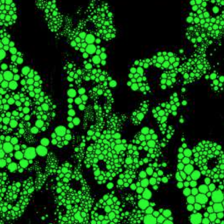
- Fredrik Skärberg: Holographic characterisation of biological nanoparticles using deep learning (@SPIE)
22 August 2022 • 5:30 PM – 7:30 PM PDT | Conv. Ctr. Exhibit Hall B1
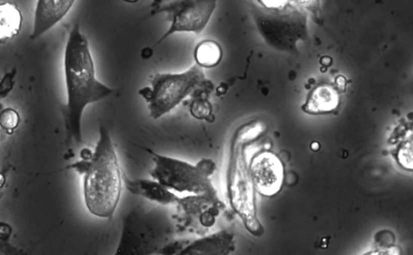
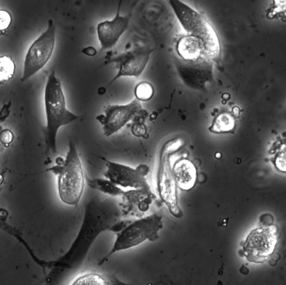
- Zofia Korczak: Dynamic virtual live/apoptotic cell assay using deep learning (@SPIE)
22 August 2022 • 5:30 PM – 7:30 PM PDT | Conv. Ctr. Exhibit Hall B1
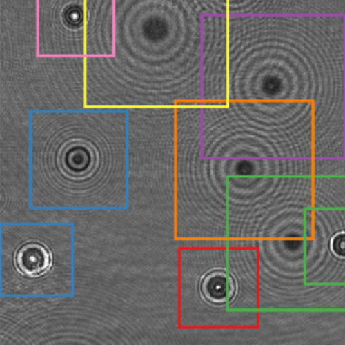
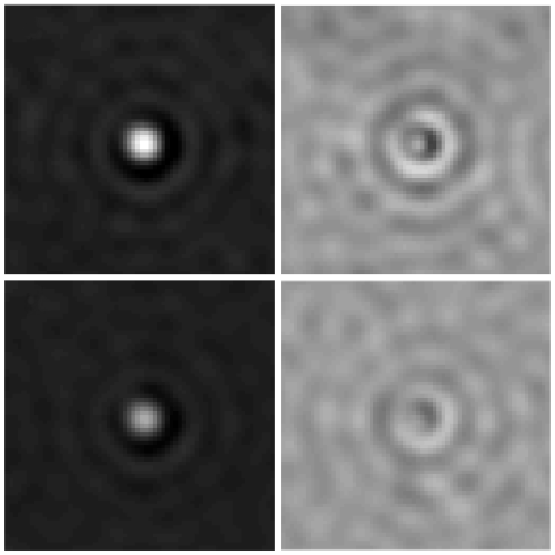
- Henrik Klein Moberg: Seeing the invisible: deep learning optical microscopy for label-free biomolecule screening in the sub-10 kDa regime (@SPIE)
23 August 2022 • 9:15 AM – 9:35 AM PDT | Conv. Ctr. Room 5A
- Jesus Pineda: Revealing the spatiotemporal fingerprint of microscopic motion using geometric deep learning (@SPIE)
23 August 2022 • 11:05 AM – 11:25 AM PDT | Conv. Ctr. Room 5A
- Agnese Callegari: Simulating intracavity optical trapping with machine learning (@SPIE)
23 August 2022 • 1:40 PM – 2:00 PM PDT | Conv. Ctr. Room 5A
- Benjamin Midtvedt: Single-shot self-supervised particle tracking (@SPIE)
23 August 2022 • 2:20 PM – 2:40 PM PDT | Conv. Ctr. Room 5A
- Yu-Wei Chang: Neural network training with highly incomplete medical datasets (@SPIE)
24 August 2022 • 8:00 AM – 8:20 AM PDT | Conv. Ctr. Room 5A
- Daniel Midtvedt: Label-free characterization of biological matter across scales (@SPIE)
24 August 2022 • 9:10 AM – 9:40 AM PDT | Conv. Ctr. Room 5A
- Harshith Bachimanchi: Quantitative microplankton tracking by holographic microscopy and deep learning (@SPIE)
24 August 2022 • 11:45 AM – 12:05 PM PDT | Conv. Ctr. Room 5A
- Pablo Emiliano Gomez Ruiz: BRAPH 2.0: brain connectivity software with multilayer network analyses and machine learning (@SPIE)
24 August 2022 • 2:05 PM – 2:25 PM PDT | Conv. Ctr. Room 5A
- Yu-Wei Chang: Deep-learning analysis in tau PET for Alzheimer’s continuum (@SPIE)
24 August 2022 • 4:40 PM – 5:00 PM PDT | Conv. Ctr. Room 5A
Giovanni Volpe is also co-author of the presentations:
- Antonio Ciarlo: Periodic feedback effect in counterpropagating intracavity optical tweezers (@SPIE)
24 August 2022 • 2:00 PM – 2:20 PM PDT | Conv. Ctr. Room 1B
- Anna Canal Garcia: Multilayer brain connectivity analysis in Alzheimer’s disease using functional MRI data (@SPIE)
24 August 2022 • 2:25 PM – 2:45 PM PDT | Conv. Ctr. Room 5A
- Mite Mijalkov: A novel method for quantifying men and women-like features in brain structure and function (@SPIE)
24 August 2022 • 3:05 PM – 3:25 PM PDT | Conv. Ctr. Room 5A
- Carlo Manzo: An anomalous competition: assessment of methods for anomalous diffusion through a community effort (@SPIE)
25 August 2022 • 9:00 AM – 9:30 AM PDT | Conv. Ctr. Room 5A
- Stefano Bo: Characterization of anomalous diffusion with neural networks (@SPIE)
25 August 2022 • 10:00 AM – 10:30 AM PDT | Conv. Ctr. Room 5A