An in vivo mimetic liver-lobule-chip (LLoC) for stem cell maturation, and zonation of hepatocyte-like cells on chip published in Lab on a Chip

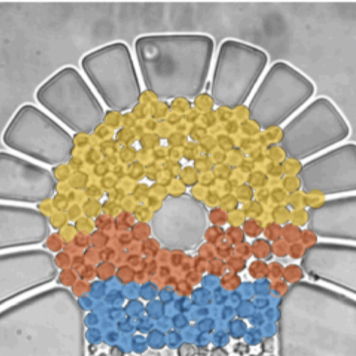
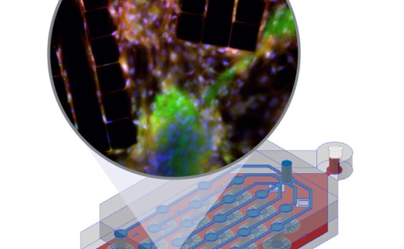
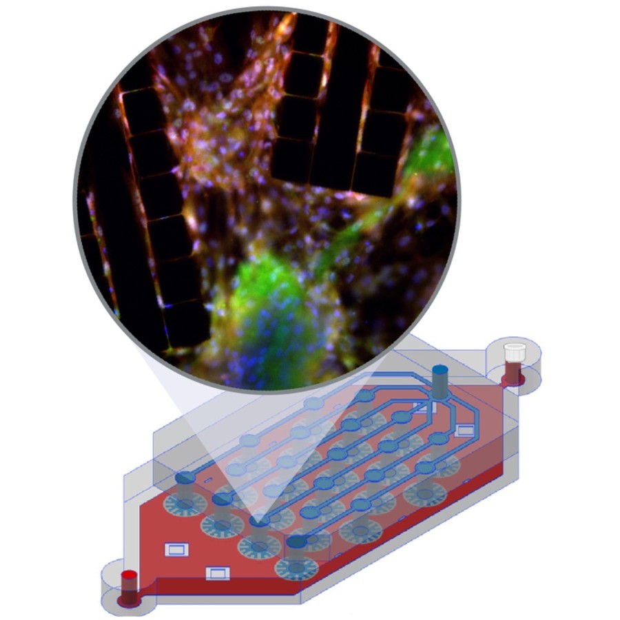
The image shows a liver-lobule-chip (LLoC) with 21 artificial lobules mimicking liver microarchitecture. Its PDMS design supports diffusion-based perfusion, shear stress, and nutrient gradients and enables iPSC-derived hepatic maturation and spatially organized, zonated function in 3D. (Image by C. Beck Adiels)
An in vivo mimetic liver-lobule-chip (LLoC) for stem cell maturation, and zonation of hepatocyte-like cells on chip
Philip Dalsbecker, Siiri Suominen, Muhammad Asim Faridi, Reza Mahdavi, Julia Johansson, Charlotte Hamngren Blomqvist, Mattias Goksör, Katriina Aalto-Setälä, Leena E. Viiri and Caroline B. Adiels
Lab on a Chip 25, 4328 – 4344 (2025)
doi: 10.1039/D4LC00509K

In vitro cell culture models play a crucial role in preclinical drug discovery. To achieve optimal culturing environments and establish physiologically relevant organ-specific conditions, it is imperative to replicate in vivo scenarios when working with primary or induced pluripotent cell types. However, current approaches to recreating in vivo conditions and generating relevant 3D cell cultures still fall short. In this study, we validate a liver-lobule-chip (LLoC) containing 21 artificial liver lobules, each representing the smallest functional unit of the human liver. The LLoC facilitates diffusion-based perfusion via sinusoid-mimetic structures, providing physiologically relevant shear stress exposure and radial nutrient concentration gradients within each lobule. We demonstrate the feasibility of long term cultures (up to 14 days) of viable and functional HepG2 cells in a 3D discoid tissue structure, serving as initial proof of concept. Thereafter, we successfully differentiate sensitive, human induced pluripotent stem cell (iPSC)-derived cells into hepatocyte-like cells over a period of 20 days on-chip, exhibiting advancements in maturity compared to traditional 2D cultures. Further, hepatocyte-like cells cultured in the LLoC exhibit zonated protein expression profiles, indicating the presence of metabolic gradients characteristic of liver lobules. Our results highlight the suitability of the LLoC for long-term discoid tissue cultures, specifically for iPSCs, and their differentiation in a perfused environment. We envision the LLoC as a starting point for more advanced in vitro models, allowing for the combination of multiple liver cell types to create a comprehensive liver model for disease-onchip studies. Ultimately, when combined with stem cell technology, the LLoC offers a promising and robust on-chip liver model that serves as a viable alternative to primary hepatocyte cultures—ideally suited for preclinical drug screening and personalized medicine applications.

Spatial clustering of molecular localizations with graph neural networks on ArXiv

MIRO employs a recurrent graph neural network to refine SMLM point clouds by compressing clusters around their center, enhancing inter-cluster distinction and background separation for efficient clustering. (Image by J. Pineda.)
Spatial clustering of molecular localizations with graph neural networks
Jesús Pineda, Sergi Masó-Orriols, Joan Bertran, Mattias Goksör, Giovanni Volpe and Carlo Manzo
arXiv: 2412.00173

Single-molecule localization microscopy (SMLM) generates point clouds corresponding to fluorophore localizations. Spatial cluster identification and analysis of these point clouds are crucial for extracting insights about molecular organization. However, this task becomes challenging in the presence of localization noise, high point density, or complex biological structures. Here, we introduce MIRO (Multimodal Integration through Relational Optimization), an algorithm that uses recurrent graph neural networks to transform the point clouds in order to improve clustering efficiency when applying conventional clustering techniques. We show that MIRO supports simultaneous processing of clusters of different shapes and at multiple scales, demonstrating improved performance across varied datasets. Our comprehensive evaluation demonstrates MIRO’s transformative potential for single-molecule localization applications, showcasing its capability to revolutionize cluster analysis and provide accurate, reliable details of molecular architecture. In addition, MIRO’s robust clustering capabilities hold promise for applications in various fields such as neuroscience, for the analysis of neural connectivity patterns, and environmental science, for studying spatial distributions of ecological data.
Deep learning for optical tweezers published in Nanophotonics

Real-time control of optical tweezers with deep learning. (Image by the Authors of the manuscript.)
Deep learning for optical tweezers
Antonio Ciarlo, David Bronte Ciriza, Martin Selin, Onofrio M. Maragò, Antonio Sasso, Giuseppe Pesce, Giovanni Volpe and Mattias Goksör
Nanophotonics, 13(17), 3017-3035 (2024)
doi: 10.1515/nanoph-2024-0013
arXiv: 2401.02321

Optical tweezers exploit light–matter interactions to trap particles ranging from single atoms to micrometer-sized eukaryotic cells. For this reason, optical tweezers are a ubiquitous tool in physics, biology, and nanotechnology. Recently, the use of deep learning has started to enhance optical tweezers by improving their design, calibration, and real-time control as well as the tracking and analysis of the trapped objects, often outperforming classical methods thanks to the higher computational speed and versatility of deep learning. In this perspective, we show how cutting-edge deep learning approaches can remarkably improve optical tweezers, and explore the exciting, new future possibilities enabled by this dynamic synergy. Furthermore, we offer guidelines on integrating deep learning with optical trapping and optical manipulation in a reliable and trustworthy way.

Dynamic live/apoptotic cell assay using phase-contrast imaging and deep learning on bioRxiv

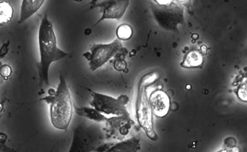
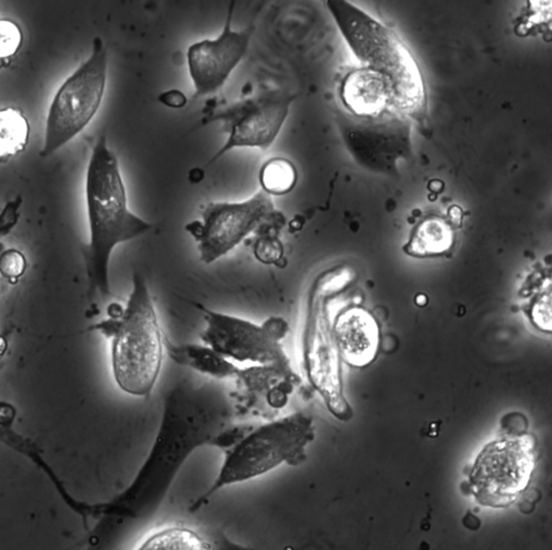
Phase-contrast image before virtual staining. (Image by the Authors.)
Dynamic live/apoptotic cell assay using phase-contrast imaging and deep learning
Zofia Korczak, Jesús Pineda, Saga Helgadottir, Benjamin Midtvedt, Mattias Goksör, Giovanni Volpe, Caroline B. Adiels
bioRxiv: 10.1101/2022.07.18.500422

Chemical live/dead assay has a long history of providing information about the viability of cells cultured in vitro. The standard methods rely on imaging chemically-stained cells using fluorescence microscopy and further analysis of the obtained images to retrieve the proportion of living cells in the sample. However, such a technique is not only time-consuming but also invasive. Due to the toxicity of chemical dyes, once a sample is stained, it is discarded, meaning that longitudinal studies are impossible using this approach. Further, information about when cells start programmed cell death (apoptosis) is more relevant for dynamic studies. Here, we present an alternative method where cell images from phase-contrast time-lapse microscopy are virtually-stained using deep learning. In this study, human endothelial cells are stained live or apoptotic and subsequently counted using the self-supervised single-shot deep-learning technique (LodeSTAR). Our approach is less labour-intensive than traditional chemical staining procedures and provides dynamic live/apoptotic cell ratios from a continuous cell population with minimal impact. Further, it can be used to extract data from dense cell samples, where manual counting is unfeasible.

Intercellular Communication Induces Glycolytic Synchronisation Waves published in PNAS

Intercellular communication induces glycolytic synchronization waves between individually oscillating cells
Martin Mojica-Benavides, David D. van Niekerk, Mite Mijalkov, Jacky L. Snoep, Bernhard Mehlig, Giovanni Volpe, Caroline B. Adiels & Mattias Goksör
PNAS 118(6), e2010075118 (2021)
doi: 10.1073/pnas.2010075118
arXiv: 1909.05187

Metabolic oscillations in single cells underlie the mechanisms behind cell synchronization and cell-cell communication. For example, glycolytic oscillations mediated by biochemical communication between cells may synchronize the pulsatile insulin secretion by pancreatic tissue, and a link between glycolytic synchronization anomalies and type-2 diabetes has been hypotesized. Cultures of yeast cells have provided an ideal model system to study synchronization and propagation waves of glycolytic oscillations in large populations. However, the mechanism by which synchronization occurs at individual cell-cell level and overcome local chemical concentrations and heterogenic biological clocks, is still an open question because of experimental limitations in sensitive and specific handling of single cells. Here, we show how the coupling of intercellular diffusion with the phase regulation of individual oscillating cells induce glycolytic synchronization waves. We directly measure the single-cell metabolic responses from yeast cells in a microfluidic environment and characterize a discretized cell-cell communication using graph theory. We corroborate our findings with simulations based on a kinetic detailed model for individual yeast cells. These findings can provide insight into the roles cellular synchronization play in biomedical applications, such as insulin secretion regulation at the cellular level.

Press release on joint research on intercellular communication mechanism by Biological Physics Lab and Soft Matter Lab

The article Intercellular Communication Induces Glycolytic Synchronisation Waves published in PNAS has been featured in the News of the Faculty of Science of Gothenburg University.

Here the links to the press releases:
Swedish: Forskare har knäckt koden för cellkommunikation
English: Researchers have broken the code for cell communication