Illustration of three different experiments autonomously performed by the SmartTrap system: DNA pulling experiments (top), red blood cell stretching (bottom left), and particle-particle interaction measurements (bottom right). (Image by M. Selin.)SmartTrap: automated precision experiments with optical tweezers
Martin Selin, Antonio Ciarlo, Giuseppe Pesce, Lars Bengtsson, Joan Camunas-Soler, Vinoth Sundar Rajan, Fredrik Westerlund, L. Marcus Wilhelmsson, Isabel Pastor, Felix Ritort, Steven B. Smith, Carlos Bustamante, Giovanni Volpe
Nature Methods (2026)
arXiv: 2505.05290
doi: 10.1038/s41592-026-03129-3
Optical tweezers are widely used in single-molecule biophysics, cell biomechanics and soft matter physics, but require a human operator, limiting throughput and repeatability. Here we present a smart optical tweezers platform, named SmartTrap, capable of performing complex experiments autonomously by integrating real-time three-dimensional particle tracking, custom electronics and a microfluidics system. Through a series of experiments, we demonstrate it can operate continuously, acquiring high-precision data over extended periods of time. By bridging the gap between manual experimentation and autonomous operation, SmartTrap establishes a robust and open-source framework for the next generation of optical tweezers research, capable of performing large-scale studies in single-molecule biophysics, cell mechanics and colloidal science with minimal experimental overhead and operator bias.
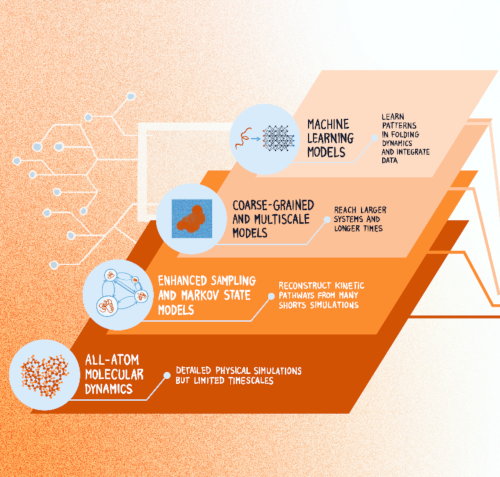
Computational advances in protein folding studies. Current approaches address multiple levels of resolution and methodological frameworks, however, none of the existing methods provides quantitative and dynamic information of the relationship between protein sequence and folding mechanism at all-atom resolution and at scale. (Graphics by J. Sacquegno.)Protein Dynamics Beyond Structure Prediction
Juliette Griffié, Sviatlana Shashkova, Antonio Ciarlo, Sreekanth K. Manikandan, Claes Andréasson, Malin Bäckström, Tristan Bereau, Hjalmar Brismar, Carlos Bustamante, Marta Carroni, Roberto Covino, Andreas Dahlin, Sebastian Deindl, Lucie Delemotte, Arne Elofsson, John Eriksson, Giovanna Fragneto, Anders Gunnarsson, Per Hammarström, Caroline Ingre, Christian Kaiser, Petronella Kettunen, Mark C. Leake, Benjamin Loos, Anna Månberg, Antonia S. J. S. Mey, Richard Neutze, Thomas Nyström, Karl Palmås, Charley Schaefer, Markus J. Tamás, Nicola Ticozzi, Tomás S. Pilvelic, Jacopo Sacquegno, B.M. (Betty)Tijms, Gunnar von Heijne, Björn Wallner, Vitali Zhaunerchyk, Simon Olsson, Joana B. Pereira, Julia Fernandez-Rodriguez, Fredrik Westerlund, Giovanni Volpe
arXiv: 2606.08647
How microorganisms respond to and interact with their environment can vary significantly from individual to individual, which can have important microbiological and ecological implications. However, most microscopy techniques can only observe motile microorganisms for short times because of their limited fields of view. Using Lagrangian tracking, a single microorganism can be followed in 3D, potentially indefinitely, allowing to decipher individual phenotypical traits. Current Lagrangian tracking methods use the fluorescence signal emitted by the microorganism as feedback to keep it in focus. However, over long times, epifluorescent imaging can induce photobleaching and photodamage, and importantly, not all microorganisms can easily be made fluorescent. Additionally, traditional algorithms used in feedback loops to determine microorganism position are prone to errors, especially in optically complex media. Here, we present a faster, more reliable, and versatile Lagrangian tracking method that uses deep learning to determine the 3D position of the microorganism. This new method demonstrates enhanced accuracy and speed in tracking fluorescent bacteria with fluorescence microscopy also in optically complex media. Furthermore, we track bacteria with other microscopy modalities, such as brightfield microscopy — for example, this enables us to track magnetotactic bacteria, which cannot be made fluorescent without degrading their magnetotactic properties. These novel capabilities allow to extract previously inaccessible quantitative information, significantly advancing the study of microorganism behavior — and thus opening new avenues for research in complex biological and ecological systems.
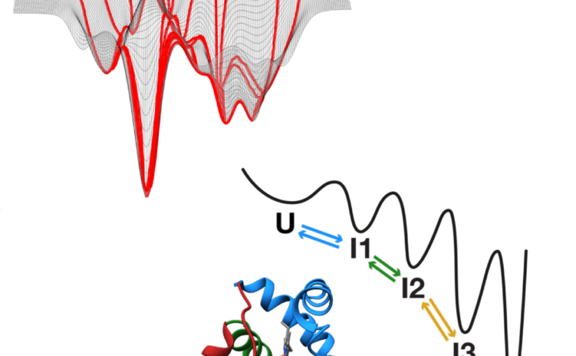
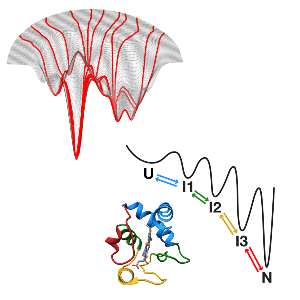
Conceptual models of protein folding: funnel versus foldon. (Figure from the Authors of the manuscript.)How Do Proteins Fold?
Carlos Bustamante, Christian Kaiser, Erik Lindahl, Robert Sosa, Giovanni Volpe
arXiv: 2510.27074
How proteins fold remains a central unsolved problem in biology. While the idea of a folding code embedded in the amino acid sequence was introduced more than 6 decades ago, this code remains undefined. While we now have powerful predictive tools to predict the final native structure of proteins, we still lack a predictive framework for how sequences dictate folding pathways. Two main conceptual models dominate as explanations of folding mechanism: the funnel model, in which folding proceeds through many alternative routes on a rugged, hyperdimensional energy landscape; and the foldon model, which proposes a hierarchical sequence of discrete intermediates. Recent advances on two fronts are now enabling folding studies in unprecedented ways. Powerful experimental approaches; in particular, single-molecule force spectroscopy and hydrogen (deuterium exchange assays) allow time-resolved tracking of the folding process at high resolution. At the same time, computational breakthroughs culminating in algorithms such as AlphaFold have revolutionized static structure prediction, opening opportunities to extend machine learning toward dynamics. Together, these developments mark a turning point: for the first time, we are positioned to resolve how proteins fold, why they misfold, and how this knowledge can be harnessed for biology and medicine.
Carlos Bustamante. (Photo by H. P. Thanabalan.)Fundamentals and Applications of Single Molecule Force Spectroscopy – Waernska Professorship lectures Professor Carlos Bustamante, who is visiting the Soft Matter Lab between 28 April and 27 May and is a winner of the Waernska Professorship, will be giving a series of lectures on Fundamentals and Applications of Single Molecule Force Spectroscopy.
Professor Carlos Bustamante from UC Berkeley is a pioneer in the use of optical tweezers for the biomechanical study of single molecules. He will explain the basics of how and why you can perform single-molecule experiments with them.
Here is the schedule and location of the lectures:
– 29 April 2025 from 13:00 to 17:00 in Gustaf Dalén-salen;
– 05 May 2025 from 13:00 to 17:00 in FL71;
– 06 May 2025 from 13:00 to 17:00 in Lecture Hall FL71;
– 07 May 2025 from 13:00 to 17:00 in Lecture Hall KB.
Carlos Bustamante. (Photo by H. P. Thanabalan.)We are delighted to welcome Professor Carlos Bustamante to the Soft Matter Lab as the winner of the prestigious Waernska Professorship.
Professor Bustamante is a world-renowned expert in single-molecule biophysics and a Full Professor of Molecular and Cell Biology, Chemistry and Physics at the University of California, Berkeley, USA.
His pioneering work has significantly advanced our understanding of the physical behaviour of biological molecules. Using techniques such as optical tweezers, atomic force microscopy and fluorescence microscopy, Professor Bustamante has provided key insights into molecular motors, protein and RNA folding, and the mechanisms of gene expression and regulation.
We are honoured to host Professor Bustamante at the Soft Matter Lab and look forward to exciting scientific exchanges and collaborations during his visit.
His visit is currently planned between 28 April and 27 May 2025.
An additional visit, yet to be confirmed, might take place during the fall of 2025.