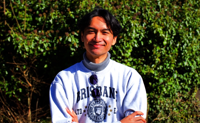
Brian is a master student in Physics at the Physics Department of Gothenburg University.
During his time at the Soft Matter Lab, he will work on a project about deep learning based particle tracking and trajectory linking.

Brian is a master student in Physics at the Physics Department of Gothenburg University.
During his time at the Soft Matter Lab, he will work on a project about deep learning based particle tracking and trajectory linking.

Harshith Bachimanchi, Giovanni Volpe
Journal of Physics: Photonics 7, 013001 (2025)
doi: 10.1088/2515-7647/ada101
arXiv: 2409.16488
Diffusion models have emerged as a prominent technique in generative modeling with neural networks, making their mark in tasks like text-to-image translation and super-resolution. In this tutorial, we provide a comprehensive guide to build denoising diffusion probabilistic models from scratch, with a specific focus on transforming low-resolution microscopy images into their corresponding high-resolution versions in the context of super-resolution microscopy. We provide the necessary theoretical background, the essential mathematical derivations, and a detailed Python code implementation using PyTorch. We discuss the metrics to quantitatively evaluate the model, illustrate the model performance at different noise levels of the input low-resolution images, and briefly discuss how to adapt the tutorial for other applications. The code provided in this tutorial is also available as a Python notebook in the supplementary information.

The defense will take place in PJ, Institutionen för fysik, Origovägen 6b, Göteborg, at 13:00.
Title: Annotation-free deep learning for quantitative microscopy
Abstract: Quantitative microscopy is an essential tool for studying and understanding microscopic structures. However, analyzing the large and complex datasets generated by modern microscopes presents significant challenges. Manual analysis is time-intensive and subjective, rendering it impractical for large datasets. While automated algorithms offer faster and more consistent results, they often require careful parameter tuning to achieve acceptable performance, and struggle to interpret the more complex data produced by modern microscopes. As such, there is a pressing need to develop new, scalable analysis methods for quantitative microscopy. In recent years, deep learning has transformed the field of computer vision, achieving superhuman performance in tasks ranging from image classification to object detection. However, this success depends on large, annotated datasets, which are often unavailable in microscopy. As such, to successfully and efficiently apply deep learning to microscopy, new strategies that bypass the dependency on extensive annotations are required. In this dissertation, I aim to lower the barrier for applying deep learning in microscopy by developing methods that do not rely on manual annotations and by providing resources to assist researchers in using deep learning to analyze their own microscopy data. First, I present two cases where training annotations are generated through alternative means that bypass the need for human effort. Second, I introduce a deep learning method that leverages symmetries in both the data and the task structure to train a statistically optimal model for object detection without any annotations. Third, I propose a method based on contrastive learning to estimate nanoparticle sizes in diffraction-limited microscopy images, without requiring annotations or prior knowledge of the optical system. Finally, I deliver a suite of resources that empower researchers in applying deep learning to microscopy. Through these developments, I aim to demonstrate that deep learning is not merely a “black box” tool. Instead, effective deep learning models should be designed with careful consideration of the data, assumptions, task structure, and model architecture, encoding as much prior knowledge as possible. By structuring these interactions with care, we can develop models that are more efficient, interpretable, and generalizable, enabling them to tackle a wider range of microscopy tasks.
Thesis: https://hdl.handle.net/2077/84178
Supervisor: Giovanni Volpe
Examiner: Dag Hanstorp
Opponent: Ivo Sbalzarini
Committee: Susan Cox, Maria Arrate Munoz Barrutia, Ignacio Arganda-Carreras
Alternate board member: Måns Henningson


fluorescence microscopy imaging
Ivo Sbalzarini
Max Planck Institute of Molecular Cell Biology and Genetics
Center for Systems Biology Dresden
https://sbalzarini-lab.org/
Date: 9 January 2025
Time: 11:00
Place: Nexus
Anqi Lyu starts her PhD at the Physics Department of the University of Gothenburg on 8 January 2025.
Anqi has a Master degree in Medical Bioinformatics from University of Verona, Italy.
In her PhD, she will focus on delineating how plasma factors globally influence endothelial cells, with emphasis on their roles in health, ageing and disease, by utilizing computational tools in combination with interdisciplinary approaches.

Viktor Martvall, Henrik Klein Moberg, Athanasios Theodoridis, David Tomeček, Pernilla Ekborg-Tanner, Sara Nilsson, Giovanni Volpe, Paul Erhart, Christoph Langhammer
ACS Sensors 10, 376–386 (2025)
arXiv: 2312.15372
doi: 10.1021/acssensors.4c02616
Rapidly detecting hydrogen leaks is critical for the safe large-scale implementation of hydrogen technologies. However, to date, no technically viable sensor solution exists that meets the corresponding response time targets under technically relevant conditions. Here, we demonstrate how a tailored long short-term transformer ensemble model for accelerated sensing (LEMAS) speeds up the response of an optical plasmonic hydrogen sensor by up to a factor of 40 and eliminates its intrinsic pressure dependence in an environment emulating the inert gas encapsulation of large-scale hydrogen installations by accurately predicting its response value to a hydrogen concentration change before it is physically reached by the sensor hardware. Moreover, LEMAS provides a measure for the uncertainty of the predictions that are pivotal for safety-critical sensor applications. Our results advertise the use of deep learning for the acceleration of sensor response, also beyond the realm of plasmonic hydrogen detection.

Gerhard Jung, Rinske M. Alkemade, Victor Bapst, Daniele Coslovich, Laura Filion, François P. Landes, Andrea J. Liu, Francesco Saverio Pezzicoli, Hayato Shiba, Giovanni Volpe, Francesco Zamponi, Ludovic Berthier & Giulio Biroli
Nature Review Physics (2025)
doi: 10.1038/s42254-024-00791-4
arxiv: 2311.14752
Unravelling the connections between microscopic structure, emergent physical properties and slow dynamics has long been a challenge when studying the glass transition. The absence of clear visible structural order in amorphous configurations complicates the identification of the key physical mechanisms underpinning slow dynamics. The difficulty in sampling equilibrated configurations at low temperatures hampers thorough numerical and theoretical investigations. We explore the potential of machine learning (ML) techniques to face these challenges, building on the algorithms that have revolutionized computer vision and image recognition. We present both successful ML applications and open problems for the future, such as transferability and interpretability of ML approaches. To foster a collaborative community effort, we also highlight the ‘GlassBench’ dataset, which provides simulation data and benchmarks for both 2D and 3D glass formers. We compare the performance of emerging ML methodologies, in line with benchmarking practices in image and text recognition. Our goal is to provide guidelines for the development of ML techniques in systems displaying slow dynamics and inspire new directions to improve our theoretical understanding of glassy liquids.

Shuyang Yao, Arvid Harder, Fahimeh Darki, Yu-Wei Chang , Ang Li, Kasra Nikouei, Giovanni Volpe, Johan N Lundström, Jian Zeng , Naomi Wray, Yi Lu, Patrick F Sullivan, Jens Hjerling-Leffler
Nature Communications 16, 395 (2025)
doi: 10.1038/s41467-024-55611-1
medRxiv: 10.1101/2024.01.18.24301478
Identifying cell types and brain regions critical for psychiatric disorders and brain traits is essential for targeted neurobiological research. By integrating genomic insights from genome-wide association studies with a comprehensive single-cell transcriptomic atlas of the adult human brain, we prioritized specific neuronal clusters significantly enriched for the SNP-heritabilities for schizophrenia, bipolar disorder, and major depressive disorder along with intelligence, education, and neuroticism. Extrapolation of cell-type results to brain regions reveals the whole-brain impact of schizophrenia genetic risk, with subregions in the hippocampus and amygdala exhibiting the most significant enrichment of SNP-heritability. Using functional MRI connectivity, we further confirmed the significance of the central and lateral amygdala, hippocampal body, and prefrontal cortex in distinguishing schizophrenia cases from controls. Our findings underscore the value of single-cell transcriptomics in understanding the polygenicity of psychiatric disorders and suggest a promising alignment of genomic, transcriptomic, and brain imaging modalities for identifying common biological targets.

He received his Ph.D. in Theoretical Physics in 2020 from Stockholm University under the supervision of Supriya Krishnamurthy. His thesis, titled “Nonequilibrium Thermodynamics at the Microscopic Scales,” focused on finite and short-time fluctuations in non-equilibrium systems, as opposed to the large-time asymptotic properties studied within the framework of large deviation theory. One of the key outcomes of his Ph.D. research was the development of a method to infer entropy production rates directly from experimentally accessible trajectories in a model-independent manner.
Following his PhD, Sreekanth received the NORDITA postdoctoral fellowship for independent research. During this time, he expanded on his earlier work by developing generalizations of the inference scheme for entropy production and integrating it with machine-learning tools for practical inference of dissipative forces and entropy production from experimental data. Later, in 2022, he was awarded the Wallenberg Scholarship for postdoctoral research at Stanford, where he developed machine-learning-based non-equilibrium control techniques for targeted self-assembly and transport of biomolecular systems.
Currently he is interested in combining methods from Non-equilibrium Physics and Machine Learning to quantitatively characterize and control nanoscale biophysical processes.

Jesús Pineda, Sergi Masó-Orriols, Joan Bertran, Mattias Goksör, Giovanni Volpe and Carlo Manzo
arXiv: 2412.00173
Single-molecule localization microscopy (SMLM) generates point clouds corresponding to fluorophore localizations. Spatial cluster identification and analysis of these point clouds are crucial for extracting insights about molecular organization. However, this task becomes challenging in the presence of localization noise, high point density, or complex biological structures. Here, we introduce MIRO (Multimodal Integration through Relational Optimization), an algorithm that uses recurrent graph neural networks to transform the point clouds in order to improve clustering efficiency when applying conventional clustering techniques. We show that MIRO supports simultaneous processing of clusters of different shapes and at multiple scales, demonstrating improved performance across varied datasets. Our comprehensive evaluation demonstrates MIRO’s transformative potential for single-molecule localization applications, showcasing its capability to revolutionize cluster analysis and provide accurate, reliable details of molecular architecture. In addition, MIRO’s robust clustering capabilities hold promise for applications in various fields such as neuroscience, for the analysis of neural connectivity patterns, and environmental science, for studying spatial distributions of ecological data.