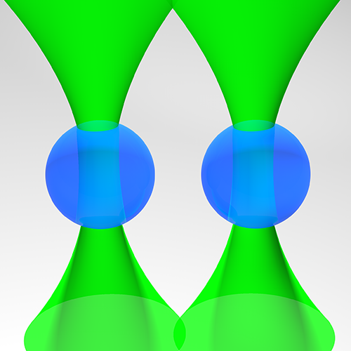
Schematic of the experimental setup used in the experiment. (Image by A. Magazzù.)Controlling the dynamics of colloidal particles by critical Casimir forces Alessandro Magazzù
729. WE-Heraeus Stiftung Seminar on Fluctuation-induced Forces
15 February 2022, 14:50 CET
Critical Casimir forces can play an important role for applications in nano-science and nano-technology, owing to their piconewton strength, nanometric action range, fine tunability as a function of temperature, and exquisite dependence on the surface properties of the involved objects. Here, we investigate the effects of critical Casimir forces on the free dynamics of a pair of colloidal particles dispersed in the bulk of a near-critical binary liquid solvent, using blinking optical tweezers. In particular, we measure the time evolution of the distance between the two colloids to determine their relative diffusion and drift velocity.
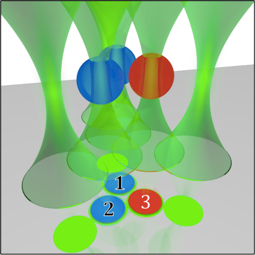
Sketch of the experimental setup for the measurement of nonadditivity of critical Casimir forces. (Image by S. Paladugu.)Experimental Study of Critical Fluctuations and Critical Casimir Forces Giovanni Volpe
729. WE-Heraeus Stiftung Seminar on Fluctuation-induced Forces
14 February 2022, 16:35 CET
Critical Casimir forces (CCF) are a powerful tool to control the self-assembly and complex behavior of microscopic and nanoscopic colloids. While CCF were theoretically predicted in 1978 [1], their first direct experimental evidence was provided only in 2008, using total internal reflection microscopy (TIRM) [2]. Since then, these forces have been investigated under various conditions, for example, by varying the properties of the involved surfaces or with moving boundaries. In addition, a number of studies of the phase behavior of colloidal dispersions in a critical mixture indicate critical Casimir forces as candidates for tuning the self-assembly of nanostructures and quantum dots, while analogous fluctuation-induced effects have been investigated, for example, at the percolation transition of a chemical sol, in the presence of temperature gradients, and even in granular fluids and active matter. In this presentation, I’ll give an overview of this field with a focus on recent results on the measurement of many-body forces in critical Casimir forces [3], the realization of micro- and nanoscopic engines powered by critical fluctuations [4, 5], and the creation of light-controllable colloidal molecules [6] and active droploids [7].
References
[1] ME Fisher and PG de Gennes. Phenomena at the walls in a critical binary mixture. C. R. Acad. Sci. Paris B 287, 207 (1978).
[2] C Hertlein, L Helden, A Gambassi, S Dietrich and C Bechinger. Direct measurement of critical Casimir forces. Nature 451, 172 (2008).
[3] S Paladugu, A Callegari, Y Tuna, L Barth, S Dietrich, A Gambassi and G Volpe. Nonadditivity of critical Casimir forces. Nat. Commun. 7, 11403 (2016).
[4] F Schmidt, A Magazzù, A Callegari, L Biancofiore, F Cichos and G Volpe. Microscopic engine powered by critical demixing. Phys. Rev. Lett. 120, 068004 (2018).
[5] F Schmidt, H Šípová-Jungová, M Käll, A Würger and G Volpe. Non-equilibrium properties of an active nanoparticle in a harmonic potential. Nat. Commun. 12, 1902 (2021).
[6] F Schmidt, B Liebchen, H Löwen and G Volpe. Light-controlled assembly of active colloidal molecules. J. Chem. Phys. 150, 094905 (2019).
[7] J Grauer, F Schmidt, J Pineda, B Midtvedt, H Löwen, G Volpe and B Liebchen. Active droploids. Nat. Commun. 12, 6005 (2021).
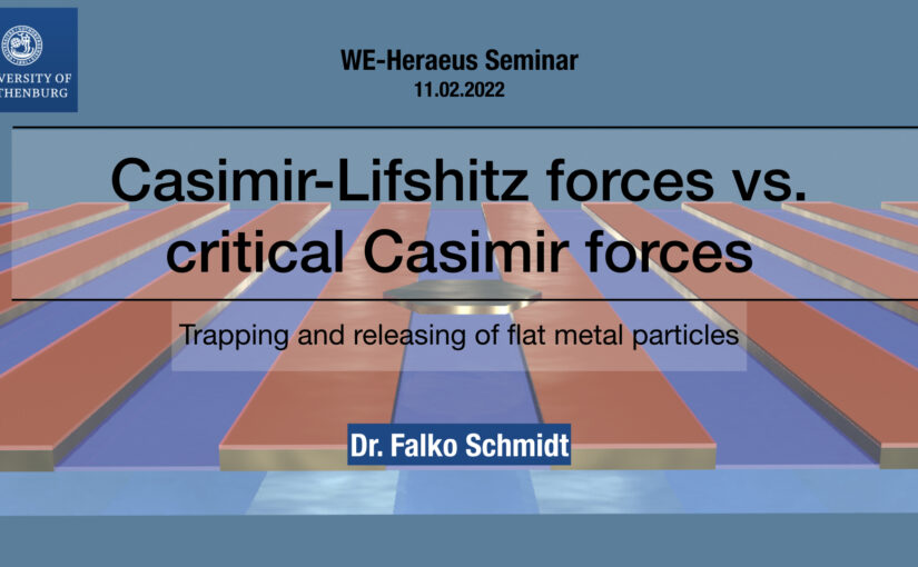
Title slide of the presentation. (Image by F. Schmidt.)Casimir-Lifshitz forces vs. Critical Casimir forces: Trapping and releasing of flat metallic particles Falko Schmidt
729. WE-Heraeus Stiftung Seminar on Fluctuation-induced Forces
16 February 2022, 14:50 CET
Casimir forces in quantum electrodynamics emerge between microscopic metallic objects because of the confinement of the vacuum electromagnetic fluctuations occuring even at zero temperature. Their generalization at finite temperature and in material media are referred to as Casimir-Lifshitz forces. These forces are typically attractive, leading to the widespread problem of stiction between the metallic parts of micro- and nanodevices. Recently, repulsive Casimir forces have been experimentally realized but their use of specialized materials stills means that the system can not be controlled dynamically and thus limits further implementation to real-world applications. Here, we experimentally demonstrate that repulsive critical Casimir forces, which emerge in a critical binary liquid mixture upon approaching the critical temperature, can be used to prevent stiction due to Casimir-Lifshitz forces. We show that critical Casimir forces can be dynamically tuned via temperature, eventually overcoming Casimir-Lifshitz attraction. We study a microscopic gold flake above a flat gold-coated substrate immersed in a critical mixture. Far from the critical temperature, stiction occurs because of Casimir-Lifshitz forces. Upon approaching the critical temperature, however, we observe the emergence of repulsive critical Casimir forces that are sufficiently strong to counteract stiction. By removing one of the key limitations to their deployment, this experimental demonstration can accelerate the development of micro- and nanodevices for a broad range of applications.
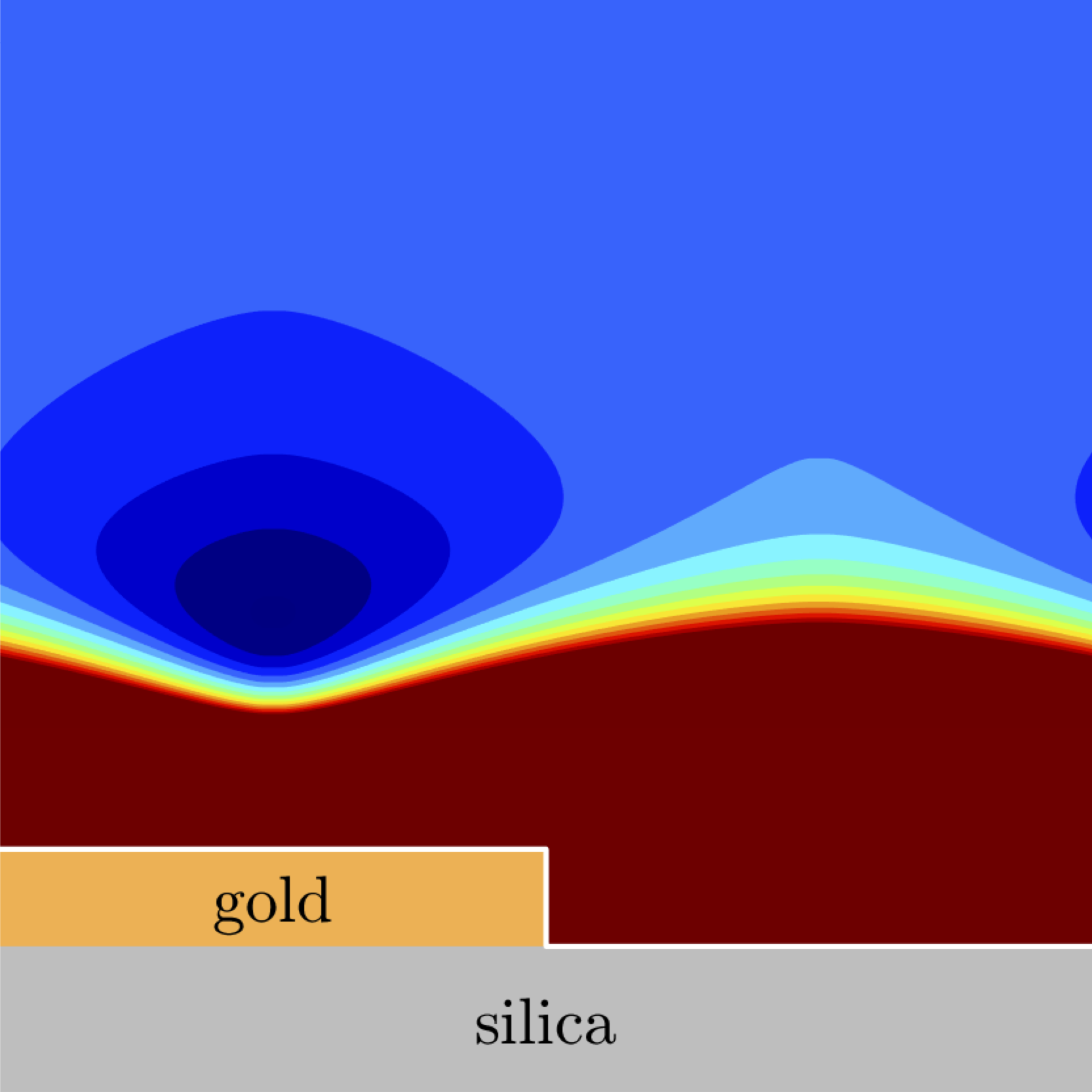
Potential energy landscape for a flake suspended on a patterned substrate. (Image by A. Callegari.)Theoretical and numerical study of the interplay of Casimir-Lifshitz and critical Casimir force for a metallic flake suspended on a metal-coated substrate Agnese Callegari
729. WE-Heraeus Stiftung Seminar on Fluctuation-induced Forces
14 February 2022, 14:50 CET
Casimir-Lifshitz forces arise between uncharged metallic objects because of the confinement of the electromagnetic fluctuations. Typically, these forces are attractive, and they are the main cause of stiction between microscopic metallic parts of micro- and nanodevices. Critical Casimir forces emerge between objects suspended in a critical binary liquid mixture upon approaching the critical temperature, can be made either attractive or repulsive by choosing the appropriate boundary conditions, and dynamically tuned via the temperature.
Experiments show that repulsive critical Casimir forces can be used to prevent stiction due to Casimir-Lifshitz forces. In a recent work, a microscopic metallic flake was suspended in a liquid solution above a metal-coated substrate [1]. By suspending the flake in a binary critical mixture and tuning the temperature we can control the flake’s hovering height above the substrate and, in the case of repulsive critical Casimir forces, prevent stiction.
Here, we present the model for the system of the metallic flake suspended above a metal-coated substrate in a binary critical mixture and show that repulsive critical Casimir forces can effectively counteract Casimir-Lifshitz forces and can be used to control dynamically the height of the flake above the surface. This provides a validation of the experimental results and a base to explore and design the behavior of similar systems in view of micro- and nanotechnological applications.
References
[1] F. Schmidt, A. Callegari, A. Daddi-Moussa-Ider, B. Munkhbat, R. Verre, T. Shegai, M. Käll, H. Löwen, A. Gambassi and G. Volpe, to be submitted (2022)
After a brief overview of artificial intelligence, machine learning and deep learning, I will present a series of recent works in which we have employed deep learning for applications in experimental soft matter.
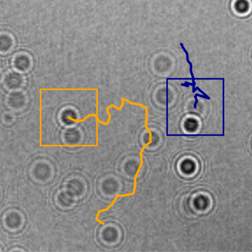
Tracking of the planktons. (Image by H. Bachimanchi.)Characterising plankton behaviours using deep learning powered inline holography Harshith Bachimanchi
Presentation at Prof. Ralf Metzler’s Theoretical Physics group at University of Potsdam (Online)
4 February 2022, 14:15 CET
Digital holographic microscopy is a powerful label-free imaging technique for studying biological specimens. The complex optical fields of microscopic objects can be stored in the form of interference patterns and can be reconstructed by using the principles of holography. Recently, we have developed a digital inline holographic microscope with a deep learning powered analysis to track planktons through generations, and continuously measure their three-dimensional position and dry mass. By bringing planktons of different trophic levels together, we were able to perform a quantitative assessment of trophic interactions between planktons such as feeding events, biomass transfer from cell to cell, etc. In this talk, I will be giving a short overview of our method and present some of our recent results.
Deep learning for microscopy, optical trapping, and active matter Giovanni Volpe
Keynote Talk at IUPAP conference on Condensed Matter Physics and Optics
Online
20 January 2022
15:00 PST
After a brief overview of artificial intelligence, machine learning and deep learning, I will present a series of recent works in which we have employed deep learning for applications in photonics and active matter. In particular, I will explain how we employed deep learning to enhance digital video microscopy, to estimate the properties of anomalous diffusion, to characterize microscopic force fields, to improve the calculation of optical forces, and to characterize nanoparticles. Finally, I will provide an outlook for the application of deep learning in photonics and active matter.
Claus Roll is visiting the Soft Matter Lab on the 19 November 2021.
Claus is the director in Europe of OPTICA (former OSA) and he will be in Gothenburg for an hybrid event organised together with the local OPTICA student chapter and the FFF (Föreningen för Forskarstuderande i Fysik) group.
The visit starts with a tour of different labs including the Soft matter and Biophysics lab. The tour is followed by an hybrid career seminar by Claus Roll, both in person and online starting at 10:30. The presentation is followed by a social lunch and networking session.
Thermoplasmonic Tweezers: Probing single-molecules and more
G. V. Pavan Kumar
IISER, Pune, India.
24 November 2021
Online
In this presentation, we will discuss two specific issues: How to perform single-molecule surface enhanced Raman scattering (SERS) in an optothermal trap? and how to design optothermal fields to trap and interrogate molecules and colloids in a fluid?
In recent years, performing SERS in optical traps has emerged as an important development in nano- and bio-photonics. To this end, tweezer techniques based on surface-plasmons facilitate deeper optical potentials at sub-wavelength scales, and simultaneously provide enhanced electric and optothermal fields. In this
presentation, we will discuss various strategies developed in my laboratory to perform single-molecule SERS in optical and plasmonic tweezer platforms. Specifically, we will highlight some thermoplasmonic effects and directionality aspects of the tweezer platforms in metallic thin film and some plasmonic nano-architectures.
Short bio:
G.V. Pavan Kumar is an associate professor of physics at the Indian Institute of Science Education and Research (IISER), Pune, India.
He obtained his PhD from JNCASR, Bangalore. Subsequently he was a postdoctoral fellow at ICFO-Barcelona and Purdue University, before joining IISER in 2010.
His current research interests are optical, optothermal and nanophotonic forces and their utility in probing single molecules and soft-matter systems at micro and nanoscale.
To this end, his lab has been interfacing optical tweezer platforms with a variety of optical spectroscopy and microscopy tools.
He blogs on topics related to science: https://backscattering.wordpress.com/
We present DeepTrack 2.0, a software to design, train, and validate deep-learning solutions for digital microscopy. We demonstrate it for applications from particle localization, tracking, and characterization, to cell counting and classification, to virtual staining.