Active Matter
Giovanni Volpe
6 May 2022, 13:00 (CEST)
Nordita workshop: Are There Universal Laws in Non-Equilibrium Statistical Physics?
Albano Building 3
Stockholm, 2-13 May 2022
Category: Presentation
Presentation by Gille Claude Vanwalleghem, 4 May 2022
Imaging large neuronal circuits from the Brain to the Gut
Gilles Claude Vanwalleghem
4 May 2022, 12:30 CET
As a transparent animal and with powerful light-based tools to monitor the brain, the larval zebrafish offers a perfect window into functioning neural circuits. We can image the whole brain of zebrafish with cellular resolution, as they respond to various stimuli and record the activity of thousands of neurons. I will focus on two recent studies, one in collaboration with optical physicists, using optical tweezers to move otolith in the inner ear and simulate acceleration. We identified several salient response types, and showed the fish can respond to unnatural stimuli. The other used a microfluidics device to apply water flow to the fish and stimulate the lateral line. The fish’s brain could encode the speed, duration and direction of the water flow, but we also showed that the circuit was biased towards one specific direction of flow. Finally, I will briefly present the new focus of my lab, the gut-brain axis is a physiological communication network between the microbiome, enteric and central nervous system. We are using light sheet microscopy to image the activity of the ENS neurons from 3 to 7 days post fertilization fish. We observed that the spontaneous neuronal activity increases from 3 to 5 dpf, before dropping suddenly at day 7.
Bio
I received my PhD in 2012 from the Universite Libre de Bruxelles where I worked on the Trypanosoma brucei parasite. We discovered a key role of Trypanosoma brucei adenylate cyclases in host-pathogen interactions, as well as the mechanisms through which the human APOL1 can trigger the parasite’s death. In 2014, I was awarded an EMBO long-term fellowship, to shift my focus to neuroscience and the use of optogenetics in larval zebrafish. My work since has spanned several sensory modalities in the zebrafish, including optical traps for vestibular stimulation, visual loom responses, auditory processing, and water flow perception.
I am an assistant professor at Aarhus University since October 2021, where I will focus on the gut-brain axis, I am especially interested in the interactions between neurons, bacteria and the immune system.
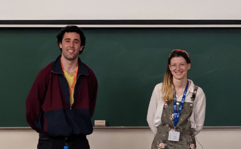
Laura Natali and David Bronte Ciriza presented an effective communication activity in Lisbon
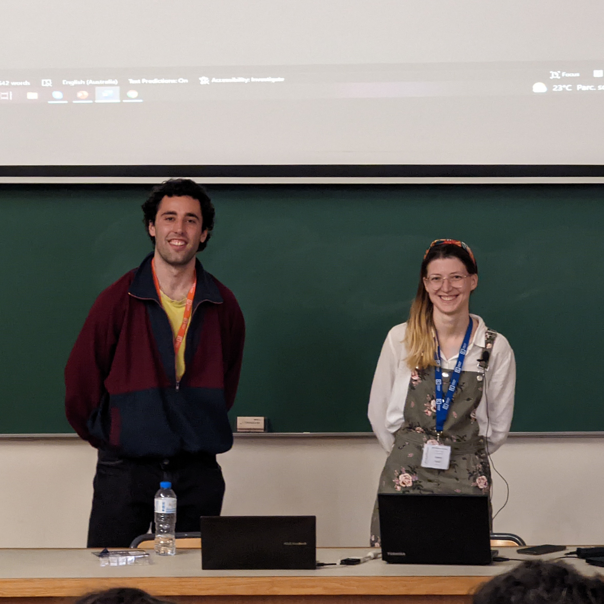
Then, the ESRs briefly described their research in a popular science style, so addressed to a broader public. The first hour concluded with a presentation about rules to keep in mind while communicating both in oral and written form.
Afterwards, a few examples among the written texts were selected and discussed with all the participants. The aim was to exchange feedback and suggestions on how to make the communication more effective. The feedback was the inspiration for everyone to review their communication example, and the final versions are being uploaded on the official twitter account @ActiveMatterITN.
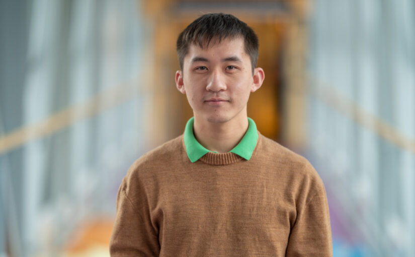
Presentation by Hang Zhao, 3 May 2022
Medical image segmentation using deep learning.
Hang Zhao
3 May 2022, 11:00
Image segmentation and synthesis of CT image based on deep learning: Deep learning methods for medical image segmentation are hindered by the lack of training data. This thesis aims to develop a method that overcomes this problem. Basic U-net trained on XCAT phantom data was tested first. The segmentation results were unsatisfactory even when artificial quantum noise was added. As a workaround, CycleGAN was used to add tissue textures to the XCAT phantom images by analyzing patient CT images. The generated images were used to train the network. The textures introduced by CycleGAN improved the segmentation, but some errors remained. Basic U-net was replaced with Attention U-net, which further improved the segmentation. More work is needed to fine-tune and thoroughly evaluate the method. The results obtained so far demonstrate the potential of this method for the segmentation of medical images. The proposed algorithms may be used in iterative image reconstruction algorithms in multi-energy computed tomography.
3D Cell nuclei segmentation using digital nuclei phantom and 3D deep learning methods : The analysis of microscopy image is helpful to pathological analysis. Nowadays, deep learning has shown the capabilities of processing the medical imaging data. However, developing deep learning methods in microscopy image analysis can be challenging because of the lack of ground truth and various resolution of microscopy image data. This project aims to build a digital nuclei phantom that simulates the actual microscopy images, including mitotic rate, nucleus size, noise, point spread function, and diverse resolutions. The phantom images were used to train 3D deep neural network for nuclei segmentation. The trained neural network was tested for segmentation on datasets with different resolutions. The neural network successfully performed segmentation on most resolutions in our dataset, and the segmentation results reflect the morphology and density of nuclei in microscopy images. The future work will focus on improving the nuclei phantom to generate more realistic phantom images, thereby further helping with segmentation.
Bio:
My name is Hang, I took my bachelor’s in China on radiophysics, and master’s at Linköping University on biomedical imaging. After I graduate, I joined Karolinska as a research assistant on 3D microscopy image processing. Now, I am working at Linköping university as a full time research engineer, contributing to cardiovascular MR image processing, supervised by Petter Dyverfeldt.
Presentation by Timo Betz, 20 April 2022
How fundamental physics leads the way to a better understanding of life’s complexity
Timo Betz
Georg August University Göttingen
20 April 2022, 12:30 CEST
Online
Many biological systems rely on fundamental physical principles for their proper function. Here, mechanical processes such as force generation and adaptation of stiffness and viscosity have been very successfully used to explain complex biomedical questions with physical concepts. Such advances have been largely driven by new methods that allow to quantify biological processes and to construct theoretical models with high predictive power. I will present our recent approaches that allow to study active force generation and mobility in different biological systems over several length scales. Starting with active motion of membranes and intracellular particles in oocytes followed by cytosolic fluidification during cell division we will construct a surprisingly general description of active motion inside the cytoplasm. In a further direction, similar principles are used to study the flow of whole tissue. Here we analyze pressure driven outbursts of cancer cells from model tumors, but also the collective motion during zebrafish development that eventually shapes a whole new animal. The tools we use are largely based on continuum mechanics and statistical mechanics, and give deep insights into the physical principles that are exploited by cells and living objects to perform their intriguing function.
Links: BetzLab
Presentation by Cynthia Reichhardt, 6 April 2022
Clogging, Dynamics and Reentrant Fluid for Active Matter on Periodic Substrates
Cynthia Reichhardt
Theoretical Division, Los Alamos National Laboratory, Los Alamos, NM, USA
6 April 2022, 16:00 CEST
Online
We explore the interactions between substrate length scales and correlation length scales of run-and-tumble active matter disks. For the case of a Casimir geometry of two plates placed a distance d apart, we show that an effective active-matter-mediated attraction arises due to a geometric shadowing effect [1]. Next we shrink the plates down to columns and consider connections to jamming [2] and clogging effects [3] found in passive granular matter. The active particles are driven with an external force through columns placed in a square periodic array [4]. When the drive is applied along a symmetry direction of the array, we find a clog-free uniform liquid state for low activity, while at higher activity, the density becomes increasingly heterogeneous and an active clogged state emerges in which the mobility is strongly reduced. For driving along non-symmetry or incommensurate directions, there are two different clogging behaviors consisting of a drive dependent clogged state in the low activity thermal limit and a drive independent clogged state at high activity. These regimes are separated by a uniform flowing liquid at intermediate activity. There is a critical activity level above which the thermal clogged state does not occur, as well as an optimal activity level that maximizes the disk mobility. Thermal clogged states are dependent on the driving direction while active clogged states are not [5].
References:
[1] D. Ray, C. Reichhardt, and C.J.O. Reichhardt, Phys. Rev. E 90, 013019 (2014).
[2] J.A. Drocco, M.B. Hastings, C.J.O. Reichhardt, and C. Reichhardt, Phys. Rev. Lett. 95, 088001 (2005).
[3] H. Peter, A. Libal, C. Reichhardt, and C.J.O. Reichhardt, Sci. Rep. 8, 10252 (2018).
[4] C. Reichhardt and C.J.O. Reichhardt, Phys. Rev. E 102, 042616 (2020).
[5] C. Reichhardt and C.J.O. Reichhardt, Phys. Rev. E 103, 062603 (2021).
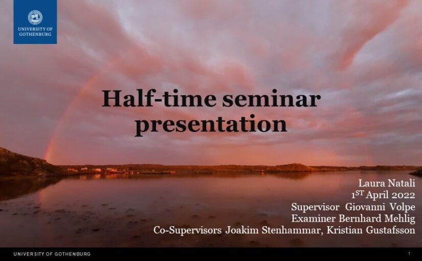
Laura Natali presented her half-time seminar on 1 April 2022
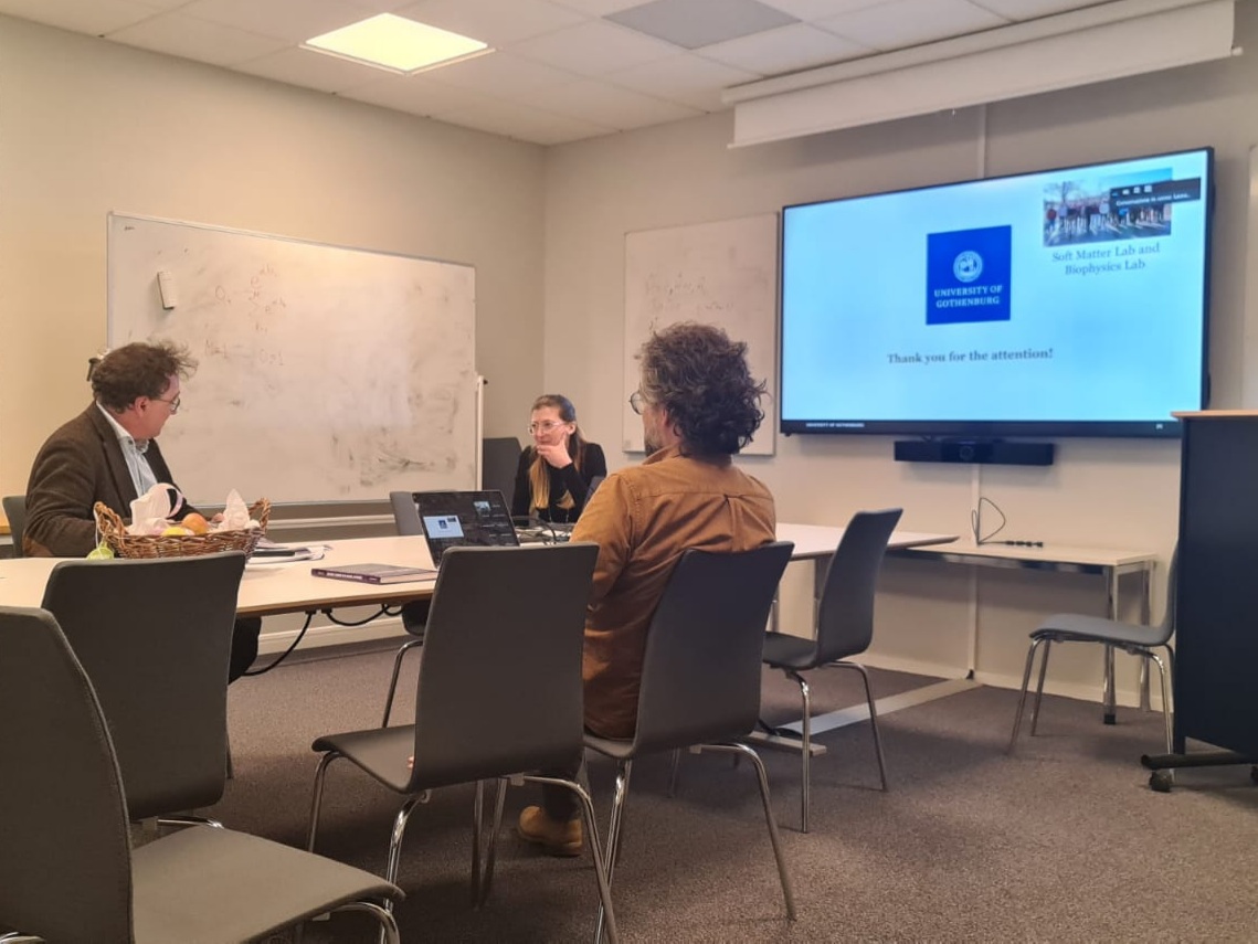
The presentation was held in hybrid format, with part of the audience in the Von Bahr room and the rest connected through zoom. The half-time consisted in a presentation about her past and planned projects and it was followed by a discussion and questions proposed by her opponent Bernhard Mehlig.
The presentation started with a description of her concluded projects about employing neural networks in an epidemic agent-based model, published in Improving epidemic testing and containment strategies using machine learning accepted in Machine Learning: Science and Technology. It continued with her second project, about handling incomplete medical datasets with neural networks, available online as a preprint Neural Network Training with Highly Incomplete Datasets on ArXiv. In the last section, she outlined the proposed continuation of her PhD, with an ongoing project for combining artificial active matter with neural networks.
Invited Lecture by G. Volpe at the Geilo school, 21-31 March 2022
Machine learning and active matter
Giovanni Volpe
22 March 2022
10:30-11:30 and 15:30-17:30
The Geilo School 2022 – The Physics of Evolving Matter: Memory, Learning, and Evolution
Invited Talk by G. Volpe at Complex Lagrangian Problems of Particles in Flows, 15 March 2022
Giovanni Volpe
Complex Lagrangian Problems of Particles in Flows
Online, 15 March 2022, 10:15 CET
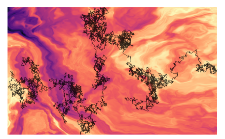
Deviations from Brownian motion leading to anomalous diffusion are found in transport dynamics from quantum physics to life sciences. The characterization of anomalous diffusion from the measurement of an individual trajectory is a challenging task, which traditionally relies on calculating the trajectory mean squared displacement. However, this approach breaks down for cases of practical interest, e.g., short or noisy trajectories, heterogeneous behaviour, or non-ergodic processes. Recently, several new approaches have been proposed, mostly building on the ongoing machine-learning revolution. To perform an objective comparison of methods, we gathered the community and organized an open competition, the Anomalous Diffusion challenge (AnDi). Participating teams applied their algorithms to a commonly-defined dataset including diverse conditions. Although no single method performed best across all scenarios, machine-learning-based approaches achieved superior performance for all tasks. The discussion of the challenge results provides practical advice for users and a benchmark for developers.
Links:
Complex Lagrangian Problems of Particles in Flows program
Plenary Talk by G. Volpe at Physics Days 2022 – Future Leaders, 3 March 2022
Giovanni Volpe
3 March 2022, 13:15
Plenary talk for Physics Days 2022 – Future Leaders
Online
After a brief overview of artificial intelligence, machine learning and deep learning, I will present a series of recent works in which we have employed deep learning for applications in microscopy, optical tweezers, and active matter. In particular, I will explain how we employed deep learning to enhance digital video microscopy [1,2], to perform virtual staining of [3], to estimate the properties of anomalous diffusion [4,5,6], to characterize microscopic force fields [7], to improve the calculation of optical forces [8], and to characterize nanoparticles [9]. Finally, I will provide an outlook on the future for the application of deep learning in these fields.
References
[1] S. Helgadottir, A. Argun, and G. Volpe. Digital video microscopy enhanced by deep learning. Optica 6, 506 (2019).
[2] B. Midtvedt, S. Helgadottir, A. Argun, J. Pineda, D. Midtvedt, and G. Volpe. Quantitative digital microscopy with deep learning. Appl. Phys. Rev. 8, 011310 (2021).
[3] S. Helgadottir, B. Midtvedt, J. Pineda, et al. Extracting quantitative biological information from bright-field cell images using deep learning. Biophys. Rev. 2, 031401 (2021).
[4] S. Bo, F. Schmidt, R. Eichhorn, and G. Volpe. Measurement of anomalous diffusion using recurrent neural networks. Phys. Rev. E 100, 010102 (2019).
[5] A. Argun, G. Volpe, and S. Bo. Classification, inference and segmentation of anomalous diffusion with recurrent neural networks. J. Phys. A: Math. Theor. 54, 294003 (2021).
[6] G. Muñoz-Gil, G. Volpe, M. A. Garcia-March, et al. Objective comparison of methods to decode anomalous diffusion. Nat. Commun. 12, 6253 (2021).
[7] A. Argun, T. Thalheim, S. Bo, F. Cichos, and G. Volpe. Enhanced force-field calibration via machine learning. Appl. Phys. Rev. 7, 041404 (2020).
[8] I.C.D. Lenton, G. Volpe, A.B. Stilgoe, T.A. Nieminen, and H. Rubinsztein-Dunlop. Machine learning reveals complex behaviours in optically trapped particles. Mach. Learn.: Sci. Technol. 1, 045009 (2020).
[9] B. Midtvedt, E. Olsén, F. Eklund, F. Höök, C.B. Adiels, G. Volpe, and D. Midtvedt. Fast and accurate nanoparticle characterization using deep-learning-enhanced off-axis holography. ACS Nano 15, 2240 (2021).
Link: Physics Days 2022 – Future Leaders
The Physics Days 2022 is organized by the Finnish Physical Society and the Department of Applied Physics at Aalto University.