Functional gradients of the medial parietal cortex in a healthy cohort with family history of sporadic Alzheimer’s disease published in Alzheimer’s Research & Therapy

Spatial maps depicting the strongest connections from the medial parietal cortex to other cortical and subcortical areas in the PREVENT-AD cohort. (Reproduced from the publication.)
Functional gradients of the medial parietal cortex in a healthy cohort with family history of sporadic Alzheimer’s disease
Dániel Veréb, Mite Mijalkov, Yu-Wei Chang, Anna Canal-Garcia, Emiliano Gomez-Ruis, Anne Maass, Sylvia Villeneuve, Giovanni Volpe Joana B. Pereira
Alzheimer’s Research & Therapy 15, 82 (2023)
doi: 10.1186/s13195-023-01228-3

Background
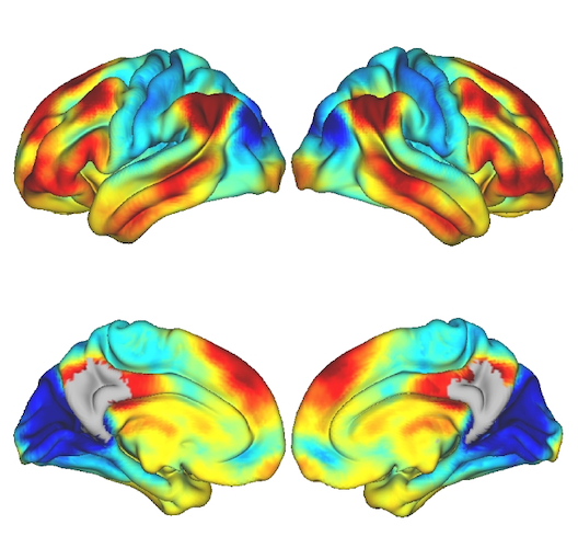
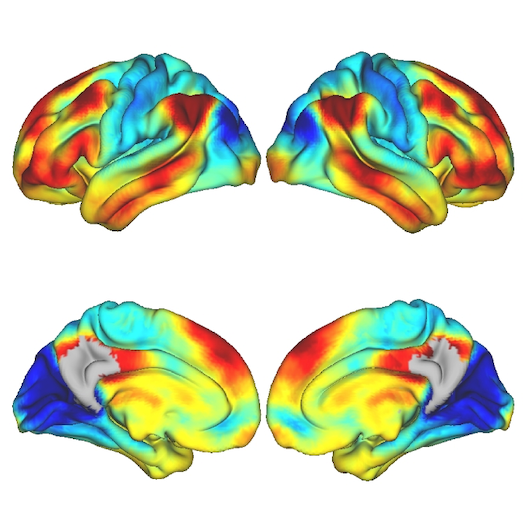
The medial parietal cortex is an early site of pathological protein deposition in Alzheimer’s disease (AD). Previous studies have identified different subregions within this area; however, these subregions are often heterogeneous and disregard individual differences or subtle pathological alterations in the underlying functional architecture. To address this limitation, here we measured the continuous connectivity gradients of the medial parietal cortex and assessed their relationship with cerebrospinal fluid (CSF) biomarkers, ApoE ε4 carriership and memory in asymptomatic individuals at risk to develop AD.

Methods
Two hundred sixty-three cognitively normal participants with a family history of sporadic AD who underwent resting-state and task-based functional MRI using encoding and retrieval tasks were included from the PREVENT-AD cohort. A novel method for characterizing spatially continuous patterns of functional connectivity was applied to estimate functional gradients in the medial parietal cortex during the resting-state and task-based conditions. This resulted in a set of nine parameters that described the appearance of the gradient across different spatial directions. We performed correlation analyses to assess whether these parameters were associated with CSF biomarkers of phosphorylated tau181 (p-tau), total tau (t-tau), and amyloid-ß1-42 (Aß). Then, we compared the spatial parameters between ApoE ε4 carriers and noncarriers, and evaluated the relationship between these parameters and memory.

Results
Alterations involving the superior part of the medial parietal cortex, which was connected to regions of the default mode network, were associated with higher p-tau, t-tau levels as well as lower Aß/p-tau levels during the resting-state condition (p < 0.01). Similar alterations were found in ApoE ε4 carriers compared to non-carriers (p < 0.003). In contrast, lower immediate memory scores were associated with changes in the middle part of the medial parietal cortex, which was connected to inferior temporal and posterior parietal regions, during the encoding task (p = 0.001). No results were found when using conventional connectivity measures.

Conclusions
Functional alterations in the medial parietal gradients are associated with CSF AD biomarkers, ApoE e4 carriership, and lower memory in an asymptomatic cohort with a family history of sporadic AD, suggesting that functional gradients are sensitive to subtle changes associated with early AD stages.

Light, Matter, Action: Shining light on active matter published in ACS Photonics

Actuation of active matter by different properties of light. (Image by M. Rey.)
Light, Matter, Action: Shining light on active matter
Marcel Rey, Giovanni Volpe, Giorgio Volpe
ACS Photonics, 10, 1188–1201 (2023)
arXiv: 2301.13034
doi: 10.1021/acsphotonics.3c00140

Light carries energy and momentum. It can therefore alter the motion of objects from atomic to astronomical scales. Being widely available, readily controllable and broadly biocompatible, light is also an ideal tool to propel microscopic particles, drive them out of thermodynamic equilibrium and make them active. Thus, light-driven particles have become a recent focus of research in the field of soft active matter. In this perspective, we discuss recent advances in the control of soft active matter with light, which has mainly been achieved using light intensity. We also highlight some first attempts to utilize light’s additional degrees of freedom, such as its wavelength, polarization, and momentum. We then argue that fully exploiting light with all of its properties will play a critical role to increase the level of control over the actuation of active matter as well as the flow of light itself through it. This enabling step will advance the design of soft active matter systems, their functionalities and their transfer towards technological applications.

Roadmap for Optical Tweezers published in Journal of Physics: Photonics

Illustration of an optical tweezers holding a particle. (Image by A. Magazzù.)
Roadmap for optical tweezers
Giovanni Volpe, Onofrio M Maragò, Halina Rubinsztein-Dunlop, Giuseppe Pesce, Alexander B Stilgoe, Giorgio Volpe, Georgiy Tkachenko, Viet Giang Truong, Síle Nic Chormaic, Fatemeh Kalantarifard, Parviz Elahi, Mikael Käll, Agnese Callegari, Manuel I Marqués, Antonio A R Neves, Wendel L Moreira, Adriana Fontes, Carlos L Cesar, Rosalba Saija, Abir Saidi, Paul Beck, Jörg S Eismann, Peter Banzer, Thales F D Fernandes, Francesco Pedaci, Warwick P Bowen, Rahul Vaippully, Muruga Lokesh, Basudev Roy, Gregor Thalhammer-Thurner, Monika Ritsch-Marte, Laura Pérez García, Alejandro V Arzola, Isaac Pérez Castillo, Aykut Argun, Till M Muenker, Bart E Vos, Timo Betz, Ilaria Cristiani, Paolo Minzioni, Peter J Reece, Fan Wang, David McGloin, Justus C Ndukaife, Romain Quidant, Reece P Roberts, Cyril Laplane, Thomas Volz, Reuven Gordon, Dag Hanstorp, Javier Tello Marmolejo, Graham D Bruce, Kishan Dholakia, Tongcang Li, Oto Brzobohatý, Stephen H Simpson, Pavel Zemánek, Felix Ritort, Yael Roichman, Valeriia Bobkova, Raphael Wittkowski, Cornelia Denz, G V Pavan Kumar, Antonino Foti, Maria Grazia Donato, Pietro G Gucciardi, Lucia Gardini, Giulio Bianchi, Anatolii V Kashchuk, Marco Capitanio, Lynn Paterson, Philip H Jones, Kirstine Berg-Sørensen, Younes F Barooji, Lene B Oddershede, Pegah Pouladian, Daryl Preece, Caroline Beck Adiels, Anna Chiara De Luca, Alessandro Magazzù, David Bronte Ciriza, Maria Antonia Iatì, Grover A Swartzlander Jr
Journal of Physics: Photonics 2(2), 022501 (2023)
arXiv: 2206.13789
doi: 110.1088/2515-7647/acb57b

Optical tweezers are tools made of light that enable contactless pushing, trapping, and manipulation of objects, ranging from atoms to space light sails. Since the pioneering work by Arthur Ashkin in the 1970s, optical tweezers have evolved into sophisticated instruments and have been employed in a broad range of applications in the life sciences, physics, and engineering. These include accurate force and torque measurement at the femtonewton level, microrheology of complex fluids, single micro- and nano-particle spectroscopy, single-cell analysis, and statistical-physics experiments. This roadmap provides insights into current investigations involving optical forces and optical tweezers from their theoretical foundations to designs and setups. It also offers perspectives for applications to a wide range of research fields, from biophysics to space exploration.

Geometric deep learning reveals the spatiotemporal fingerprint of microscopic motion published in Nature Machine Intelligence

Input graph structure including a redundant number of edges. (Image by J. Pineda.)
Geometric deep learning reveals the spatiotemporal fingerprint of microscopic motion
Jesús Pineda, Benjamin Midtvedt, Harshith Bachimanchi, Sergio Noé, Daniel Midtvedt, Giovanni Volpe, Carlo Manzo
Nature Machine Intelligence 5, 71–82 (2023)
arXiv: 2202.06355
doi: 10.1038/s42256-022-00595-0

The characterization of dynamical processes in living systems provides important clues for their mechanistic interpretation and link to biological functions. Thanks to recent advances in microscopy techniques, it is now possible to routinely record the motion of cells, organelles, and individual molecules at multiple spatiotemporal scales in physiological conditions. However, the automated analysis of dynamics occurring in crowded and complex environments still lags behind the acquisition of microscopic image sequences. Here, we present a framework based on geometric deep learning that achieves the accurate estimation of dynamical properties in various biologically-relevant scenarios. This deep-learning approach relies on a graph neural network enhanced by attention-based components. By processing object features with geometric priors, the network is capable of performing multiple tasks, from linking coordinates into trajectories to inferring local and global dynamic properties. We demonstrate the flexibility and reliability of this approach by applying it to real and simulated data corresponding to a broad range of biological experiments.

Faster and more accurate geometrical-optics optical force calculation using neural networks published in ACS Photonics

Focused rays scattered by an ellipsoidal particles (left). Optical torque along y calculated in the x-y plane using ray scattering with a grid of 1600 rays (up, right) and using a trained neural network (down, right). (Image by the Authors of the manuscript.)
Faster and more accurate geometrical-optics optical force calculation using neural networks
David Bronte Ciriza, Alessandro Magazzù, Agnese Callegari, Gunther Barbosa, Antonio A. R. Neves, Maria A. Iatì, Giovanni Volpe, Onofrio M. Maragò
ACS Photonics 10, 234–241 (2023)
doi: 10.1021/acsphotonics.2c01565
arXiv: 2209.04032

Optical forces are often calculated by discretizing the trapping light beam into a set of rays and using geometrical optics to compute the exchange of momentum. However, the number of rays sets a trade-off between calculation speed and accuracy. Here, we show that using neural networks permits one to overcome this limitation, obtaining not only faster but also more accurate simulations. We demonstrate this using an optically trapped spherical particle for which we obtain an analytical solution to use as ground truth. Then, we take advantage of the acceleration provided by neural networks to study the dynamics of an ellipsoidal particle in a double trap, which would be computationally impossible otherwise.

Corneal endothelium assessment in specular microscopy images with Fuchs’ dystrophy via deep regression of signed distance maps published in Biomedical Optics Express

Example of final segmentation with the UNet-dm of the specular microscopy image of a severe case of cornea guttata. (Image by the Authors of the manuscript.)
Corneal endothelium assessment in specular microscopy images with Fuchs’ dystrophy via deep regression of signed distance maps
Juan S. Sierra, Jesus Pineda, Daniela Rueda, Alejandro Tello, Angelica M. Prada, Virgilio Galvis, Giovanni Volpe, Maria S. Millan, Lenny A. Romero, Andres G. Marrugo
Biomedical Optics Express 14, 335-351 (2023)
doi: 10.1364/BOE.477495
arXiv: 2210.07102

Specular microscopy assessment of the human corneal endothelium (CE) in Fuchs’ dystrophy is challenging due to the presence of dark image regions called guttae. This paper proposes a UNet-based segmentation approach that requires minimal post-processing and achieves reliable CE morphometric assessment and guttae identification across all degrees of Fuchs’ dystrophy. We cast the segmentation problem as a regression task of the cell and gutta signed distance maps instead of a pixel-level classification task as typically done with UNets. Compared to the conventional UNet classification approach, the distance-map regression approach converges faster in clinically relevant parameters. It also produces morphometric parameters that agree with the manually-segmented ground-truth data, namely the average cell density difference of -41.9 cells/mm2 (95% confidence interval (CI) [-306.2, 222.5]) and the average difference of mean cell area of 14.8 um2 (95% CI [-41.9, 71.5]). These results suggest a promising alternative for CE assessment.

Single-shot self-supervised object detection in microscopy published in Nature Communications

LodeSTAR tracks the plankton Noctiluca scintillans. (Image by the Authors of the manuscript.)
Single-shot self-supervised particle tracking
Benjamin Midtvedt, Jesús Pineda, Fredrik Skärberg, Erik Olsén, Harshith Bachimanchi, Emelie Wesén, Elin K. Esbjörner, Erik Selander, Fredrik Höök, Daniel Midtvedt, Giovanni Volpe
Nature Communications 13, 7492 (2022)
arXiv: 2202.13546
doi: 10.1038/s41467-022-35004-y

Object detection is a fundamental task in digital microscopy, where machine learning has made great strides in overcoming the limitations of classical approaches. The training of state-of-the-art machine-learning methods almost universally relies on vast amounts of labeled experimental data or the ability to numerically simulate realistic datasets. However, experimental data are often challenging to label and cannot be easily reproduced numerically. Here, we propose a deep-learning method, named LodeSTAR (Localization and detection from Symmetries, Translations And Rotations), that learns to detect microscopic objects with sub-pixel accuracy from a single unlabeled experimental image by exploiting the inherent roto-translational symmetries of this task. We demonstrate that LodeSTAR outperforms traditional methods in terms of accuracy, also when analyzing challenging experimental data containing densely packed cells or noisy backgrounds. Furthermore, by exploiting additional symmetries we show that LodeSTAR can measure other properties, e.g., vertical position and polarizability in holographic microscopy.

Active matter in space published in npj Microgravity

Effect of gravity on matter: Sedimentation and creaming. Fv and Fg represent the viscous force and gravitational force, respectively. (Image by Authors.)
Active matter in space
Giorgio Volpe, Clemens Bechinger, Frank Cichos, Ramin Golestanian, Hartmut Löwen, Matthias Sperl and Giovanni Volpe
npj Microgravity, 8, 54 (2022)
doi: 10.1038/s41526-022-00230-7

In the last 20 years, active matter has been a highly dynamic field of research, bridging fundamental aspects of non-equilibrium thermodynamics with applications to biology, robotics, and nano-medicine. Active matter systems are composed of units that can harvest and harness energy and information from their environment to generate complex collective behaviours and forms of self-organisation. On Earth, gravity-driven phenomena (such as sedimentation and convection) often dominate or conceal the emergence of these dynamics, especially for soft active matter systems where typical interactions are of the order of the thermal energy. In this review, we explore the ongoing and future efforts to study active matter in space, where low-gravity and microgravity conditions can lift some of these limitations. We envision that these studies will help unify our understanding of active matter systems and, more generally, of far-from-equilibrium physics both on Earth and in space. Furthermore, they will also provide guidance on how to use, process and manufacture active materials for space exploration and colonisation.

Tunable critical Casimir forces counteract Casimir-Lifshitz attraction published in Nature Physics

Gold flake suspended over a functionalized gold-coated substrate. (Image by F. Schmidt.)
Tunable critical Casimir forces counteract Casimir-Lifshitz attraction
Falko Schmidt, Agnese Callegari, Abdallah Daddi-Moussa-Ider, Battulga Munkhbat, Ruggero Verre, Timur Shegai, Mikael Käll, Hartmut Löwen, Andrea Gambassi and Giovanni Volpe
Nature Physics 19, 271-278 (2023)
arXiv: 2202.10926
doi: 10.1038/s41567-022-01795-6

Casimir forces in quantum electrodynamics emerge between microscopic metallic objects because of the confinement of the vacuum electromagnetic fluctuations occurring even at zero temperature. Their generalization at finite temperature and in material media are referred to as Casimir-Lifshitz forces. These forces are typically attractive, leading to the widespread problem of stiction between the metallic parts of micro- and nanodevices. Recently, repulsive Casimir forces have been experimentally realized but their reliance on specialized materials prevents their dynamic control and thus limits their further applicability. Here, we experimentally demonstrate that repulsive critical Casimir forces, which emerge in a critical binary liquid mixture upon approaching the critical temperature, can be used to actively control microscopic and nanoscopic objects with nanometer precision. We demonstrate this by using critical Casimir forces to prevent the stiction caused by the Casimir-Lifshitz forces. We study a microscopic gold flake above a flat gold-coated substrate immersed in a critical mixture. Far from the critical temperature, stiction occurs because of dominant Casimir-Lifshitz forces. Upon approaching the critical temperature, however, we observe the emergence of repulsive critical Casimir forces that are sufficiently strong to counteract stiction. This experimental demonstration can accelerate the development of micro- and nanodevices by preventing stiction as well as providing active control and precise tunability of the forces acting between their constituent parts.

Microplankton life histories revealed by holographic microscopy and deep learning published in eLife

Tracking of microplankton by holographic optical microscopy and deep learning. (Image by H. Bachimanchi.)
Microplankton life histories revealed by holographic microscopy and deep learning
Harshith Bachimanchi, Benjamin Midtvedt, Daniel Midtvedt, Erik Selander, and Giovanni Volpe
eLife 11, e79760 (2022)
arXiv: 2202.09046
doi: 10.7554/eLife.79760

The marine microbial food web plays a central role in the global carbon cycle. Our mechanistic understanding of the ocean, however, is biased towards its larger constituents, while rates and biomass fluxes in the microbial food web are mainly inferred from indirect measurements and ensemble averages. Yet, resolution at the level of the individual microplankton is required to advance our understanding of the oceanic food web. Here, we demonstrate that, by combining holographic microscopy with deep learning, we can follow microplanktons throughout their lifespan, continuously measuring their three dimensional position and dry mass. The deep learning algorithms circumvent the computationally intensive processing of holographic data and allow rapid measurements over extended time periods. This permits us to reliably estimate growth rates, both in terms of dry mass increase and cell divisions, as well as to measure trophic interactions between species such as predation events. The individual resolution provides information about selectivity, individual feeding rates and handling times for individual microplanktons. This method is particularly useful to explore the flux of carbon through micro-zooplankton, the most important and least known group of primary consumers in the global oceans. We exemplify this by detailed descriptions of micro-zooplankton feeding events, cell divisions, and long term monitoring of single cells from division to division.