Piotr Nowakowski, Nima Farahmand Bafi, Giovanni Volpe, Svyatoslav Kondrat, S. Dietrich
The Journal of Chemical Physics 161, 214114 (2024)
arXiv: 2409.08366
doi: 10.1063/5.0235449
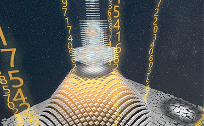
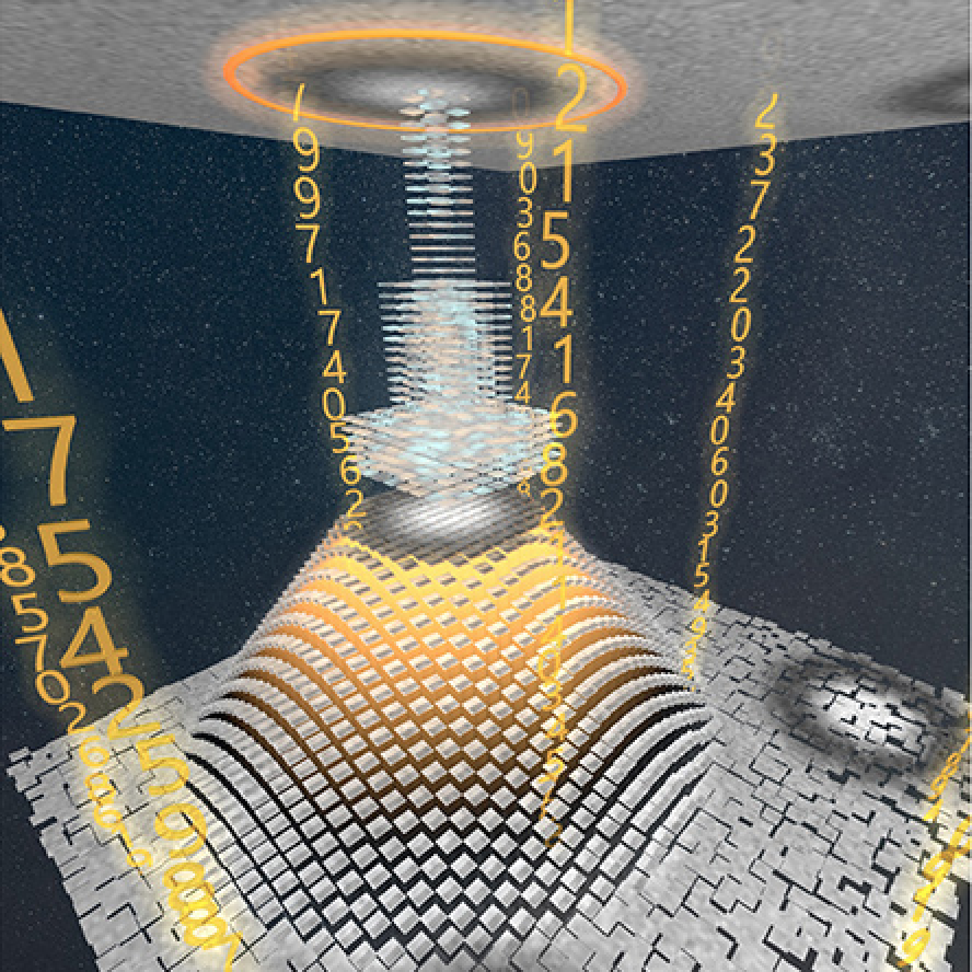
Critical Casimir forces emerge among particles or surfaces immersed in a near-critical fluid, with the sign of the force determined by surface properties and with its strength tunable by minute temperature changes. Here, we show how such forces can be used to trap a colloidal particle and levitate it above a substrate with a bull’s-eye pattern consisting of a ring with surface properties opposite to the rest of the substrate. Using the Derjaguin approximation and mean-field calculations, we find a rich behavior of spherical colloids at such a patterned surface, including sedimentation toward the ring and levitation above the ring (ring levitation) or above the bull’s-eye’s center (point levitation). Within the Derjaguin approximation, we calculate a levitation diagram for point levitation showing the depth of the trapping potential and the height at which the colloid levitates, both depending on the pattern properties, the colloid size, and the solution temperature. Our calculations reveal that the parameter space associated with point levitation shrinks if the system is driven away from a critical point, while, surprisingly, the trapping force becomes stronger. We discuss the application of critical Casimir levitation for sorting colloids by size and for determining the thermodynamic distance to criticality. Our results show that critical Casimir forces provide rich opportunities for controlling the behavior of colloidal particles at patterned surfaces.