The defense took place in PJ, Institutionen för fysik, Origovägen 6b, Göteborg, at 09:00.
Title: Quantitative Optical Microscopy of Microscale Soft Matter Systems
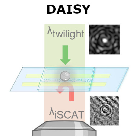
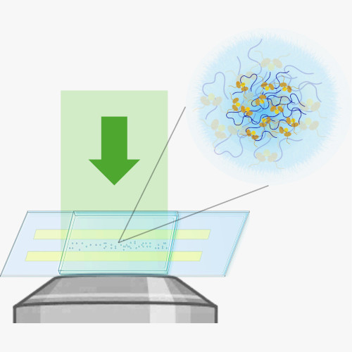
Abstract: Many biological and soft-matter particles operate at sizes below the diffraction limit and scatter light only weakly, making them hard to study with conventional microscopy. This thesis introduces two complementary, label-free interferometric methods that enable single-particle characterization across the meso–microscale. By combining optical scattering, off-axis holography, and particle tracking, these approaches quantify size, refractive index, internal structure, and mobility of individual rigid nanoparticles and soft biomolecular condensates. Together, this work provides new tools for probing the physical principles of nanoscale soft matter and phase-separated biological assemblies.
Thesis: https://hdl.handle.net/2077/90110
Supervisor: Daniel Midtvedt
Examiner: Bernhard Mehlig
Opponent: Balpreet Singh Ahluwalia
Committee: Per Augustsson, Arrate Muñoz Barrutia, Alexandra Stubelius
Alternate board member: Kristian Gustafsson