News

Presentation by A. Callegari at OSA-OMA-2021

Simulation of clustering of Janus partices in an optical potential due to hydrodynamic fluxes.
Clustering of Janus Particles Under the Effect of Optical Forces Driven by Hydrodynamic Fluxes
Agnese Callegari, S. Masoumeh Mousavi, Iryna Kasianiuk, Denis Kasyanyuk, Sabareesh K P Velu, Luca Biancofiore, Giovanni Volpe
Submitted as: OSA-OMA-2021, AM1D.3 Contribution
Date: 12 April
Time: 15 CEST

Short Abstract
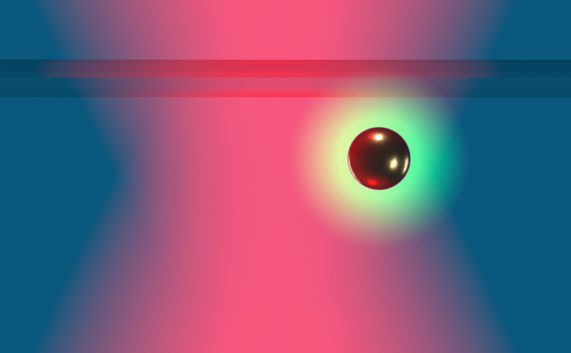
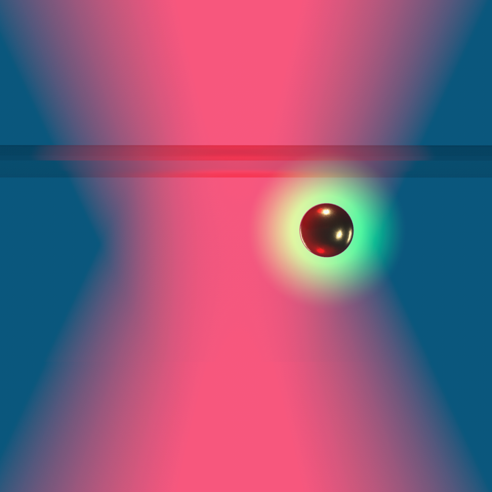
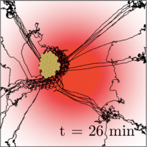
Hydrodynamic fluxes generated by Janus particles in an optical potential drive reversible clustering of colloids.

Extended Abstract

Self-organization entails the emergence of complex patterns and structures from relatively simple constituting building blocks. Phenomena such as flocking of birds and growth of bacterial colonies are examples of self-organization in nature. Also artificial microscopic systems feature similar forms of organization with the emergence of clusters, sometimes referred to as “living crystals”. In the past two decades, studies on self-organization focused on systems made of complex colloids with anisotropic surface, such as Janus particles. Depending on their surface material properties, Janus particles have been used in different fields for various applications such as self-assembly, microrheology and emulsion stabilization. Under certain conditions, Janus particles have the ability of self-propelling and behave as active Brownian particles; these active Janus particles might be used in future biomedical nano-devices for diagnostics, drug delivery and microsurgery. Studies on clustering of Janus particles have been performed by Palacci et al., who have shown the formation of living crystals in systems of light-activated Janus particles (Fe2O3-TPM) in hydrogen peroxide solution. Similarly, Buttinoni et al. demonstrated the clustering of light-activated Janus particles (carbon-SiO2) in a water-lutidine binary mixture. Other research groups have shown self-assembly and controlled crystal formations in a mixed system of light-activated Janus particles and passive colloids. In all these studies, a necessary ingredient for the clustering is the active nature of the particles. In systems of passive colloidal particles, crystallization was observed at the bottom of an attractive optical potential, close to the hard boundary during electrophoretic deposition, and in the presence of an external temperature gradient.

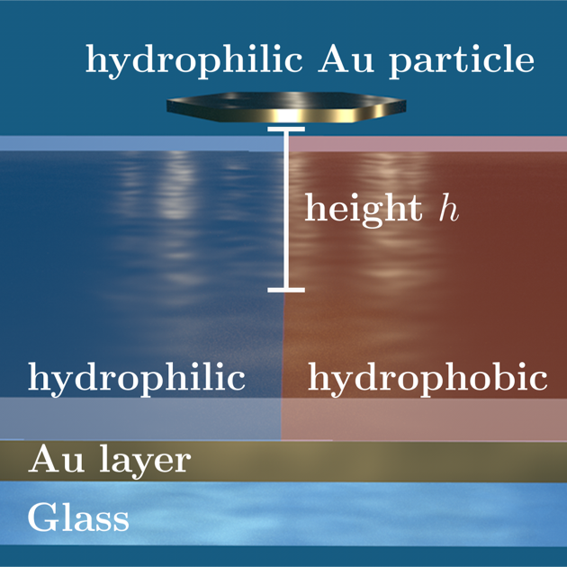
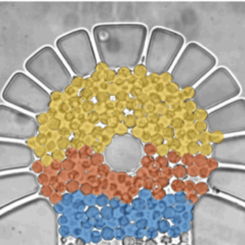
Here, we investigate the behavior of a system composed of Janus particles (silica microspheres half-coated with gold) close to a planar surface in the presence of an optical potential, and we experimentally demonstrate reversible clustering triggered by the presence of the optical field. Experimental results are compared and validated by numerical simulations, where the key ingredient for clustering is the presence of an attractive potential of hydrodynamic nature. In fact, the temperature gradient generated by the light absorption at the metallic patches on the Janus particles induces a local force field tangential to the surface of the Janus particle, which causes the fluid to slip at the surface of the particle. Because of the proximity of a planar surface, the flow pattern around the Janus particle is squeezed and results in a flow with a horizontal incoming radial component (parallel to the planar boundary) and outgoing vertical components (directed upwards from the wall). This thermophoretically-induced flow field affects the motion of other neighboring particles, so that a second nearby particle experiences an attractive hydrodynamic drag force toward the particle originating the flux. Clustering is confirmed also in mixtures of Janus particles and passive colloids (silica microspheres), where the hydrodynamic flux due to the Janus particles causes the clustering of the particles in the hybrid system and the formation of living crystals. As a further confirmation that the presence of Janus particles in the optical potential is crucial for the clustering, we show that a system with only non-Janus particles does not give rise to any clustering. We show experimentally that the clustering process is reversible, since the cluster starts to disassemble as soon as the optical potential is switched off.

Beyond their fundamental interest, the reported results are potentially relevant for various applications in the fields of self-assembly, targeted drug-delivery and bioremediation. For example, the possibility of forming clusters at a controllable distance from the minimum of a potential well offers a new route towards self-assembly near a target. Future work will be devoted to understanding how the clustering behavior can be controlled or altered by using more complex optical potentials.

Non-equilibrium properties of an active nanoparticle in a harmonic potential published in Nature Commun.

Non-spherical nanoparticle held by optical tweezers. The particle is trapped against the cover slide.

Non-equilibrium properties of an active nanoparticle in a harmonic potential
Falko Schmidt, Hana Šípová-Jungová, Mikael Käll, Alois Würger & Giovanni Volpe
Nature Communications 12, 1902 (2021)
doi: 10.1038/s41467-021-22187-z
arXiv: 2009.08393

Active particles break out of thermodynamic equilibrium thanks to their directed motion, which leads to complex and interesting behaviors in the presence of confining potentials. When dealing with active nanoparticles, however, the overwhelming presence of rotational diffusion hinders directed motion, leading to an increase of their effective temperature, but otherwise masking the effects of self-propulsion. Here, we demonstrate an experimental system where an active nanoparticle immersed in a critical solution and held in an optical harmonic potential features far-from-equilibrium behavior beyond an increase of its effective temperature. When increasing the laser power, we observe a cross-over from a Boltzmann distribution to a non-equilibrium state, where the particle performs fast orbital rotations about the beam axis. These findings are rationalized by solving the Fokker-Planck equation for the particle’s position and orientation in terms of a moment expansion. The proposed self-propulsion mechanism results from the particle’s non-sphericity and the lower critical point of the solute.

Optical Tweezers: A Comprehensive Tutorial from Calibration to Applications accepted on Advances in Optics and Photonics

Schematic of a bistable potential generated with a double-beam optical tweezers.

Optical Tweezers: A Comprehensive Tutorial from Calibration to Applications
Jan Gieseler, Juan Ruben Gomez-Solano, Alessandro Magazzù, Isaac Pérez Castillo, Laura Pérez García, Marta Gironella-Torrent, Xavier Viader-Godoy, Felix Ritort, Giuseppe Pesce, Alejandro V. Arzola, Karen Volke-Sepulveda & Giovanni Volpe
Advances in Optics and Photonics, 13(1), 74-241 (2021)
doi: https://doi.org/10.1364/AOP.394888
arXiv: 2004.05246

Since their invention in 1986 by Arthur Ashkin and colleagues, optical tweezers have become an essential tool in several fields of physics, spectroscopy, biology, nanotechnology, and thermodynamics. In this Tutorial, we provide a primer on how to calibrate optical tweezers and how to use them for advanced applications. After a brief general introduction on optical tweezers, we focus on describing and comparing the various available calibration techniques. Then, we discuss some cutting-edge applications of optical tweezers in a liquid medium, namely to study single-molecule and single-cell mechanics, microrheology, colloidal interactions, statistical physics, and transport phenomena. Finally, we consider optical tweezers in vacuum, where the absence of a viscous medium offers vastly different dynamics and presents new challenges. We conclude with some perspectives for the field and the future application of optical tweezers. This Tutorial provides both a step-by-step guide ideal for non-specialists entering the field and a comprehensive manual of advanced techniques useful for expert practitioners. All the examples are complemented by the sample data and software necessary to reproduce them.

Quantitative Digital Microscopy with Deep Learning published in Applied Physics Reviews

Particle tracking and characterization in terms of radius and refractive index.

Quantitative Digital Microscopy with Deep Learning
Benjamin Midtvedt, Saga Helgadottir, Aykut Argun, Jesús Pineda, Daniel Midtvedt, Giovanni Volpe
Applied Physics Reviews 8, 011310 (2021)
doi: 10.1063/5.0034891
arXiv: 2010.08260

Video microscopy has a long history of providing insights and breakthroughs for a broad range of disciplines, from physics to biology. Image analysis to extract quantitative information from video microscopy data has traditionally relied on algorithmic approaches, which are often difficult to implement, time consuming, and computationally expensive. Recently, alternative data-driven approaches using deep learning have greatly improved quantitative digital microscopy, potentially offering automatized, accurate, and fast image analysis. However, the combination of deep learning and video microscopy remains underutilized primarily due to the steep learning curve involved in developing custom deep-learning solutions. To overcome this issue, we introduce a software, DeepTrack 2.0, to design, train and validate deep-learning solutions for digital microscopy. We use it to exemplify how deep learning can be employed for a broad range of applications, from particle localization, tracking and characterization to cell counting and classification. Thanks to its user-friendly graphical interface, DeepTrack 2.0 can be easily customized for user-specific applications, and, thanks to its open-source object-oriented programming, it can be easily expanded to add features and functionalities, potentially introducing deep-learning-enhanced video microscopy to a far wider audience.

Invited talk by L. Pérez at SPIE Photonics West OPTO

Stable, unstable and saddle points in a speckle optical potential.
FORMA: expanding applications of optical tweezers
Laura Pérez García
Invited talk at SPIE Photonics West OPTO
6 March 2021
Online

In this presentation, Laura Pérez will talk about FORMA  (force reconstruction via maximum-likelihood-estimator analysis) which addresses the need of measuring the force fields acting on microscopic particles. Compared to alternative established methods, FORMA is faster, simpler, more accurate, and more precise. Furthermore, FORMA can also measure non-conservative and out-of-equilibrium force fields. Here, after a brief introduction to FORMA, I will present its use, advantages, and limitations. I will conclude with some recent work where we exploit Bayesian inference to expand the scope of application of FORMA.

References:
Laura Pérez García, Jaime Donlucas Pérez, Giorgio Volpe, Alejandro V. Arzola & Giovanni Volpe, High-Performance Reconstruction of Microscopic Force Fields from Brownian Trajectories, Nature Communications 9, 5166 (2018)

Time: 6 March 2021
Place: Online
Link: FORMA: expanding applications of optical tweezers at SPIE Photonics West OPTO

Optical trapping and critical Casimir forces published in EPJP

Measuring the dynamics of colloids interacting with critical Casimir interaction via blinking optical tweezers: graphical representation of the optical traps.

Optical trapping and critical Casimir forces
Agnese Callegari, Alessandro Magazzù, Andrea Gambassi & Giovanni Volpe
The European Physical Journal Plus (EPJP), 136, 213 (2021)
doi: 10.1140/epjp/s13360-020-01020-4
arXiv: 2008.01537

Critical Casimir forces emerge between objects, such as colloidal particles, whenever their surfaces spatially confine the fluctuations of the order parameter of a critical liquid used as a solvent. These forces act at short but microscopically large distances between these objects, reaching often hundreds of nanometers. Keeping colloids at such distances is a major experimental challenge, which can be addressed by the means of optical tweezers. Here, we review how optical tweezers have been successfully used to quantitatively study critical Casimir forces acting on particles in suspensions. As we will see, the use of optical tweezers to experimentally study critical Casimir forces can play a crucial role in developing nano-technologies, representing an innovative way to realize self-assembled devices at the nano- and microscale.

Nils Jacobson defended his Master thesis on 16 February 2021. Congrats!

Nils Jacobson defended his Master thesis in MPCAS at the Chalmers University of Technology on 16 February 2021. Congrats!

Screenshot of Nils Jacobson’s Master Thesis defence.
Title: Vascular Bifurcation Detection in Cerebral CT Angiography Using CNN and Frangi Filters

Segmentation and feature extraction are important tools for analysing and visualizing information in medical image data, particularly in vascular image data which relates to widely spread vascular diseases. Vessel segmentation is extensively featured in research, recently adapting trends in deep learning image processing. This paper aims to develop a vessel bifurcation detection method to support a seed point based segmentation approach. The suggested approach is a combination of classification, with a convolutional neural network (DenseNet), local vessel segmentation, with Frangi filters, and 3D morphological skeletonization. A small data set is produced for network training and evaluation. Results indicate a high classification accuracy which filters problematic samples for the Frangi filter. Thus the combination is able to suggest quality branch seed points under most circumstances. Next step would be to expand the data set to enable further optimization and more rigid evaluation. In any case a combination of a high performance classifier followed by qualitative assessment of local samples show potential.​

​Name of the master programme: MPCAS – Complex Adaptive Systems
Supervisor: Jonna Hellström and Giovanni Volpe
Examiner: Giovanni Volpe, Department of Physics, University of Gothenburg
Opponent: Eva Škvor

Place: Online via Zoom
Time: 16 February, 2021, 16:00

Link: Master thesis presentation Nils Jacobson

Presentation by F. Schmidt on QED Casimir vs Critical Casimir at MPI Stuttgart, 11 February 2021

Schematic of the experiment with a suspended metallic flake-like particle on a gold-coated substrate.
QED Casimir vs Critical Casimir Forces: Trapping and Releasing of metal flake particles

Falko Schmidt, Agnese Callegari, Giovanni Volpe
(online at) MPI Stuttgart, Germany
11 February 2021, 14:30-16.00

We propose a mechanism for restoration of collapsed structures using critical Casimir forces by investigating the diffusion of metal flake-like particles. By tuning temperature near-criticality and employing selective self-assembled monolayers the resulting repulsive critical Casimir force is large enough to lift off particle and enable transitions previously impeded by QED Casimir attraction.

Intercellular Communication Induces Glycolytic Synchronisation Waves published in PNAS

Intercellular communication induces glycolytic synchronization waves between individually oscillating cells
Martin Mojica-Benavides, David D. van Niekerk, Mite Mijalkov, Jacky L. Snoep, Bernhard Mehlig, Giovanni Volpe, Caroline B. Adiels & Mattias Goksör
PNAS 118(6), e2010075118 (2021)
doi: 10.1073/pnas.2010075118
arXiv: 1909.05187

Metabolic oscillations in single cells underlie the mechanisms behind cell synchronization and cell-cell communication. For example, glycolytic oscillations mediated by biochemical communication between cells may synchronize the pulsatile insulin secretion by pancreatic tissue, and a link between glycolytic synchronization anomalies and type-2 diabetes has been hypotesized. Cultures of yeast cells have provided an ideal model system to study synchronization and propagation waves of glycolytic oscillations in large populations. However, the mechanism by which synchronization occurs at individual cell-cell level and overcome local chemical concentrations and heterogenic biological clocks, is still an open question because of experimental limitations in sensitive and specific handling of single cells. Here, we show how the coupling of intercellular diffusion with the phase regulation of individual oscillating cells induce glycolytic synchronization waves. We directly measure the single-cell metabolic responses from yeast cells in a microfluidic environment and characterize a discretized cell-cell communication using graph theory. We corroborate our findings with simulations based on a kinetic detailed model for individual yeast cells. These findings can provide insight into the roles cellular synchronization play in biomedical applications, such as insulin secretion regulation at the cellular level.

Press release on joint research on intercellular communication mechanism by Biological Physics Lab and Soft Matter Lab

The article Intercellular Communication Induces Glycolytic Synchronisation Waves published in PNAS has been featured in the News of the Faculty of Science of Gothenburg University.

Here the links to the press releases:
Swedish: Forskare har knäckt koden för cellkommunikation
English: Researchers have broken the code for cell communication