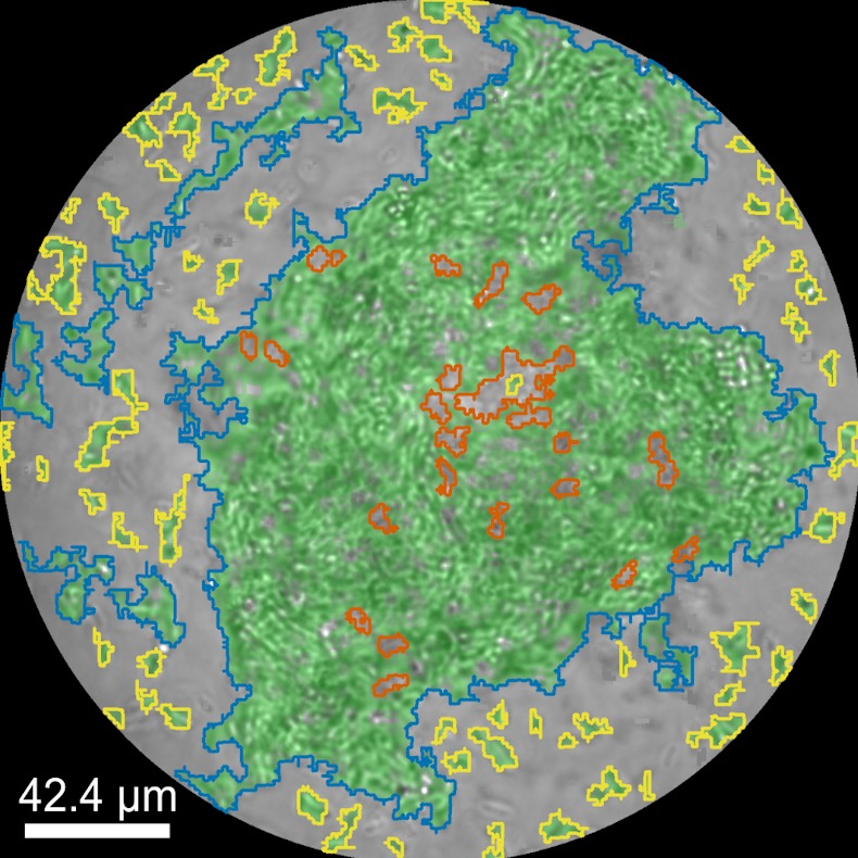
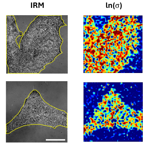
Brightfield image of biofilm inside the droplet where motile aggregates are highlighted in yellow, biofilm mass in blue, and open patches in the structures due to dispersal in orange. (Image from D. Pérez and J. Domínguez)Quantitative Analysis of Dynamic Biofilm Structures via Time-Resolved Droplet Microfluidics and Artificial Intelligence
Daniela Pérez Guerrero, Jesús Manuel Antúnez Domínguez, Aurélie Vigne, Daniel Midtvedt, Wylie Ahmed, Lisa Muiznieks, Giovanni Volpe and Caroline Beck Adiels Date: 11th March 2026 Time: 18:00 – 20:00 Place: Aula Medica, Karolinska Institute, Solna
Conference Protein Folding in Real Time, 11-13 March 2026, Stockholm, Sweden
Droplet Microfluidics offers a powerful approach to study the spatiotemporal dynamics of biofilm formation at high resolution and throughput. By encapsulating microbial communities within controlled microenvironments, it becomes possible to monitor biofilm development continuously using time-lapse imaging, capturing transitions from initial attachment to maturation and dispersal. To make sense of these complex, high-dimensional datasets, we are developing an unsupervised variational autoencoder framework that can automatically identify and separate distinct stages of biofilm growth without prior labeling. This approach enables the extraction of latent features that characterize structural and behavioral shifts within the biofilm over time. In this context, protein folding may play a critical role in regulating both the establishment and dispersal of biofilms, as the conformational states of key structural and regulatory proteins can influence adhesion, matrix production, and the transition back to planktonic states.
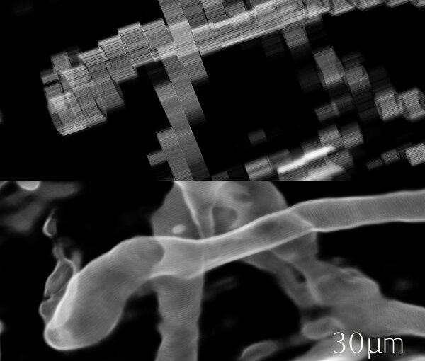
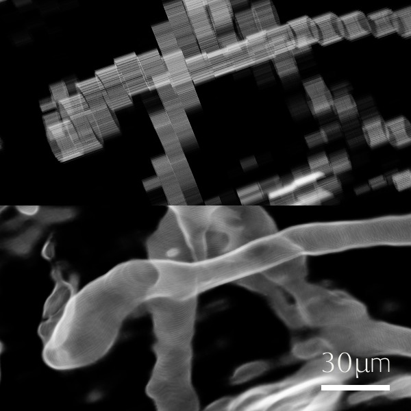
Comparison of anisotropic light-sheet microscopy data and super-resolved reconstruction, illustrating improved continuity and structural clarity of volumetric features. (Image by J. Tember.)FIREFLY – Framework for Interactive Rendering, Exploration and Feature Extraction for Light-Sheet Microscopy
John Tember, Harshith Bachimanchi, Tsz Long Chu, Xin Tian, Andrei Chagin, Giovanni Volpe Date: 11th March 2026 Time: 18:00 – 20:00 Place: Aula Medica, Karolinska Institute, Solna Conference:Protein Folding in Real Time, 11-13 March 2026, Stockholm, Sweden
We present FIREFLY (Framework for Interactive Rendering, Exploration and Feature Extraction for Light-Sheet Microscopy), a platform for scalable computational tools. Modern 3D microscopy datasets are increasingly large and complex, posing challenges for efficient visualization, processing, and quantitative analysis. Our work focuses on developing tools and methods to support interactive and high-quality analysis workflows.
We are building a Unity-based application for real-time, multi-channel 3D rendering, enabling interactive inspection and quantitative analysis of large volumetric datasets. In parallel, we explore self-supervised machine learning approaches for enhancing anisotropic microscopy volumes and improving resolution without requiring additional training data.
Together, these efforts aim to provide an integrated and scalable pipeline for visualization and analysis in light-sheet microscopy.
3D model of the integrated setup combining optical tweezers, light-sheet microscopy, and microfluidics to manipulate the gut microbiome in vivo in zebrafish. (Image by N. C. Palmero Cruz.)Optical Manipulation of Gut Microbiome and Neural Responses in Zebrafish
Norma Caridad Palmero Cruz Date: 11 March 2026 Time: 18.00-20.00 Place: Aula Medica, Stockholm Sweden
Conference Protein Folding in Real Time, 11-13 March 2026, Stockholm, Sweden
The gut-brain axis is a complex, bidirectional network linking the microbiome to the central nervous system, significantly affecting physiological processes and neurological health, including conditions like autism and depression. Due to the genetic similarities between zebrafish and humans, the zebrafish serves as a valuable model for investigating the bidirectional relationship between the gut and brain, offering insights into how it compares with human behaviors. Research on the connection between gut and brain development typically involves using germ-free lab animals, where the microbiome is eliminated, and comparing them to those with restored microbiomes. However, this method does not capture the complexity of microbiome-nervous system communication due to its all-or-nothing approach. This work presents a setup that combines microfluidic techniques, optical tweezers, and light sheet microscopy to precisely manipulate the microbiome in larval zebrafish in situ and in vivo. This approach offers deeper insights into gut-brain connectivity and its impact on neurological health.
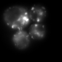
Heat-induced aggregates in Saccharomyces cerevisiae on Slimfield SMLM. Hsp104-mGFP binds to misfolded regions enabling aggregate visualisation for tracking. (Image by L. Viaene.)A single-molecule approach to study the spatial protein quality control system
Linde Viaene Date: 11 March 2026 Time: 18.00-20.00 Place: Aula Medica, Stockholm Sweden
Conference Protein Folding in Real Time, 11-13 March 2026, Stockholm, Sweden
Cell populations are inherently diverse, and averaging measurements across them can mask subtle or rare cellular behaviours. In this work, we use live Slimfield single-molecule microscopy to study the role of Hsp104 in clearing misfolded and aggregated proteins after stress. By analysing endogenously tagged Hsp104, we quantify molecular diffusion and stoichiometry before and after heat stress. Our results show a transition from faster, more mobile molecules to larger, more static assemblies following stress, consistent with Hsp104 functionally engaging with protein aggregates. These measurements provide molecular-level insight into how cells respond to proteotoxic stress.
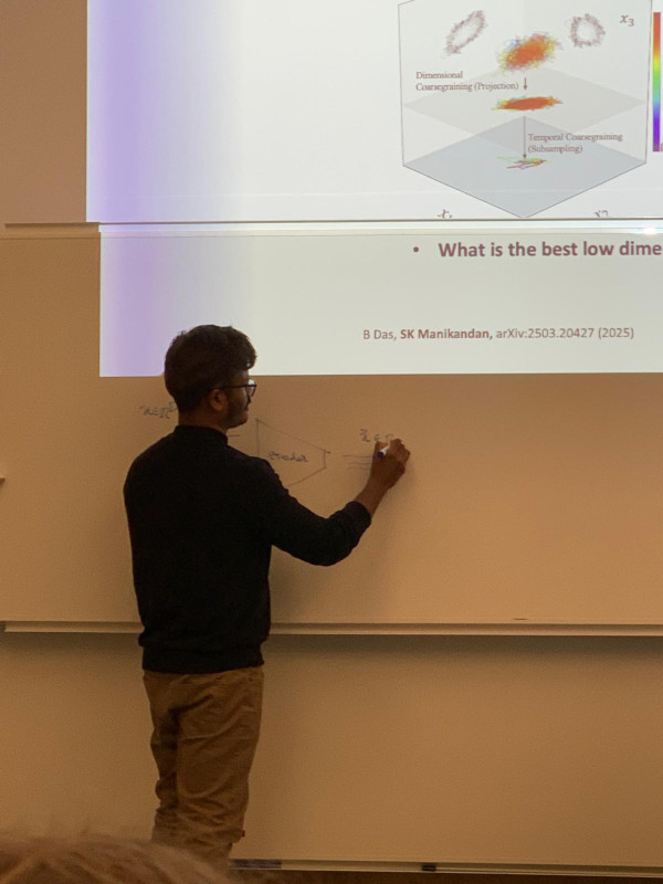
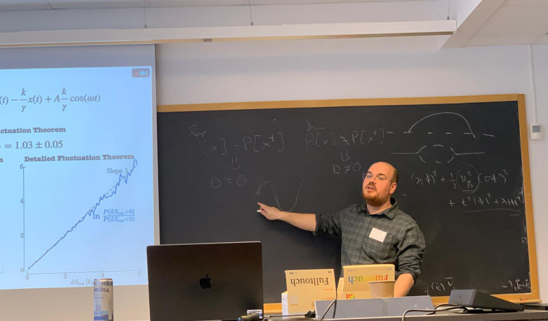
Identifying whether a process is in equilibrium, quantifying how far it lies from equilibrium, and determining optimal reduced descriptions of non-equilibrium processes remain challenging open problems. Here, we discuss how novel data-driven techniques grounded in stochastic thermodynamics can be used to efficiently learn these features directly from experimental data. In particular, we show how entropy production can be localized in space and time, and how maximally dissipative coordinates can be consistently inferred as effective low-dimensional descriptions of non-equilibrium processes. We further discuss applications to experimental biophysical systems and outline key challenges and limitations.
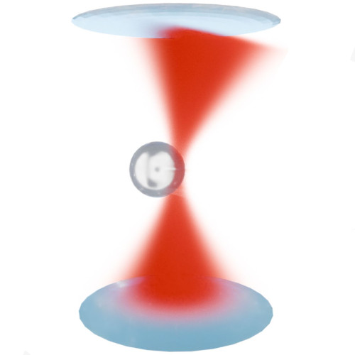
Schematic illustration of the light-momentum detection principle underlying SmartTrap. The momentum change of the trapping laser, induced by its interaction with the trapped particle, is measured to directly quantify optical forces with high precision, enabling real-time feedback and autonomous control in non-equilibrium experiments. (Figure by A. Ciarlo.)SmartTrap: Autonomous Optical Tweezers for Statistical Physics of Non-Equilibrium Systems
Antonio Ciarlo Date: 26th February 2026 Time: 13.30 Place: NORDITA, Stockholm, Sweden The 15th Nordic Workshop on Statistical Physics: Biological, Complex and Non-equilibrium Systems
Optical tweezers are a key tool in non-equilibrium statistical physics, allowing direct measurements of forces, work, and fluctuations in single-molecule and soft matter systems. However, manual operation limits throughput and the systematic study of rare events.
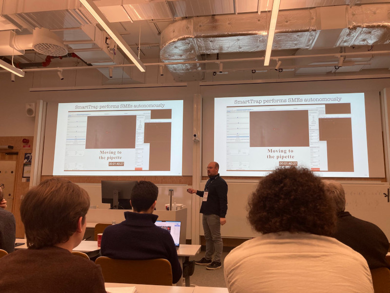
In this talk, Antonio Ciarlo will present SmartTrap, a fully autonomous optical tweezers platform integrating deep learning–based 3D tracking, adaptive feedback control, and automated microfluidics. The system operates without human intervention, executing complete force spectroscopy protocols.
Demonstrated with high-throughput DNA pulling experiments on λ-DNA, SmartTrap enables precise measurements of force–extension curves and folding kinetics. The platform also opens new possibilities for studies of colloids, single cells, and quantitative tests of non-equilibrium statistical physics.
Active Matter: Model Systems and Experimental Tests
Agnese Callegari, Antonio Ciarlo, Sreekanth Manikandan Dates and times:
23 Feb 14:00-15:00 (Agnese)
24 Feb 11:30-12:30 (Antonio)
24 Feb 14:00-15:00 (Sreekanth) Place: PJ Winter school on Geometry of nonequilibrium critical phenomena
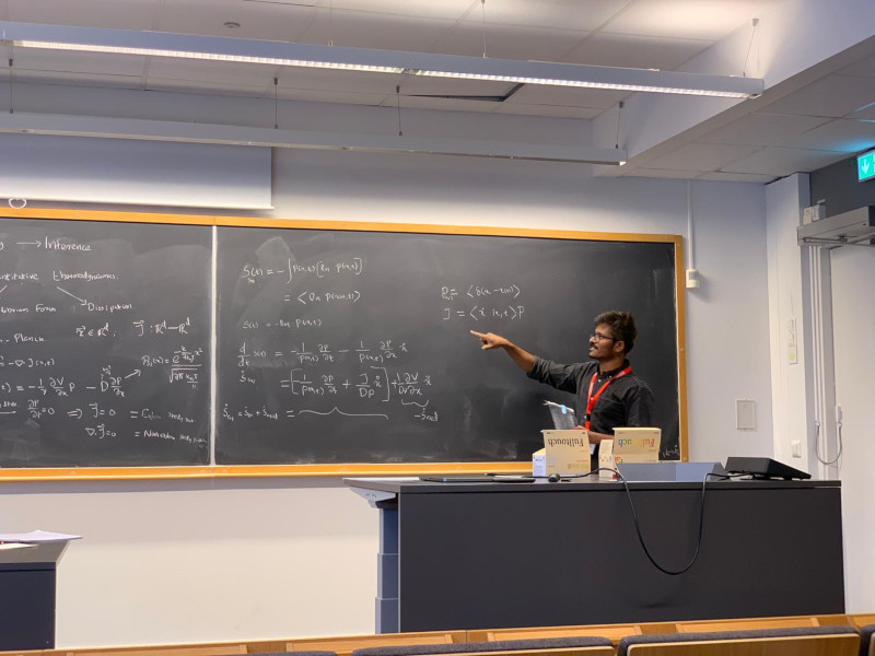
Active matter is a broad class of systems that operate intrinsically out of equilibrium. It spans multiple length scales—from macroscopic to micro- and nanoscopic—and includes both biological and artificial realizations, often displaying rich and emerging collective behaviors. The study of active matter aims to explain and interpret these phenomena using concepts and tools from physics. As such, understanding active and non-equilibrium systems requires a combination of theoretical, computational, and experimental approaches.
In the first part of the lecture, we introduce the concept of an active particle and demonstrate how it can be embodied in a macroscopic, self-propelled toy robot (a Hexbug). Despite their simplicity, such systems reproduce characteristic—and sometimes counterintuitive—features of microscopic active matter. These experiments have a strong pedagogical value and are designed to help bridge a gap in traditional physics curricula at the primary and secondary education levels.
The second part of the lecture focuses on active matter and non-equilibrium phenomena at the microscopic scale, where advanced experimental tools are essential. Optical tweezers provide precise control over microscopic systems and access to key physical observables. We introduce their operating principles and illustrate how they can be used to construct a minimal, well-controlled experimental model for studying non-equilibrium dynamics at the single-particle level.
In the final part of the lecture, we turn to the theoretical and computational tools required to analyze active matter systems. We discuss how non-equilibrium dynamics can be quantitatively characterized directly from experimental data in a model-independent framework. This naturally leads to an introduction to machine-learning–based inference techniques, which extract dynamical and thermodynamic information from data without relying on a priori assumptions about the underlying physical model.
References:
[1] A. Barona Balda, A. Argun, A. Callegari, G. Volpe. Playing with Active Matter, Am. J. Phys. 92, 847–858 (2024). https://doi.org/10.1119/5.0125111
[2] Martins, T.T., Malavazi, A.H.A., Kamizaki, L.P. et al. Fluctuation theorems with optical tweezers: theory and practice. Eur. Phys. J. Plus 141, 71 (2026). https://doi.org/10.1140/epjp/s13360-025-07181-4
[3] Manikandan, Sreekanth K. and Ghosh, T. and Mandal, T. and Biswas, A. and Sinha, B. and Mitra, D. Estimate of entropy production rate can spatiotemporally resolve the active nature of cell flickering. Phys. Rev. Res. 6, 023310 (2024). https://doi.org/10.1103/PhysRevResearch.6.023310
Photos
Antonio, presenting. (Photo by M. Orsino)Sreekanth, presenting. (Photo by A. Ciarlo)
Hang Zhao, supervised by Giovanni Volpe and Joana Pereira, will present his halftime seminar under the topic “Brain connectome revealed neuro-degenerative disease” on 9-10 am, 22nd Jan. 2026 in Nexus and through Zoom (https://gu-se.zoom.us/j/7726618257). The seminar starts from his presentation about the past and planned project, followed by a discussion and questions by his opponent, Professor Mattias Göksor.
Quantifying the spatiotemporal forces, affinities, and dissipative costs of cellular-scale non-equilibrium processes from experimental data and localizing it in space and time remain a significant open challenge. Here, I explore how principles from stochastic thermodynamics, combined with machine learning techniques, offer a promising approach to addressing this issue. I will present preliminary results from experiments on fluctuating cell membranes and simulations of non-equilibrium systems in stationary and time-dependently driven states. These studies reveal potential strategies for localizing entropy production in experimental biophysical contexts while also highlighting key challenges and limitations that must be addressed.
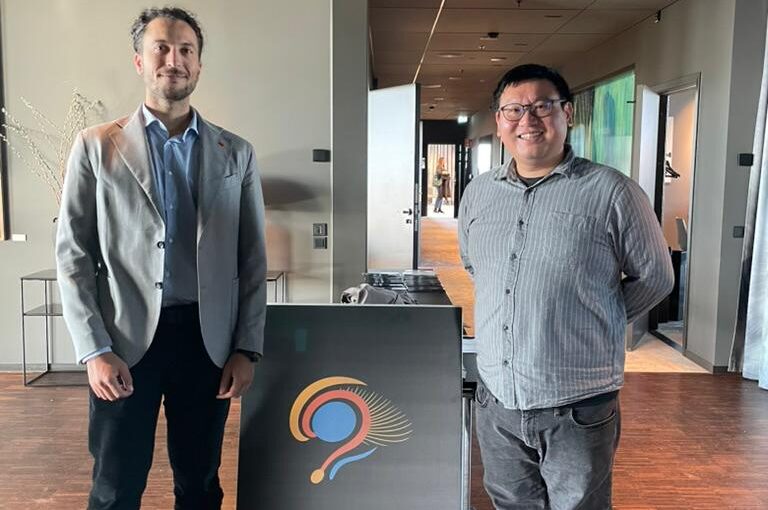
Massimiliano Passaretti (left) and Yu-Wei Chang (right) at NEME 2025. (Photo courtesy of Clarion Hotel Draken.)Graph theory and deep learning pipelines
Yu-Wei Chang, Massimiliano Passaretti NEMES 2025, 24-26 September, 2025 Date: 25 September 2025 Time: 12:45 – 14:00 Place: Clarion Hotel Draken
This workshop begins with a practical introduction to graph theory, then guides participants through BRAPH 2 to build connectomes, compute graph measures, and run group comparisons, followed by a hands-on deep-learning pipeline. It demonstrates a unified GUI/command-line workflow, a unique architecture of BRAPH 2, helping participants move smoothly from the GUI to scripts. This workshop also guides participants to reproduce multiplex and deep-learning results on their computers from the BRAPH 2 bioRxiv preprint.