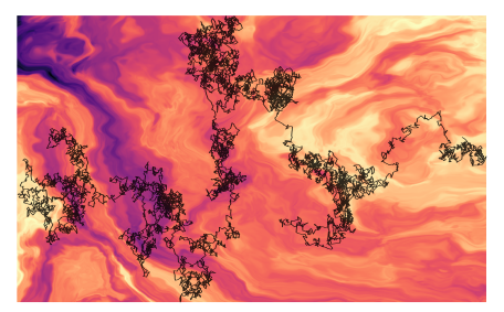
An illustration of anomalous diffusion. (Image by Gorka Muñoz-Gil.)
CORDIS, the Community Research and Development Information Service of the European Commission, recently covered Giovanni Volpe’s ComplexSwimmers ERC-StG grant in a news: Throwing down the scientific gauntlet to assess methods for anomalous diffusion.
The article highlights the joint results obtained by three EU-backed research projects (NOQIA, OPTOlogic and ComplexSwimmers) dealing with anomalous diffusion.
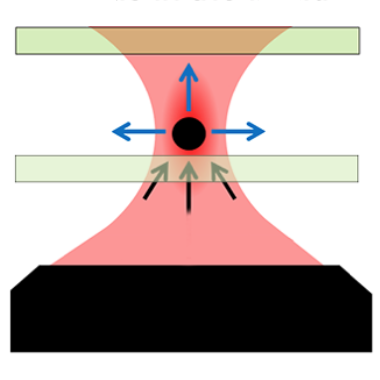
Optical beam focused into the liquid: the tire particles are pushed away from the laser focus.
Raman Tweezers for Tire and Road Wear Micro- and Nanoparticles Analysis
Pietro Giuseppe Gucciardi, Gillibert Raymond, Alessandro Magazzù, Agnese Callegari, David Bronte Ciriza, Foti Antonino, Maria Grazia Donato, Onofrio M. Maragò, Giovanni Volpe, Marc Lamy de La Chapelle & Fabienne Lagarde
Environmental Science: Nano 9, 145 – 161 (2022)
ChemRxiv: https://doi.org/10.33774/chemrxiv-2021-h59n1
doi: https://doi.org/10.1039/D1EN00553G
Tire and Road Wear Particles (TRWP) are non-exhaust particulate matter generated by road transport means during the mechanical abrasion of tires, brakes and roads. TRWP accumulate on the roadsides and are transported into the aquatic ecosystem during stormwater runoffs. Due to their size (sub-millimetric) and rubber content (elastomers), TRWP are considered microplastics (MPs). While the amount of the MPs polluting the water ecosystem with sizes from ~ 5 μm to more than 100 μm is known, the fraction of smaller particles is unknown due to the technological gap in the detection and analysis of < 5 μm MPs. Here we show that Raman Tweezers, a combination of optical tweezers and Raman spectroscopy, can be used to trap and chemically analyze individual TWRPs in a liquid environment, down to the sub-micrometric scale. Using tire particles mechanically grinded from aged car tires in water solutions, we show that it is possible to optically trap individual sub-micron particles, in a so-called 2D trapping configuration, and acquire their Raman spectrum in few tens of seconds. The analysis is then extended to samples collected from a brake test platform, where we highlight the presence of sub-micrometric agglomerates of rubber and brake debris, thanks to the presence of additional spectral features other than carbon. Our results show the potential of Raman Tweezers in environmental pollution analysis and highlight the formation of nanosized TRWP during wear.
We present DeepTrack 2.0, a software to design, train, and validate deep-learning solutions for digital microscopy. We demonstrate it for applications from particle localization, tracking, and characterization, to cell counting and classification, to virtual staining.
The study, published in Nature Communications and co-written by researchers at the Soft Matter Lab of the Department of Physics at the University of Gothenburg, originates from the AnDi Challenge, a competition co-organised by Giovanni Volpe with researchers from University of Vic – Central University of Catalunya, Institute of Photonic Sciences in Barcelona, University of Potsdam, and Valencia Polytechnic University.
The challenge was held during March–November 2020 and consisted of three main tasks concerning anomalous exponent inference, model classification, and trajectory segmentation. The goal was to provide an objective assessment of the performance of methods to characterise anomalous diffusion from single trajectories.
An illustration of anomalous diffusion. (Image by Gorka Muñoz-Gil.)Objective comparison of methods to decode anomalous diffusion
Gorka Muñoz-Gil, Giovanni Volpe, Miguel Angel Garcia-March, Erez Aghion, Aykut Argun, Chang Beom Hong, Tom Bland, Stefano Bo, J. Alberto Conejero, Nicolás Firbas, Òscar Garibo i Orts, Alessia Gentili, Zihan Huang, Jae-Hyung Jeon, Hélène Kabbech, Yeongjin Kim, Patrycja Kowalek, Diego Krapf, Hanna Loch-Olszewska, Michael A. Lomholt, Jean-Baptiste Masson, Philipp G. Meyer, Seongyu Park, Borja Requena, Ihor Smal, Taegeun Song, Janusz Szwabiński, Samudrajit Thapa, Hippolyte Verdier, Giorgio Volpe, Arthur Widera, Maciej Lewenstein, Ralf Metzler, and Carlo Manzo
Nat. Commun. 12, Article number: 6253 (2021)
doi: 10.1038/s41467-021-26320-w
arXiv: 2105.06766
Deviations from Brownian motion leading to anomalous diffusion are found in transport dynamics from quantum physics to life sciences. The characterization of anomalous diffusion from the measurement of an individual trajectory is a challenging task, which traditionally relies on calculating the trajectory mean squared displacement. However, this approach breaks down for cases of practical interest, e.g., short or noisy trajectories, heterogeneous behaviour, or non-ergodic processes. Recently, several new approaches have been proposed, mostly building on the ongoing machine-learning revolution. To perform an objective comparison of methods, we gathered the community and organized an open competition, the Anomalous Diffusion challenge (AnDi). Participating teams applied their algorithms to a commonly-defined dataset including diverse conditions. Although no single method performed best across all scenarios, machine-learning-based approaches achieved superior performance for all tasks. The discussion of the challenge results provides practical advice for users and a benchmark for developers.
Video microscopy has a long history of providing insights and breakthroughs for a broad range of disciplines, from physics to biology. Image analysis to extract quantitative information from video microscopy data has traditionally relied on algorithmic approaches, which are often difficult to implement, time consuming, and computationally expensive. Recently, alternative data-driven approaches using deep learning have greatly improved quantitative digital microscopy, potentially offering automatized, accurate, and fast image analysis. However, the combination of deep learning and video microscopy remains underutilized primarily due to the steep learning curve involved in developing custom deep-learning solutions. To overcome this issue, we introduce a software, DeepTrack 2.0, to design, train and validate deep- learning solutions for digital microscopy. We use it to exemplify how deep learning can be employed for a broad range of applications, from particle localization, tracking and characterization to cell counting and classification. Thanks to its user- friendly graphical interface, DeepTrack 2.0 can be easily customized for user-specific applications, and, thanks to its open-source object-oriented programming, it can be easily expanded to add features and functionalities, potentially introducing deep-learning-enhanced video microscopy to a far wider audience.
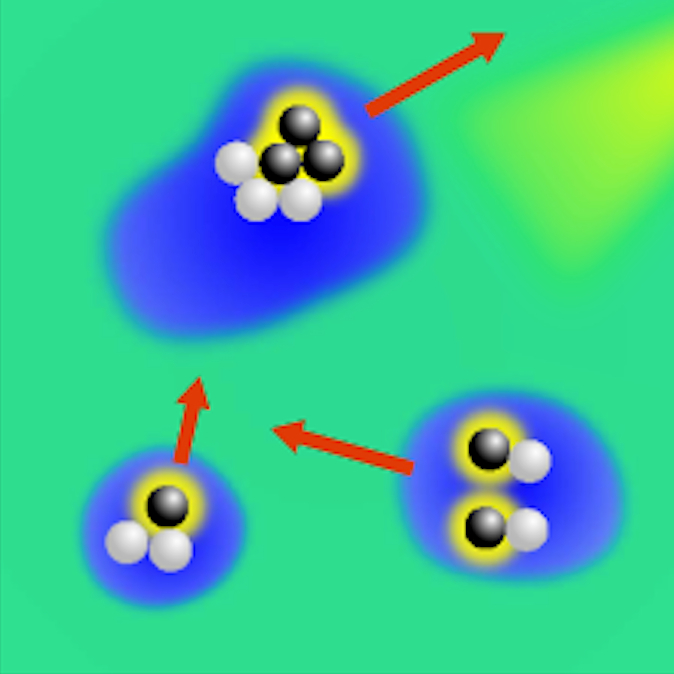
The article Active Droploids has been featured in a press release of the University of Gothenburg.
The study, published in Nature Communications, examines a special system of colloidal particles and demonstrates a new kind of active matter, which interacts with and modifies its environment. In the long run, the result of the study can be used for drug delivery inside the human body or to perform sensing of environmental pollutants and their clean-up.
Video microscopy has a long history of providing insights and breakthroughs for a broad range of disciplines, from physics to biology. Image analysis to extract quantitative information from video microscopy data has traditionally relied on algorithmic approaches, which are often difficult to implement, time consuming, and computationally expensive. Recently, alternative data-driven approaches using deep learning have greatly improved quantitative digital microscopy, potentially offering automatized, accurate, and fast image analysis. However, the combination of deep learning and video microscopy remains underutilized primarily due to the steep learning curve involved in developing custom deep-learning solutions. To overcome this issue, we introduce a software, DeepTrack 2.0, to design, train and validate deep-learning solutions for digital microscopy. We use it to exemplify how deep learning can be employed for a broad range of applications, from particle localization, tracking and characterization to cell counting and classification. Thanks to its user-friendly graphical interface, DeepTrack 2.0 can be easily customized for user-specific applications, and, thanks to its open-source object-oriented programming, it can be easily expanded to add features and functionalities, potentially introducing deep-learning-enhanced video microscopy to a far wider audience.
After a brief overview of artificial intelligence, machine learning and deep learning, I will present a series of recent works in which we have employed deep learning for applications in photonics and active matter. In particular, I will explain how we employed deep learning to enhance digital video microscopy, to estimate the properties of anomalous diffusion, to characterize microscopic force fields, to improve the calculation of optical forces, and to characterize nanoparticles. Finally, I will provide an outlook for the application of deep learning in photonics and active matter.
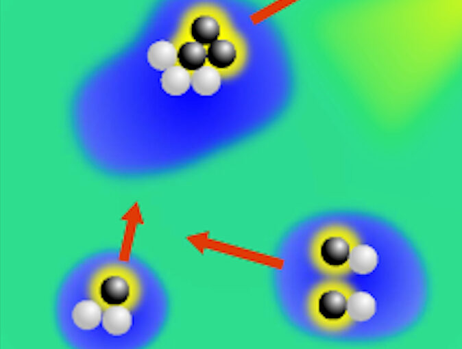
Active droploids. (Image taken from the article.)Active droploids
Jens Grauer, Falko Schmidt, Jesús Pineda, Benjamin Midtvedt, Hartmut Löwen, Giovanni Volpe & Benno Liebchen
Nat. Commun. 12, 6005 (2021)
doi: 10.1038/s41467-021-26319-3
arXiv: 2109.10677
Active matter comprises self-driven units, such as bacteria and synthetic microswimmers, that can spontaneously form complex patterns and assemble into functional microdevices. These processes are possible thanks to the out-of-equilibrium nature of active-matter systems, fueled by a one-way free-energy flow from the environment into the system. Here, we take the next step in the evolution of active matter by realizing a two-way coupling between active particles and their environment, where active particles act back on the environment giving rise to the formation of superstructures. In experiments and simulations we observe that, under light-illumination, colloidal particles and their near-critical environment create mutually-coupled co-evolving structures. These structures unify in the form of active superstructures featuring a droplet shape and a colloidal engine inducing self-propulsion. We call them active droploids—a portmanteau of droplet and colloids. Our results provide a pathway to create active superstructures through environmental feedback.