Giovanni Volpe
BPPB Seminar 2023
Date: 19 May 2023
Time: 17:00
Author: Agnese Callegari
Seminar by B. Roy, 24 May 2023
Basudev Roy
Indian Institute of Technology Madras, India
Date: 24 May 2023
Time: 12:30
Place: Nexus
Abstract:
A rigid body can have 6 degrees of freedom, namely the three translational degrees of freedom and the three rotational degrees of freedom. Of these, the translational degrees have been well explored in optical tweezers community. However, only the in-plane rotational degree of freedom has been explored. We call this in-plane degree of rotational freedom, the yaw motion in the nomenclature of the airlines. The pitch and roll degrees are only beginning to be explored recently.
In this talk, I will show you 4 ways of generating pitch rotation using the optical tweezers. I will also show you one way of detection of pitch rotation at high resolution using birefringent particles. Further, I will discuss some applications of this pitch rotation in soft matter systems and biology. I will also show you a few other projects that we are working on inside the lab.
Bio:
Basudev Roy got his MSc from Indian Institute of Technology Kharagpur and MS from the University of Maryland, College Park. He got his PhD from Indian Institute of Science Education and Research, Kolkata in 2015. He was an Alexander von Humboldt fellow at the University of Tuebingen, Germany for his postdoctoral research from 2015 to 2017. He joined Indian Institute of Technology Madras, India since 2017 where he is now an Associate Professor.
Invited Seminar by G. Volpe at LOMA, Bordeaux, 2 May 2023
Giovanni Volpe
Seminar at LOMA, Bordeaux
2 May 2023, 14:00
Video microscopy has a long history of providing insights and breakthroughs for a broad range of disciplines, from physics to biology. Image analysis to extract quantitative information from video microscopy data has traditionally relied on algorithmic approaches, which are often difficult to implement, time consuming, and computationally expensive. Recently, alternative data-driven approaches using deep learning have greatly improved quantitative digital microscopy, potentially offering automatized, accurate, and fast image analysis. However, the combination of deep learning and video microscopy remains underutilized primarily due to the steep learning curve involved in developing custom deep-learning solutions. To overcome this issue, we have introduced a software, currently at version DeepTrack 2.1, to design, train and validate deep-learning solutions for digital microscopy.
Edoardo Grasso joins the Soft Matter Lab
Edoardo Grasso joined the Soft Matter Lab on April 29, 2023.
Edoardo is a Master student at the Physics Department of the University of Turin, Italy.
During his time at the Soft Matter Lab, he will be working on application of deep learning.
He will stay in our lab till July 2, 2023.
Edoardo Manoni joins the Soft Matter Lab
Edoardo is a Master student at Chalmers University of Technology.
During his internship at the Soft Matter Lab, he will be working on the modelling of smart metavehicles.
Plenary Lecture by G. Volpe at SPIE Optics + Optoelectronics, Prague, 25 April 2023
Giovanni Volpe
SPIE Optics + Optoelectronics, Prague, 25 April 2023
Time: 09:45
Video microscopy has a long history of providing insights and breakthroughs for a broad range of disciplines, from physics to biology. Image analysis to extract quantitative information from video microscopy data has traditionally relied on algorithmic approaches, which are often difficult to implement, time consuming, and computationally expensive. Recently, alternative data-driven approaches using deep learning have greatly improved quantitative digital microscopy, potentially offering automatized, accurate, and fast image analysis. However, the combination of deep learning and video microscopy remains underutilized primarily due to the steep learning curve involved in developing custom deep-learning solutions.
To overcome this issue, we have introduced a software, currently at version DeepTrack 2.1, to design, train and validate deep-learning solutions for digital microscopy. We use it to exemplify how deep learning can be employed for a broad range of applications, from particle localization, tracking and characterization to cell counting and classification. Thanks to its user-friendly graphical interface, DeepTrack 2.1 can be easily customized for user-specific applications, and, thanks to its open-source object-oriented programming, it can be easily expanded to add features and functionalities, potentially introducing deep-learning-enhanced video microscopy to a far wider audience.
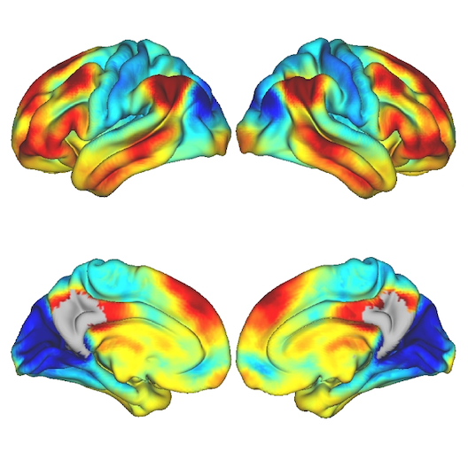
Functional gradients of the medial parietal cortex in a healthy cohort with family history of sporadic Alzheimer’s disease published in Alzheimer’s Research & Therapy
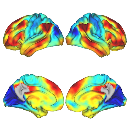
Dániel Veréb, Mite Mijalkov, Yu-Wei Chang, Anna Canal-Garcia, Emiliano Gomez-Ruis, Anne Maass, Sylvia Villeneuve, Giovanni Volpe Joana B. Pereira
Alzheimer’s Research & Therapy 15, 82 (2023)
doi: 10.1186/s13195-023-01228-3
Background
The medial parietal cortex is an early site of pathological protein deposition in Alzheimer’s disease (AD). Previous studies have identified different subregions within this area; however, these subregions are often heterogeneous and disregard individual differences or subtle pathological alterations in the underlying functional architecture. To address this limitation, here we measured the continuous connectivity gradients of the medial parietal cortex and assessed their relationship with cerebrospinal fluid (CSF) biomarkers, ApoE ε4 carriership and memory in asymptomatic individuals at risk to develop AD.
Methods
Two hundred sixty-three cognitively normal participants with a family history of sporadic AD who underwent resting-state and task-based functional MRI using encoding and retrieval tasks were included from the PREVENT-AD cohort. A novel method for characterizing spatially continuous patterns of functional connectivity was applied to estimate functional gradients in the medial parietal cortex during the resting-state and task-based conditions. This resulted in a set of nine parameters that described the appearance of the gradient across different spatial directions. We performed correlation analyses to assess whether these parameters were associated with CSF biomarkers of phosphorylated tau181 (p-tau), total tau (t-tau), and amyloid-ß1-42 (Aß). Then, we compared the spatial parameters between ApoE ε4 carriers and noncarriers, and evaluated the relationship between these parameters and memory.
Results
Alterations involving the superior part of the medial parietal cortex, which was connected to regions of the default mode network, were associated with higher p-tau, t-tau levels as well as lower Aß/p-tau levels during the resting-state condition (p < 0.01). Similar alterations were found in ApoE ε4 carriers compared to non-carriers (p < 0.003). In contrast, lower immediate memory scores were associated with changes in the middle part of the medial parietal cortex, which was connected to inferior temporal and posterior parietal regions, during the encoding task (p = 0.001). No results were found when using conventional connectivity measures.
Conclusions
Functional alterations in the medial parietal gradients are associated with CSF AD biomarkers, ApoE e4 carriership, and lower memory in an asymptomatic cohort with a family history of sporadic AD, suggesting that functional gradients are sensitive to subtle changes associated with early AD stages.
Light, Matter, Action: Shining light on active matter published in ACS Photonics
Marcel Rey, Giovanni Volpe, Giorgio Volpe
ACS Photonics, 10, 1188–1201 (2023)
arXiv: 2301.13034
doi: 10.1021/acsphotonics.3c00140
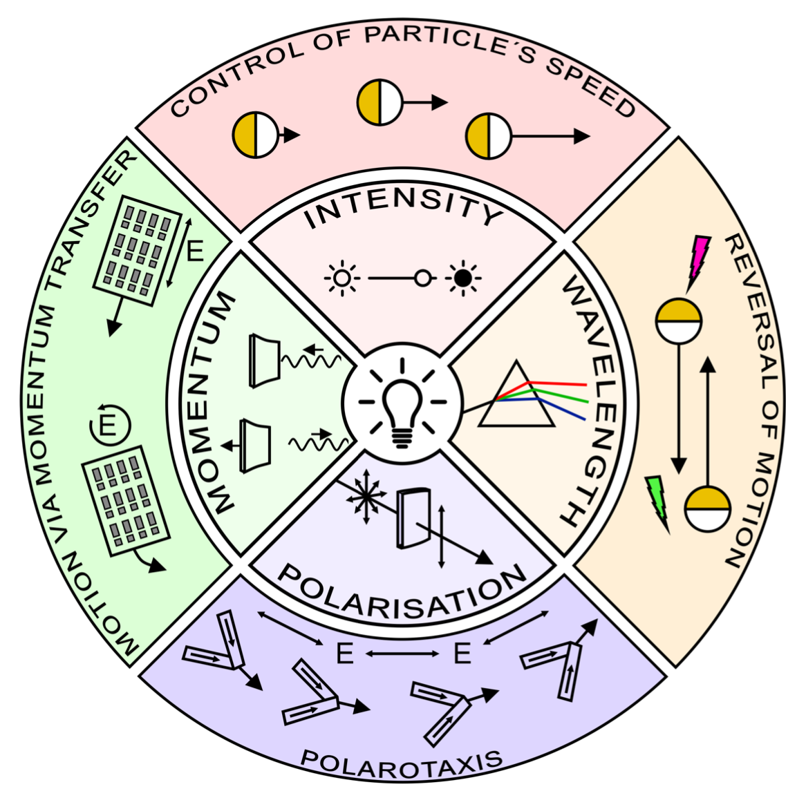
Light carries energy and momentum. It can therefore alter the motion of objects from atomic to astronomical scales. Being widely available, readily controllable and broadly biocompatible, light is also an ideal tool to propel microscopic particles, drive them out of thermodynamic equilibrium and make them active. Thus, light-driven particles have become a recent focus of research in the field of soft active matter. In this perspective, we discuss recent advances in the control of soft active matter with light, which has mainly been achieved using light intensity. We also highlight some first attempts to utilize light’s additional degrees of freedom, such as its wavelength, polarization, and momentum. We then argue that fully exploiting light with all of its properties will play a critical role to increase the level of control over the actuation of active matter as well as the flow of light itself through it. This enabling step will advance the design of soft active matter systems, their functionalities and their transfer towards technological applications.
Roadmap for Optical Tweezers published in Journal of Physics: Photonics
Giovanni Volpe, Onofrio M Maragò, Halina Rubinsztein-Dunlop, Giuseppe Pesce, Alexander B Stilgoe, Giorgio Volpe, Georgiy Tkachenko, Viet Giang Truong, Síle Nic Chormaic, Fatemeh Kalantarifard, Parviz Elahi, Mikael Käll, Agnese Callegari, Manuel I Marqués, Antonio A R Neves, Wendel L Moreira, Adriana Fontes, Carlos L Cesar, Rosalba Saija, Abir Saidi, Paul Beck, Jörg S Eismann, Peter Banzer, Thales F D Fernandes, Francesco Pedaci, Warwick P Bowen, Rahul Vaippully, Muruga Lokesh, Basudev Roy, Gregor Thalhammer-Thurner, Monika Ritsch-Marte, Laura Pérez García, Alejandro V Arzola, Isaac Pérez Castillo, Aykut Argun, Till M Muenker, Bart E Vos, Timo Betz, Ilaria Cristiani, Paolo Minzioni, Peter J Reece, Fan Wang, David McGloin, Justus C Ndukaife, Romain Quidant, Reece P Roberts, Cyril Laplane, Thomas Volz, Reuven Gordon, Dag Hanstorp, Javier Tello Marmolejo, Graham D Bruce, Kishan Dholakia, Tongcang Li, Oto Brzobohatý, Stephen H Simpson, Pavel Zemánek, Felix Ritort, Yael Roichman, Valeriia Bobkova, Raphael Wittkowski, Cornelia Denz, G V Pavan Kumar, Antonino Foti, Maria Grazia Donato, Pietro G Gucciardi, Lucia Gardini, Giulio Bianchi, Anatolii V Kashchuk, Marco Capitanio, Lynn Paterson, Philip H Jones, Kirstine Berg-Sørensen, Younes F Barooji, Lene B Oddershede, Pegah Pouladian, Daryl Preece, Caroline Beck Adiels, Anna Chiara De Luca, Alessandro Magazzù, David Bronte Ciriza, Maria Antonia Iatì, Grover A Swartzlander Jr
Journal of Physics: Photonics 2(2), 022501 (2023)
arXiv: 2206.13789
doi: 110.1088/2515-7647/acb57b
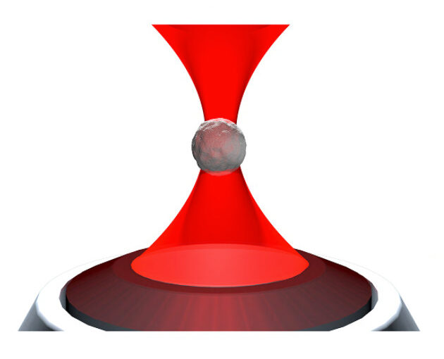
Optical tweezers are tools made of light that enable contactless pushing, trapping, and manipulation of objects, ranging from atoms to space light sails. Since the pioneering work by Arthur Ashkin in the 1970s, optical tweezers have evolved into sophisticated instruments and have been employed in a broad range of applications in the life sciences, physics, and engineering. These include accurate force and torque measurement at the femtonewton level, microrheology of complex fluids, single micro- and nano-particle spectroscopy, single-cell analysis, and statistical-physics experiments. This roadmap provides insights into current investigations involving optical forces and optical tweezers from their theoretical foundations to designs and setups. It also offers perspectives for applications to a wide range of research fields, from biophysics to space exploration.
Invited Talk by G. Volpe at 12th Nordic Workshop on Statistical Physics, Nordita, Stockholm, 15 March 2023
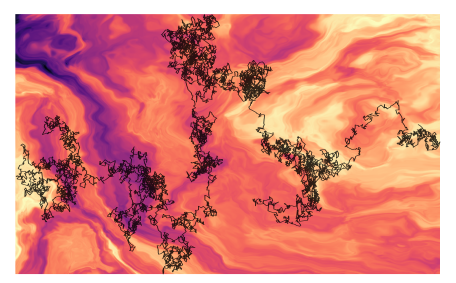
![]()
Giovanni Volpe
Nordita, Stockholm, 15 March 2023, 14:00
Deviations from the law of Brownian motion, typically referred to as anomalous diffusion, are ubiquitous in science and associated with non-equilibrium phenomena, flows of energy and information, and transport in living systems. In the last years, the booming of machine learning has boosted the development of new methods to detect and characterize anomalous diffusion from individual trajectories, going beyond classical calculations based on the mean squared displacement. We thus designed the AnDi challenge, an open community effort to objectively assess the performance of conventional and novel methods. We developed a python library for generating simulated datasets according to the most popular theoretical models of diffusion. We evaluated 16 methods over 3 different tasks and 3 different dimensions, involving anomalous exponent inference, model classification, and trajectory segmentation. Our analysis provides the first assessment of methods for anomalous diffusion in a variety of realistic conditions of trajectory length and noise. Furthermore, we compared the prediction provided by these methods for several experimental datasets. The results of this study further highlight the role that anomalous diffusion has in defining the biological function while revealing insight into the current state of the field and providing a benchmark for future developers.