Gorka Muñoz-Gil, Giovanni Volpe, Miguel Angel Garcia-March, Erez Aghion, Aykut Argun, Chang Beom Hong, Tom Bland, Stefano Bo, J. Alberto Conejero, Nicolás Firbas, Òscar Garibo i Orts, Alessia Gentili, Zihan Huang, Jae-Hyung Jeon, Hélène Kabbech, Yeongjin Kim, Patrycja Kowalek, Diego Krapf, Hanna Loch-Olszewska, Michael A. Lomholt, Jean-Baptiste Masson, Philipp G. Meyer, Seongyu Park, Borja Requena, Ihor Smal, Taegeun Song, Janusz Szwabiński, Samudrajit Thapa, Hippolyte Verdier, Giorgio Volpe, Arthur Widera, Maciej Lewenstein, Ralf Metzler, and Carlo Manzo
Nat. Commun. 12, Article number: 6253 (2021)
doi: 10.1038/s41467-021-26320-w
arXiv: 2105.06766
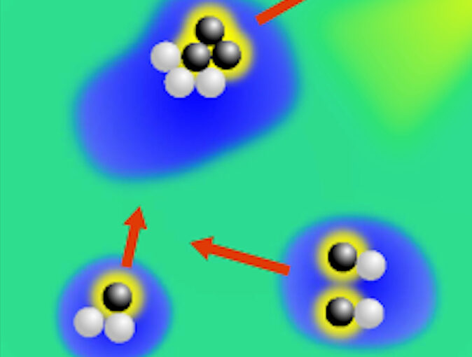
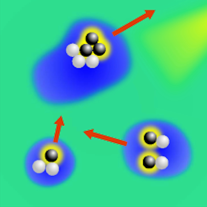
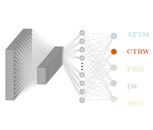
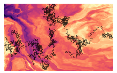
Deviations from Brownian motion leading to anomalous diffusion are found in transport dynamics from quantum physics to life sciences. The characterization of anomalous diffusion from the measurement of an individual trajectory is a challenging task, which traditionally relies on calculating the trajectory mean squared displacement. However, this approach breaks down for cases of practical interest, e.g., short or noisy trajectories, heterogeneous behaviour, or non-ergodic processes. Recently, several new approaches have been proposed, mostly building on the ongoing machine-learning revolution. To perform an objective comparison of methods, we gathered the community and organized an open competition, the Anomalous Diffusion challenge (AnDi). Participating teams applied their algorithms to a commonly-defined dataset including diverse conditions. Although no single method performed best across all scenarios, machine-learning-based approaches achieved superior performance for all tasks. The discussion of the challenge results provides practical advice for users and a benchmark for developers.