Soft Matter Lab members present at SPIE Optics+Photonics conference in San Diego, 3-7 August 2025

The Soft Matter Lab participates to the SPIE Optics+Photonics conference in San Diego, CA, USA, 3-7 August 2025, with the presentations listed below.

Giovanni Volpe, who serves as Symposium Chair for the SPIE Optics+Photonics Congress in 2025, is a coauthor of the following invited presentations:

Giovanni Volpe will also be the reference presenter of the following Poster contributions:

Presentation by D. Pérez Guerrero at SPIE-ETAI, San Diego, 6 August 2025

Automated segnmentation of bacterial structures within a droplet. The image shows a bright-field microscopy view where a large biofilm region (green, outlined in blue) has been segmented from surrounding features. Small aggregates (yellow contours) are also highlighted. This segmentation enables structural differentiation of biofilm components for downstream quantitative analysis. (Image by D. Pérez Guerrero.)
Quantitative analysis of dynamic biofilm structures via time-resolved droplet microfluidics and artificial intelligence
Daniela Pérez Guerrero, Jesús Manuel Antúnez Domínguez, Aurélie Vigne, Daniel Midtvedt, Wylie Ahmed, Lisa Muiznieks, Giovanni Volpe and Caroline Beck Adiels.
SPIE-ETAI, San Diego, CA, USA, 03 – 07 August 2025
Date: 6 August 2025
Time: 2:30 PM – 2:45 PM PDT
Place: Conv. Ctr. Room 4

Biofilms are structured communities of microorganisms that play a crucial role in medicine, biotechnology, and ecology, contributing to microbial adaptation to any environment. Despite their significance, understanding their formation, development, and behavior remains a challenge for the community. We utilize high-throughput droplet microfluidics to enable biofilm growth in miniaturized environments, generating extensive time-lapse bright-field microscopy images. To overcome experimental constraints, including dense structural heterogeneity and skewed illumination, we developed a deep learning-based segmentation approach capable of identifying biofilm structures in complex imaging conditions. Our method operates in an unsupervised manner, reducing the need for ground truth annotations and mitigating the introduced bias of manual segmentation approaches.

Our unsupervised model effectively detects and quantifies biofilm structures, even in late-stage growth, where traditional segmentation techniques fail. The neural network demonstrates robust performance across the development cycle, distinguishing biofilm boundaries and bacteria aggregates separated from the main biofilm structure despite imaging inconsistencies. Additionally, our approach reduces manual intervention, streamlining the analysis of high-throughput biofilm imaging data.

This AI-powered segmentation technique provides a reliable and scalable tool for biofilm analysis, addressing key limitations of conventional methods. By bridging the gap between microbiology research and automated image analysis, our approach facilitates more efficient and reproducible biofilm studies.

Latent Space-Driven Quantification of Biofilm Formation using Time Resolved Droplet Microfluidics on ArXiv

Automated segnmentation of bacterial structures within a droplet. The image shows a bright-field microscopy view where a large biofilm region (green, outlined in blue) has been segmented from surrounding features. Small aggregates (yellow contours) are also highlighted. This segmentation enables structural differentiation of biofilm components for downstream quantitative analysis. (Image by D. Pérez Guerrero.)
Latent Space-Driven Quantification of Biofilm Formation using Time Resolved Droplet Microfluidics
Daniela Pérez Guerrero, Jesús Manuel Antúnez Domínguez, Aurélie Vigne, Daniel Midtvedt, Wylie Ahmed, Lisa D. Muiznieks, Giovanni Volpe, Caroline Beck Adiels
arXiv: 2507.07632

Bacterial biofilms play a significant role in various fields that impact our daily lives, from detrimental public health hazards to beneficial applications in bioremediation, biodegradation, and wastewater treatment. However, high-resolution tools for studying their dynamic responses to environmental changes and collective cellular behavior remain scarce. To characterize and quantify biofilm development, we present a droplet-based microfluidic platform combined with an image analysis tool for in-situ studies. In this setup, Bacillus subtilis was inoculated in liquid Lysogeny Broth microdroplets, and biofilm formation was examined within emulsions at the water-oil interface. Bacteria were encapsulated in droplets, which were then trapped in compartments, allowing continuous optical access throughout biofilm formation. Droplets, each forming a distinct microenvironment, were generated at high throughput using flow-controlled pressure pumps, ensuring monodispersity. A microfluidic multi-injection valve enabled rapid switching of encapsulation conditions without disrupting droplet generation, allowing side-by-side comparison. Our platform supports fluorescence microscopy imaging and quantitative analysis of droplet content, along with time-lapse bright-field microscopy for dynamic observations. To process high-throughput, complex data, we integrated an automated, unsupervised image analysis tool based on a Variational Autoencoder (VAE). This AI-driven approach efficiently captured biofilm structures in a latent space, enabling detailed pattern recognition and analysis. Our results demonstrate the accurate detection and quantification of biofilms using thresholding and masking applied to latent space representations, enabling the precise measurement of biofilm and aggregate areas.

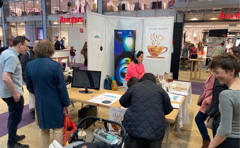
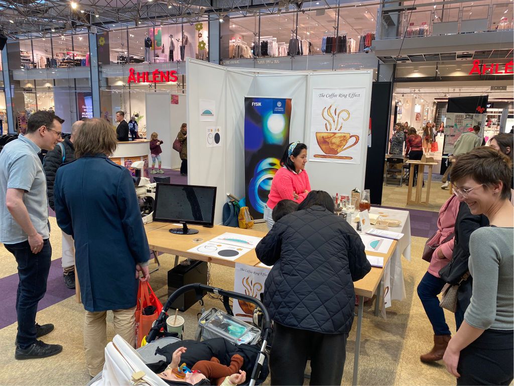
“Coffee Rings” presented at Gothenburg Science Festival 2023

Coffee Ring exposition at science festival Gothenburg. (Photo by C. Beck Adiels.)
Our recent work on “coffee rings” was presented at the Gothenburg Science Festival, which, with about 100 000 visitors each year, is one of the largest popular science events in Europe.

On Wednesday 19th April 2023, Marcel Rey, Laura Natali, Daniela Pérez Guerrero and Caroline Adiels set up a stand in Nordstan.

In this guided exhibition, visitors were able to observe the flow inside a drying droplet using optical microscopes. They learned how the suspended solid coffee particles flow from the inside towards the edge of the coffee droplet, where they accumulate and cause the characteristic coffee ring pattern after drying.

Nowadays, the coffee ring effect presents still a major challenge in ink-jet printing or coating technologies, where a uniform drying is required. We thus shared our recently developed strategies to overcome the coffee ring effect and obtain a uniform deposit of drying droplets.

And finally, visitors were also offered a freshly-brewed espresso to not only drink but also to experience the “coffee ring effect” hands on.