

Giovanni Volpe
Optics & Photonics International Congress 2025 (OPIC 2025), The 11th Optical Manipulation and Structured Materials Conference (OMC2025)
Date: 21 April 2025
Time: 13:45 JST
Place: Yokohama, Japan (Online, Pre-recorded)
News
Yan Chen joins the Soft Matter Lab
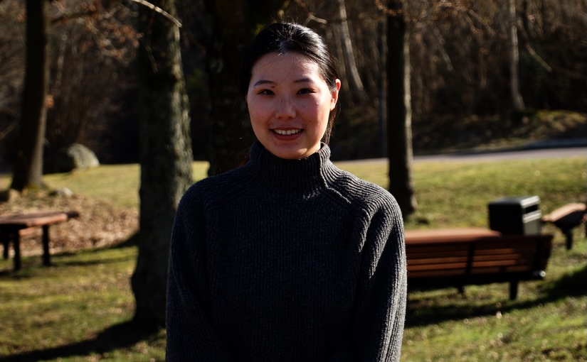
Yan received a Ph.D. degree in Materials Physics and Chemistry, Fudan University, China
In her research, she focuses on multistimuli-responsive microrobots.

Invited talk by M. Selin at University of Münster, 11 April 2025

Martin Selin
Date: 11 April 2025
Time: 10:30
Place: University of Münster, Germany
Optical tweezers are powerful tools for probing microscale forces in systems ranging from colloidal particles to single molecules. Here, we demonstrate their use in two different fields. First, by trapping individual colloidal particles, we study their adsorption dynamics at liquid–liquid interfaces, highlighting the critical role of surface chemistry and the presence of polymer shells. We also observe reversible polymer attachments and stretching. Second, we apply tweezers to study single-molecule mechanics. By automating these complex biophysical experiments, we enable high-throughput measurements of molecular dynamics. Our results suggest that, like DNA, synthetic polymers can be effectively described by the worm-like chain model.

BRAPH 2: a flexible, open-source, reproducible, community-oriented, easy-to-use framework for network analyses in neurosciences on bioRxiv

Yu-Wei Chang, Blanca Zufiria-Gerbolés, Pablo Emiliano Gómez-Ruiz, Anna Canal-Garcia, Hang Zhao, Mite Mijalkov, Joana Braga Pereira, Giovanni Volpe
bioRxiv: 10.1101/2025.04.11.648455
As network analyses in neuroscience continue to grow in both complexity and size, flexible methods are urgently needed to provide unbiased, reproducible insights into brain function. BRAPH 2 is a versatile, open-source framework that meets this challenge by offering streamlined workflows for advanced statistical models and deep learning in a community-oriented environment. Through its Genesis compiler, users can build specialized distributions with custom pipelines, ensuring flexibility and scalability across diverse research domains. These powerful capabilities will ensure reproducibility and accelerate discoveries in neuroscience.
Presentation by M. Selin at Ostwald Colloquium, 8 April 2025

Martin Selin
Date: 8 April 2025
Time: 17:40
Place: Center for Interdisciplinary Research, Bielefeld University, Germany
Colloidal particles typically require salt to overcome electrostatic barriers and adsorb to liquid-liquid interfaces. Here, we show that coating particles with polymers enables spontaneous adsorption without salt. Our model system consists of silica cores coated with poly(2-(dimethylamino)ethyl methacrylate) (PDMAEMA). Using optical tweezers, we track individual particles showing that the polymer shell makes particles jump into a dodecane–water interface. This behavior extends to other polymers. By tuning pH, we control polymer swelling and adsorption distance. At very low pH, the attachment to the interface is weak enough that the optical tweezers can pull particles out from the interface. During this desorption process we observe single polymers detaching. These findings offer new approaches for designing responsive emulsions.

Laura Natali defended her PhD thesis on March 28th, 2025. Congrats!

The defense took place in PJ, Institutionen för fysik, Origovägen 6b, Göteborg, at 10:00.
Title: Neural Networks for Complex Systems: From Epidemic Modeling to Swarm Robotics
Abstract: Deep learning models, inspired by the structure of the brain, were first developed in the last century. These models are trained to recognize patterns in large amounts of data. Recently, deep learning has made a big impact, both in research and in everyday applications, like healthcare, image recognition, and language translation.
However, despite their advancements, these models still fall short of the abilities found in biological brains, which are adaptable, energy-efficient, and have evolved over millions of years. In contrast, artificial models are specialized and struggle to adapt to new information.
To help address this gap, we have developed a robotic experiment that combines the programmability of artificial neural networks with some of the physical constraints seen in biological systems.
Thesis: https://hdl.handle.net/2077/84676
Supervisor: Giovanni Volpe
Examiner: Bernhard Mehlig
Opponent: Hamid Kellay
Committee: Maria Guix Noguera, Juliane Simmchen, Michael Felsberg
Alternate board member: Paolo Vinai

Harshith Bachimanchi defended his PhD thesis on March 26, 2025. Congrats!

The defense took place in PJ, Institutionen för fysik, Origovägen 6b, Göteborg, at 13:00.
Title: Deep Learning Enhanced Optical Methods for Single-Plankton Studies
Abstract: Among Earth’s earliest life forms, cyanobacteria reshaped the planet by oxygenating the atmosphere during the Great Oxidation Event 2.4 billion years ago. This process, which led to ozone formation and UV protection, paved the way for more complex photosynthetic organisms—phytoplankton, the eukaryotic descendants of cyanobacteria. Today, phytoplankton drive the global carbon cycle, producing 50–80% of Earth’s oxygen and fueling the marine food web. Microzooplankton consume nearly two-thirds of the organic carbon generated, yet despite their ecological significance, tracking biomass flow at the single-cell level remains a major challenge.
This thesis presents novel methodologies that integrate advanced optical techniques, deep learning, and simulated datasets to analyze microplankton dynamics with unprecedented resolution.
A key contribution is a deep-learning-enhanced holographic microscopy approach that quantifies microplankton biomass at the single-cell level while simultaneously capturing their three-dimensional swimming behavior. This method overcomes computational bottlenecks in traditional holography, enabling high-throughput analysis across diverse species and size ranges. Expanding on this, I demonstrate its application in mixed-species experiments to examine feeding interactions between phytoplankton and microzooplankton, capturing biomass transfer and behavioral shifts during predation.
Beyond direct imaging, this thesis leverages synthetic data to advance microscopy-based research. Neural networks trained on simulated microscopy datasets are used to detect, segment, and classify plankton species while reconstructing motion dynamics. To showcase the versatility of this approach, I present its application in a non-biological setting—detecting bubble-propelled artificial micromotors within complex experimental backgrounds. In addition to object detection, these methods also enable motion characterization of microscopic entities. To demonstrate this, I introduce synthetic microscopy videos that model microscopic organisms undergoing various anomalous diffusion behaviors. This framework is then used to develop a method that extracts motion characteristics without explicit trajectory linking, broadening its applications beyond plankton ecology.
Finally, I investigate how zooplankton—key players in the marine food web—respond to ocean wave-induced light patterns using an LED matrix. The results suggest that zooplankton use steady light sources, such as celestial objects, to ascend more rapidly during favorable low-turbulent conditions, offering new insights into their migratory strategies. Collectively, this thesis bridges marine ecology, microscopy, artificial intelligence, and biophysics to provide new tools for exploring the unseen dynamics that shape our planet.
Thesis: https://hdl.handle.net/2077/84857
Supervisor: Giovanni Volpe
Examiner: Raimund Feifel
Opponent: Anupam Sengupta
Committee: Elisa Berdalet, Maria Guix Noguera, Josefin Titelman
Alternate board member: Paolo Vinai
Computational memory capacity predicts aging and cognitive decline published in Nature Communications

Mite Mijalkov, Ludvig Storm, Blanca Zufiria-Gerbolés, Dániel Veréb, Zhilei Xu, Anna Canal-Garcia, Jiawei Sun, Yu-Wei Chang, Hang Zhao, Emiliano Gómez-Ruiz, Massimiliano Passaretti, Sara Garcia-Ptacek, Miia Kivipelto, Per Svenningsson, Henrik Zetterberg, Heidi Jacobs, Kathy Lüdge, Daniel Brunner, Bernhard Mehlig, Giovanni Volpe, Joana B. Pereira
Nature Communications 16, 2748 (2025)
doi: 10.1038/s41467-025-57995-0
Memory is a crucial cognitive function that deteriorates with age. However, this ability is normally assessed using cognitive tests instead of the architecture of brain networks. Here, we use reservoir computing, a recurrent neural network computing paradigm, to assess the linear memory capacities of neural-network reservoirs extracted from brain anatomical connectivity data in a lifespan cohort of 636 individuals. The computational memory capacity emerges as a robust marker of aging, being associated with resting-state functional activity, white matter integrity, locus coeruleus signal intensity, and cognitive performance. We replicate our findings in an independent cohort of 154 young and 72 old individuals. By linking the computational memory capacity of the brain network with cognition, brain function and integrity, our findings open new pathways to employ reservoir computing to investigate aging and age-related disorders.

Global graph features unveiled by unsupervised geometric deep learning on ArXiv

Mirja Granfors, Jesús Pineda, Blanca Zufiria Gerbolés, Joana B. Pereira, Carlo Manzo, Giovanni Volpe
arXiv: 2503.05560
Graphs provide a powerful framework for modeling complex systems, but their structural variability makes analysis and classification challenging. To address this, we introduce GAUDI (Graph Autoencoder Uncovering Descriptive Information), a novel unsupervised geometric deep learning framework that captures both local details and global structure. GAUDI employs an innovative hourglass architecture with hierarchical pooling and upsampling layers, linked through skip connections to preserve essential connectivity information throughout the encoding-decoding process. By mapping different realizations of a system – generated from the same underlying parameters – into a continuous, structured latent space, GAUDI disentangles invariant process-level features from stochastic noise. We demonstrate its power across multiple applications, including modeling small-world networks, characterizing protein assemblies from super-resolution microscopy, analyzing collective motion in the Vicsek model, and capturing age-related changes in brain connectivity. This approach not only improves the analysis of complex graphs but also provides new insights into emergent phenomena across diverse scientific domains.
Invited talk by S. Manikandan at the 14th Nordic Workshop on Statistical Physics, Nordita, 5 March 2025

Sreekanth Manikandan
Date: 5 Mar 2025
Time: 14:45
Place: Nordita
Part of the 14th Nordic Workshop on Statistical Physics
Quantifying the spatiotemporal forces, affinities, and dissipative costs of cellular-scale non-equilibrium processes from experimental data and localizing it in space and time remain a significant open challenge. Here, I explore how principles from stochastic thermodynamics, combined with machine learning techniques, offer a promising approach to addressing this issue. I will present preliminary results from experiments on fluctuating cell membranes and simulations of non-equilibrium systems in stationary and time-dependently driven states. These studies reveal potential strategies for localizing entropy production in experimental biophysical contexts while also highlighting key challenges and limitations that must be addressed.