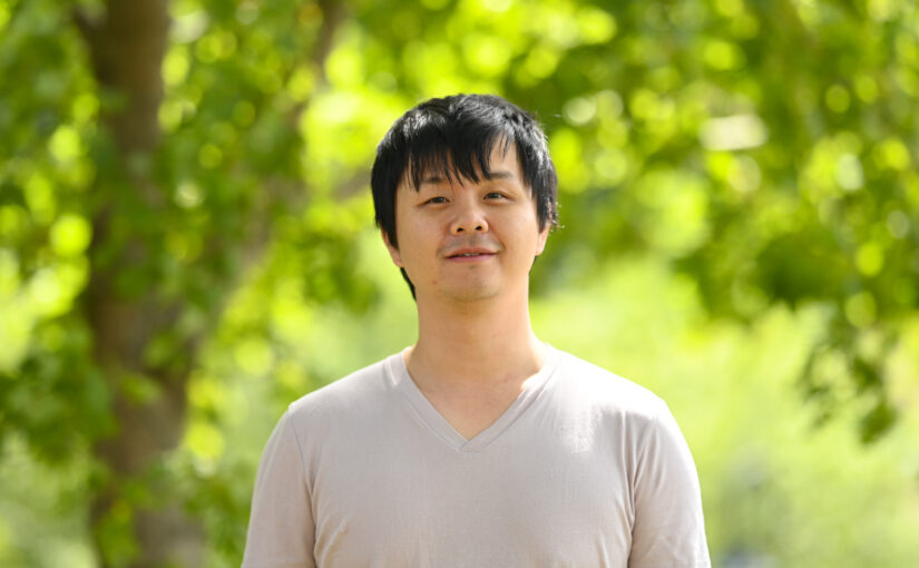
He will start his new appointment on May 6th 2024. His research will focus on nanooptics technology for electronic paper, optical neural networks, and intelligent microparticles.
He will start his new appointment on May 6th 2024. His research will focus on nanooptics technology for electronic paper, optical neural networks, and intelligent microparticles.
Correlated photons in superresolution imaging and correlated motions in biophysical interaction
Alexander Rohrbach
15 May 2024
12:30
Nexus
Abstract
Our research concentrates on light scattering at small biological structures enabling image formation and particle tracking in biophysics.
Coherent light, i.e. correlated photons enable higher scattering cross-sections than for instance incoherent fluorescence light. Thereby laser light enables to acquire images with millisecond integration times and small motion blur of dynamic particles, such as viruses in the cell periphery. The inherent speckle formation in coherent imaging is avoided by a novel technique called Rotating Coherent Scattering (ROCS) microscopy, which is the only technique that can image diffusing viruses and thereby allows to investigate their binding behavior to the cell periphery.
In the second part of my talk I discuss correlated particle motions, i.e. timescale dependent memory effects in viscoelastic media such as the cell periphery. Using a frequency decomposition of the tracked particle motions, apparently invisible binding of particles to the cell can be made visible.
Short CV
I studied physics at the university of Erlangen-Nürnberg (Germany), where I did my diploma in 1994 at the institute of optics. During my PhD in physics in Heidelberg I investigated different kinds of light scattering at the University, as well as evanescent wave microscopy at the Max-Planck-Institute for medical research. In both cases I worked on applications in cell biology. After my PhD in 1998, I continued my research as a Post-Doc at the European Molecular Biology Laboratory (EMBL) in Heidelberg. I intensified my studies on microscopy, light scattering and optical forces. In 2001 I became project leader of the photonic force microscopy group at EMBL, where I concentrated on the further technical development of this scanning probe microscopy and on applications in biophysics and soft matter physics. In 2005 I was awarded with the habilitation in physics at the university of Heidelberg. Since January 2006 I have been a full professor for Bio- and Nano-Photonics at IMTEK, Faculty of engineering and since 2007 also a member of the physics faculty, University of Freiburg.
I love mathematical models and I hate when the performance of scientists is squeezed into metric numbers.
Harshith Bachimanchi, Matthew I. M. Pinder, Chloé Robert, Pierre De Wit, Jonathan Havenhand, Alexandra Kinnby, Daniel Midtvedt, Erik Selander, Giovanni Volpe
Limnology and Oceanography Letters (2024)
doi: 10.1002/lol2.10392
arXiv: 2309.08500
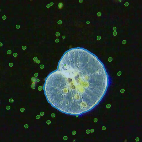
The implementation of deep learning algorithms has brought new perspectives to plankton ecology. Emerging as an alternative approach to established methods, deep learning offers objective schemes to investigate plankton organisms in diverse environments. We provide an overview of deep-learning-based methods including detection and classification of phytoplankton and zooplankton images, foraging and swimming behavior analysis, and finally ecological modeling. Deep learning has the potential to speed up the analysis and reduce the human experimental bias, thus enabling data acquisition at relevant temporal and spatial scales with improved reproducibility. We also discuss shortcomings and show how deep learning architectures have evolved to mitigate imprecise readouts. Finally, we suggest opportunities where deep learning is particularly likely to catalyze plankton research. The examples are accompanied by detailed tutorials and code samples that allow readers to apply the methods described in this review to their own data.
Emiliano Gómez will defend his PhD thesis on the 22th of May at 10:30. The defense will take place in KA (Chemistry Department, Johanneberg Campus)
Title: Development and Application of a software to analyse networks with multilayer graph theory and deep learning
Abstract:
Network theory gives us the tools necessary to produce a model of our brain, how the brain is wired will give us a new level of insight into its functionality. The brain network, the connectome, is formed by structural links such as synapses or fiber pathways in the brain. This connectome might also be interpreted from a statistical relationship between the flow of information, or activation correlation between brain regions. Mapping these networks can be achieved by using neuroimaging, which allows obtaining information on the brain in vivo. Different neuroimaging modalities will capture different properties of the brain. Statistical analysis is necessary for extracting meaningful insights regarding the network patterns obtained from neuroimages. For this, huge data banks are a byproduct of the need for enough data to be able to tackle medical and biological questions.
In this work, we present a software “Brain Analysis using Graph Theory 2” (BRAPH 2) (Paper I), which addresses the need for a toolbox designed for both complex graph theory and deep learning analyses of different imaging modalities. With BRAPH 2, we offer the neuroimaging community a tool that is open-source, flexible, and intuitive. BRAPH 2, at its core, comes with multi-graph capabilities. For Paper II, we employed the power of multiplex and multigraph capabilities of BRAPH 2 to analyze sex differences in brain connectivity for an aging healthy population. Finally, for Paper III, BRAPH 2 has been adapted to two new graph measures (global memory capacity, and nodal memory capacity), which obtain a prediction of memory capacity using Reservoir Computing and relate this new measure to biological and cognitive characteristics of the cohort.
Supervisor: Giovanni Volpe
Examiner: Raimund Feifel
Opponent: Saikat Chatterjee, KTH, Stockholm
Committee: Marija Cvijovic, Alireza Salami, Wojciech Chachólski
Alternate board member: Mats Granath

Here, young researchers get to present exciting space-related work they have been or are doing at Chalmers / Gothenburg University – in one or two minutes, with the help/support of a picture and/or a prop. This event was participated by Marcus Wandt, Sweden’s third astronaut.
In this event, Hari presented his topic titled “Annelid inspired soft robot for planetary exploration” where this project is in collaboration with the European Space Agency (ESTEC-ESA) and Gothenburg University.
Introduction to G-Research, a quantitative research and technology company
Charles Martinez
G-Research, London, UK
27 March 2024
12:30-14:30
PJ
Organized by the CHAIR theme AI for Scientific Data Analysis
We are a leading quantitative research and technology company based in London. Day to day we use a variety of quantitative techniques to predict financial markets from large data sets worldwide. Mathematics, statistics, machine learning, natural language processing and deep learning is what our business is built on. Our culture is academic and highly intellectual. In this seminar I will explain our background, current AI research applications to finance and our ongoing outreach, recruitment and grants programme.
Bio: Dr Charles Martinez is the Academic Relations Manager at G-Research. Charles started his studies as a physicist at University Portsmouth Physics department’s MPhys programme, and later completed a PhD in Phonon interactions in Gallium Nitride nanostructures at the University of Nottingham. Charles then worked on indexing and abstract databases at the Institution for Engineering and Technology (IET) before moving into sales in 2010. Charles’ previous role was as Elsevier’s Key Account Manager, managing sales and renewals for the UK Russell Group institutions, Government and Funding body accounts, including being one of the negotiators in the recent UK ScienceDirect Read and Publish agreement. Since leaving Elsevier Charles is dedicated to forming beneficial partnerships between G-Research and Europe’s top institutions, and is living in Cambridge, UK.
Learning about G-Research: thinking about strategies in quantitative finance
Charles Martinez
G-Research, London, UK
27 March 2024
10:00-11:30
FB (Fysik-Huset)
We are a leading quantitative research and technology company based in London. Day to day we use a variety of quantitative techniques to predict financial markets from large data sets worldwide. Mathematics, statistics, machine learning, natural language processing and deep learning is what our business is built on. Our culture is academic and highly intellectual. In this seminar I will explain our background, current AI research applications to finance and our ongoing outreach, recruitment and grants programme. The seminar will be aimed at students who are curious about quant finance or interested in internship opportunities. We will also play an interactive game. The game will last around 1 hour and there will be prizes for the Top 3 scores (amazon vouchers – £100). Dice will be provided.
Bio: Dr Charles Martinez is the Academic Relations Manager at G-Research. Charles started his studies as a physicist at University Portsmouth Physics department’s MPhys programme, and later completed a PhD in Phonon interactions in Gallium Nitride nanostructures at the University of Nottingham. Charles then worked on indexing and abstract databases at the Institution for Engineering and Technology (IET) before moving into sales in 2010. Charles’ previous role was as Elsevier’s Key Account Manager, managing sales and renewals for the UK Russell Group institutions, Government and Funding body accounts, including being one of the negotiators in the recent UK ScienceDirect Read and Publish agreement. Since leaving Elsevier Charles is dedicated to forming beneficial partnerships between G-Research and Europe’s top institutions, and is living in Cambridge, UK.
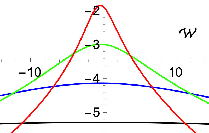
Pascal Viot, Aykut Argun, Giovanni Volpe, Alberto Imparato, Lamberto Rondoni, Gleb Oshanin
Soft Matter, 20, 3154-3160 (2024)
arxiv: 2307.05248
doi: 10.1039/D3SM01606D
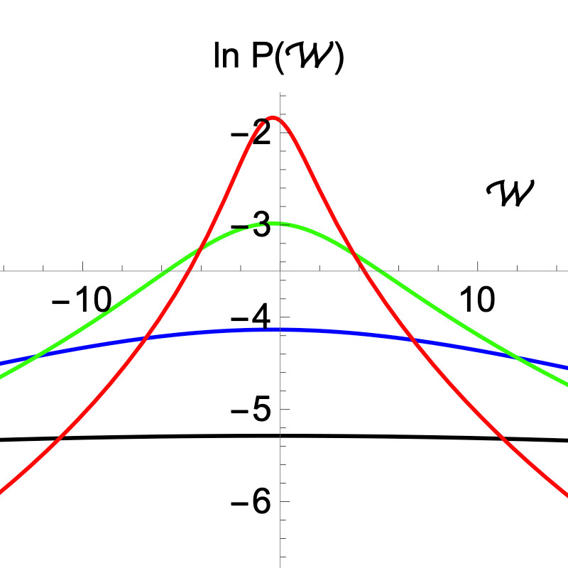
The Brownian gyrator (BG) is often called a minimal model of a nano-engine performing a rotational motion, judging solely upon the fact that in non-equilibrium conditions its torque, specific angular momentum L and specific angular velocity W have non-zero mean values. For a time-discretised (with time-step δt) model we calculate here the previously unknown probability density functions (PDFs) of L and W. We show that for finite δt, the PDF of L has exponential tails and all moments are therefore well-defined. At the same time, this PDF appears to be effectively broad – the noise-to-signal ratio is generically bigger than unity meaning that L is strongly not self-averaging. Concurrently, the PDF of W exhibits heavy power-law tails and its mean W is the only existing moment. The BG is therefore not an engine in the common sense: it does not exhibit regular rotations on each run and its fluctuations are not only a minor nuisance – on contrary, their effect is completely destructive for the performance. Our theoretical predictions are confirmed by numerical simulations and experimental data. We discuss some plausible improvements of the model which may result in a more systematic rotational motion.
Wylie Ahmed
Laboratoire de Physique Theorique, Toulouse (France) and California State University, Fullerton (USA)
13 March 2024, 12:30, Nexus
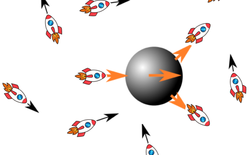
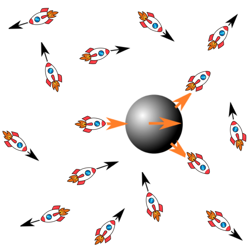
Motivated by nucleus centering in mouse oocytes, we explore a different type of biological active matter. We investigate the stochastic force fluctuations of micro swimmers in two scenarios: (1) a single swimmer navigating through a passive fluid; (2) a dense suspension of swimmers surrounding a passive tracer. By direct force measurement using optical tweezers we show that the force trajectory of an individual micro swimmer exhibits rich oscillatory dynamics that vary in time. Interestingly, when these highly fluctuating force dynamics are analyzed using the framework of stochastic thermodynamics we recover energy dissipation rates in agreement with time-averaged fluid dynamics studies. For a dense suspension of swimmers serving as an active bath for a passive tracer we observe both shear thinning and thickening, which depends on Peclet number, and enhanced diffusion of our tracer by a factor of 2. We estimate the energy transfer rate from the active bath to the passive tracer. These two scenarios allow us to explore energy exchange between an active swimmer in a passive bath and a passive tracer in an active bath.
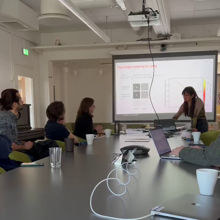
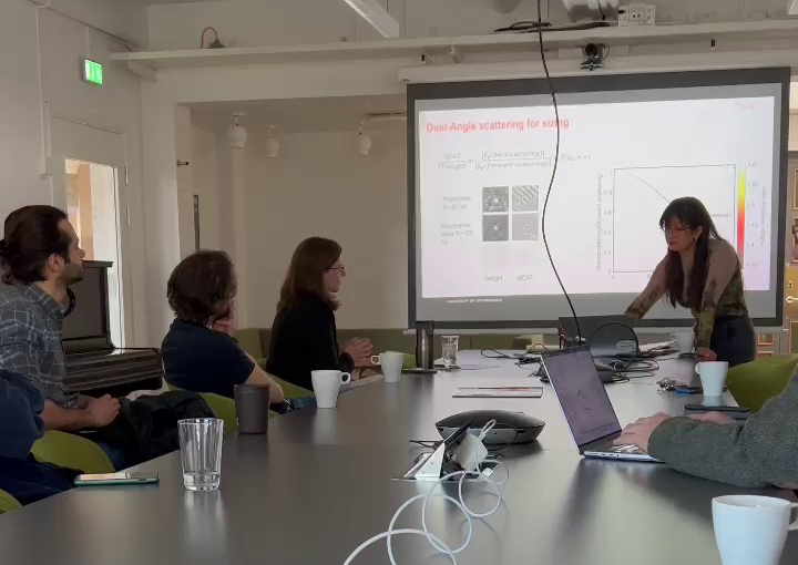
The presentation, “Quantitative Analysis of Nanoparticle Properties Using Optical Scattering Techniques,” was held in a hybrid format, with part of the audience in the Nexus room and the rest connected through Zoom. The half-time consisted of a presentation about her past and planned projects, followed by a discussion and questions proposed by her opponent, Dr. Hana Jungová.
The presentation started with a short background introduction to optical scattering techniques and nanoparticle characterization techniques, followed by an introduction and description of the first paper, “Dual-Angle Interferometric Scattering Microscopy for Optical Multiparametric Particle Characterization,” and, in the end, a brief description of the projects in which Berenice is involved.
In the last section, she outlined the proposed continuation of her PhD: quantification and characterization of biomolecular condensates and their evolution over time, monitoring lipid droplets during long timescales inside living cells, and parametrization for core-shell particles.