Sex differences in multilayer functional network topology over the course of aging in 37543 UK Biobank participants accepted on Network Neuroscience

Example of the 21 resting-state networks used as nodes and their positive (red) and negative connections (blue) for one of 140 the subjects included in the analyses. (Image by the Authors of the manuscript.)
Sex differences in multilayer functional network topology over the course of aging in 37543 UK Biobank participants
Mite Mijalkov, Dániel Veréb, Oveis Jamialahmadi, Anna Canal-Garcia, Emiliano Gómez-Ruiz, Didac Vidal-Piñeiro, Stefano Romeo, Giovanni Volpe, Joana B. Pereira
Network Neuroscience 1-40 (2022)
doi: 10.1162/netn_a_00286
medRxiv: 10.1101/2022.03.08.22272089

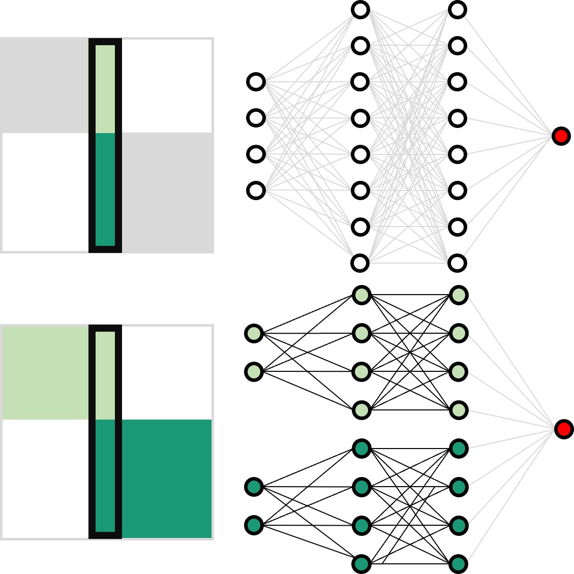
Aging is a major risk factor for cardiovascular and neurodegenerative disorders, with considerable societal and economic implications. Healthy aging is accompanied by changes in functional connectivity between and within resting-state functional networks, which have been associated with cognitive decline. However, there is no consensus on the impact of sex on these age-related functional trajectories. Here, we show that multilayer measures provide crucial information on the interaction between sex and age on network topology, allowing for better assessment of cognitive, structural, and cardiovascular risk factors that have been shown to differ between men and women, as well as providing additional insights into the genetic influences on changes in functional connectivity that occur during aging. In a large cross-sectional sample of 37543 individuals from the UK Biobank cohort, we demonstrate that such multilayer measures that capture the relationship between positive and negative connections are more sensitive to sex-related changes in the whole-brain connectivity patterns and their topological architecture throughout aging, when compared to standard connectivity and topological measures. Our findings indicate that multilayer measures contain previously unknown information on the relationship between sex and age, which opens up new avenues for research into functional brain connectivity in aging.

Multi-cohort and longitudinal Bayesian clustering study of stage and subtype in Alzheimer’s disease published in Nature Communications

Comparison of cluster-specific covariance matrixes with node strength. (Image by the Authors.)
Multi-cohort and longitudinal Bayesian clustering study of stage and subtype in Alzheimer’s disease
Konstantinos Poulakis, Joana B. Pereira, J.-Sebastian Muehlboeck, Lars-Olof Wahlund, Örjan Smedby, Giovanni Volpe, Colin L. Masters, David Ames, Yoshiki Niimi, Takeshi Iwatsubo, Daniel Ferreira, Eric Westman, Japanese Alzheimer’s Disease Neuroimaging Initiative & Australian Imaging, Biomarkers and Lifestyle study
Nature Communications 13, 4566 (2022)
doi: 10.1038/s41467-022-32202-6

Understanding Alzheimer’s disease (AD) heterogeneity is important for understanding the underlying pathophysiological mechanisms of AD. However, AD atrophy subtypes may reflect different disease stages or biologically distinct subtypes. Here we use longitudinal magnetic resonance imaging data (891 participants with AD dementia, 305 healthy control participants) from four international cohorts, and longitudinal clustering to estimate differential atrophy trajectories from the age of clinical disease onset. Our findings (in amyloid-β positive AD patients) show five distinct longitudinal patterns of atrophy with different demographical and cognitive characteristics. Some previously reported atrophy subtypes may reflect disease stages rather than distinct subtypes. The heterogeneity in atrophy rates and cognitive decline within the five longitudinal atrophy patterns, potentially expresses a complex combination of protective/risk factors and concomitant non-AD pathologies. By alternating between the cross-sectional and longitudinal understanding of AD subtypes these analyses may allow better understanding of disease heterogeneity.

Unraveling Parkinson’s disease heterogeneity using subtypes based on multimodal data published in Parkinsonism and Related Disorders

Particular of the brain in the group comparison analysis. (Image by the Authors.)
Unraveling Parkinson’s disease heterogeneity using subtypes based on multimodal data
Franziska Albrecht, Konstantinos Poulakis, Malin Freidle, Hanna Johansson, Urban Ekman, Giovanni Volpe, Eric Westman, Joana B. Pereira, Erika Franzén
Parkinsonism and Related Disorders 102, 19-29 (2022)
doi: 10.1016/j.parkreldis.2022.07.014

Background

Parkinson’s disease (PD) is a clinically and neuroanatomically heterogeneous neurodegenerative disease characterized by different subtypes. To this date, no studies have used multimodal data that combines clinical, motor, cognitive and neuroimaging assessments to identify these subtypes, which may provide complementary, clinically relevant information. To address this limitation, we subtyped participants with mild-moderate PD based on a rich, multimodal dataset of clinical, cognitive, motor, and neuroimaging variables.

Methods

Cross-sectional data from 95 PD participants from our randomized EXPANd (EXercise in PArkinson’s disease and Neuroplasticity) controlled trial were included. Participants were subtyped using clinical, motor, and cognitive assessments as well as structural and resting-state MRI data. Subtyping was done by random forest clustering. We extracted information about the subtypes by inspecting their neuroimaging profiles and descriptive statistics.

Results

Our multimodal subtyping analysis yielded three PD subtypes: a motor-cognitive subtype characterized by widespread alterations in brain structure and function as well as impairment in motor and cognitive abilities; a cognitive dominant subtype mainly impaired in cognitive function that showed frontoparietal structural and functional changes; and a motor dominant subtype impaired in motor variables without any brain alterations. Motor variables were most important for the subtyping, followed by gray matter volume in the right medial postcentral gyrus.

Conclusions

Three distinct PD subtypes were identified in our multimodal dataset. The most important features to subtype PD participants were motor variables in addition to structural MRI in the sensorimotor region. These findings have the potential to improve our understanding of PD heterogeneity, which in turn can lead to personalized interventions and rehabilitation.

Dynamic live/apoptotic cell assay using phase-contrast imaging and deep learning on bioRxiv

Phase-contrast image before virtual staining. (Image by the Authors.)
Dynamic live/apoptotic cell assay using phase-contrast imaging and deep learning
Zofia Korczak, Jesús Pineda, Saga Helgadottir, Benjamin Midtvedt, Mattias Goksör, Giovanni Volpe, Caroline B. Adiels
bioRxiv: 10.1101/2022.07.18.500422

Chemical live/dead assay has a long history of providing information about the viability of cells cultured in vitro. The standard methods rely on imaging chemically-stained cells using fluorescence microscopy and further analysis of the obtained images to retrieve the proportion of living cells in the sample. However, such a technique is not only time-consuming but also invasive. Due to the toxicity of chemical dyes, once a sample is stained, it is discarded, meaning that longitudinal studies are impossible using this approach. Further, information about when cells start programmed cell death (apoptosis) is more relevant for dynamic studies. Here, we present an alternative method where cell images from phase-contrast time-lapse microscopy are virtually-stained using deep learning. In this study, human endothelial cells are stained live or apoptotic and subsequently counted using the self-supervised single-shot deep-learning technique (LodeSTAR). Our approach is less labour-intensive than traditional chemical staining procedures and provides dynamic live/apoptotic cell ratios from a continuous cell population with minimal impact. Further, it can be used to extract data from dense cell samples, where manual counting is unfeasible.

Neural Network Training with Highly Incomplete Datasets published in Machine Learning: Science and Technology

Working principles for training neural networks with highly incomplete dataset: vanilla (upper panel) vs GapNet (lower panel) (Image by Yu-Wei Chang.)
Neural Network Training with Highly Incomplete Datasets
Yu-Wei Chang, Laura Natali, Oveis Jamialahmadi, Stefano Romeo, Joana B. Pereira, Giovanni Volpe
Machine Learning: Science and Technology 3, 035001 (2022)
arXiV: 2107.00429
doi: 10.1088/2632-2153/ac7b69

Neural network training and validation rely on the availability of large high-quality datasets. However, in many cases only incomplete datasets are available, particularly in health care applications, where each patient typically undergoes different clinical procedures or can drop out of a study. Since the data to train the neural networks need to be complete, most studies discard the incomplete datapoints, which reduces the size of the training data, or impute the missing features, which can lead to artefacts. Alas, both approaches are inadequate when a large portion of the data is missing. Here, we introduce GapNet, an alternative deep-learning training approach that can use highly incomplete datasets. First, the dataset is split into subsets of samples containing all values for a certain cluster of features. Then, these subsets are used to train individual neural networks. Finally, this ensemble of neural networks is combined into a single neural network whose training is fine-tuned using all complete datapoints. Using two highly incomplete real-world medical datasets, we show that GapNet improves the identification of patients with underlying Alzheimer’s disease pathology and of patients at risk of hospitalization due to Covid-19. By distilling the information available in incomplete datasets without having to reduce their size or to impute missing values, GapNet will permit to extract valuable information from a wide range of datasets, benefiting diverse fields from medicine to engineering.

Deep learning in light–matter interactions published in Nanophotonics

Artificial neurons can be combined in a dense neural network (DNN), where the input layer is connected to the output layer via a set of hidden layers. (Image by the Authors.)
Deep learning in light–matter interactions
Daniel Midtvedt, Vasilii Mylnikov, Alexander Stilgoe, Mikael Käll, Halina Rubinsztein-Dunlop and Giovanni Volpe
Nanophotonics, 11(14), 3189-3214 (2022)
doi: 10.1515/nanoph-2022-0197

The deep-learning revolution is providing enticing new opportunities to manipulate and harness light at all scales. By building models of light–matter interactions from large experimental or simulated datasets, deep learning has already improved the design of nanophotonic devices and the acquisition and analysis of experimental data, even in situations where the underlying theory is not sufficiently established or too complex to be of practical use. Beyond these early success stories, deep learning also poses several challenges. Most importantly, deep learning works as a black box, making it difficult to understand and interpret its results and reliability, especially when training on incomplete datasets or dealing with data generated by adversarial approaches. Here, after an overview of how deep learning is currently employed in photonics, we discuss the emerging opportunities and challenges, shining light on how deep learning advances photonics.

Label-free nanofluidic scattering microscopy of size and mass of single diffusing molecules and nanoparticles published in Nature Methods

Kymographs of DNA inside Channel II. (Image by the Authors.)
Label-free nanofluidic scattering microscopy of size and mass of single diffusing molecules and nanoparticles
Barbora Špačková, Henrik Klein Moberg, Joachim Fritzsche, Johan Tenghamn, Gustaf Sjösten, Hana Šípová-Jungová, David Albinsson, Quentin Lubart, Daniel van Leeuwen, Fredrik Westerlund, Daniel Midtvedt, Elin K. Esbjörner, Mikael Käll, Giovanni Volpe & Christoph Langhammer
Nature Methods 19, 751–758 (2022)
doi: 10.1038/s41592-022-01491-6

Label-free characterization of single biomolecules aims to complement fluorescence microscopy in situations where labeling compromises data interpretation, is technically challenging or even impossible. However, existing methods require the investigated species to bind to a surface to be visible, thereby leaving a large fraction of analytes undetected. Here, we present nanofluidic scattering microscopy (NSM), which overcomes these limitations by enabling label-free, real-time imaging of single biomolecules diffusing inside a nanofluidic channel. NSM facilitates accurate determination of molecular weight from the measured optical contrast and of the hydrodynamic radius from the measured diffusivity, from which information about the conformational state can be inferred. Furthermore, we demonstrate its applicability to the analysis of a complex biofluid, using conditioned cell culture medium containing extracellular vesicles as an example. We foresee the application of NSM to monitor conformational changes, aggregation and interactions of single biomolecules, and to analyze single-cell secretomes.

Directed Brain Connectivity Identifies Widespread Functional Network Abnormalities in Parkinson’s Disease published in Cerebral Cortex

Visual display of the nodes that show significant differences between controls and participants with PD in network measures using the anti-symmetric correlation method. (Image by the Authors.)
Directed Brain Connectivity Identifies Widespread Functional Network Abnormalities in Parkinson’s Disease
Mite Mijalkov, Giovanni Volpe, Joana B Pereira
Cerebral Cortex 32(3), 593–607 (2022)
doi: 10.1093/cercor/bhab237

Parkinson’s disease (PD) is a neurodegenerative disorder characterized by topological abnormalities in large-scale functional brain networks, which are commonly analyzed using undirected correlations in the activation signals between brain regions. This approach assumes simultaneous activation of brain regions, despite previous evidence showing that brain activation entails causality, with signals being typically generated in one region and then propagated to other ones. To address this limitation, here, we developed a new method to assess whole-brain directed functional connectivity in participants with PD and healthy controls using antisymmetric delayed correlations, which capture better this underlying causality. Our results show that whole-brain directed connectivity, computed on functional magnetic resonance imaging data, identifies widespread differences in the functional networks of PD participants compared with controls, in contrast to undirected methods. These differences are characterized by increased global efficiency, clustering, and transitivity combined with lower modularity. Moreover, directed connectivity patterns in the precuneus, thalamus, and cerebellum were associated with motor, executive, and memory deficits in PD participants. Altogether, these findings suggest that directional brain connectivity is more sensitive to functional network differences occurring in PD compared with standard methods, opening new opportunities for brain connectivity analysis and development of new markers to track PD progression.

Multiplex Connectome Changes across the Alzheimer’s Disease Spectrum Using Gray Matter and Amyloid Data published in Cerebral Cortex

Brain nodes. (Image taken from the article.)
Multiplex Connectome Changes across the Alzheimer’s Disease Spectrum Using Gray Matter and Amyloid Data
Mite Mijalkov, Giovanni Volpe, Joana B Pereira
Anna Canal-Garcia, Emiliano Gómez-Ruiz, Mite Mijalkov, Yu-Wei Chang, Giovanni Volpe, Joana B Pereira, Alzheimer’s Disease Neuroimaging Initiative
Cerebral Cortex, bhab429 (2022)
doi: 10.1093/cercor/bhab429

The organization of the Alzheimer’s disease (AD) connectome has been studied using graph theory using single neuroimaging modalities such as positron emission tomography (PET) or structural magnetic resonance imaging (MRI). Although these modalities measure distinct pathological processes that occur in different stages in AD, there is evidence that they are not independent from each other. Therefore, to capture their interaction, in this study we integrated amyloid PET and gray matter MRI data into a multiplex connectome and assessed the changes across different AD stages. We included 135 cognitively normal (CN) individuals without amyloid-β pathology (Aβ−) in addition to 67 CN, 179 patients with mild cognitive impairment (MCI) and 132 patients with AD dementia who all had Aβ pathology (Aβ+) from the Alzheimer’s Disease Neuroimaging Initiative. We found widespread changes in the overlapping connectivity strength and the overlapping connections across Aβ-positive groups. Moreover, there was a reorganization of the multiplex communities in MCI Aβ + patients and changes in multiplex brain hubs in both MCI Aβ + and AD Aβ + groups. These findings offer a new insight into the interplay between amyloid-β pathology and brain atrophy over the course of AD that moves beyond traditional graph theory analyses based on single brain networks.

Comparison of Two-Dimensional- and Three-Dimensional-Based U-Net Architectures for Brain Tissue Classification in One-Dimensional Brain CT published in Frontiers of Computational Neuroscience

CT is split into smaller patches. (Image by the Authors.)
Comparison of Two-Dimensional- and Three-Dimensional-Based U-Net Architectures for Brain Tissue Classification in One-Dimensional Brain CT
Meera Srikrishna, Rolf A. Heckemann, Joana B. Pereira, Giovanni Volpe, Anna Zettergren, Silke Kern, Eric Westman, Ingmar Skoog and Michael Schöll
Frontiers of Computational Neuroscience 15, 785244 (2022)
doi: 10.3389/fncom.2021.785244

Brain tissue segmentation plays a crucial role in feature extraction, volumetric quantification, and morphometric analysis of brain scans. For the assessment of brain structure and integrity, CT is a non-invasive, cheaper, faster, and more widely available modality than MRI. However, the clinical application of CT is mostly limited to the visual assessment of brain integrity and exclusion of copathologies. We have previously developed two-dimensional (2D) deep learning-based segmentation networks that successfully classified brain tissue in head CT. Recently, deep learning-based MRI segmentation models successfully use patch-based three-dimensional (3D) segmentation networks. In this study, we aimed to develop patch-based 3D segmentation networks for CT brain tissue classification. Furthermore, we aimed to compare the performance of 2D- and 3D-based segmentation networks to perform brain tissue classification in anisotropic CT scans. For this purpose, we developed 2D and 3D U-Net-based deep learning models that were trained and validated on MR-derived segmentations from scans of 744 participants of the Gothenburg H70 Cohort with both CT and T1-weighted MRI scans acquired timely close to each other. Segmentation performance of both 2D and 3D models was evaluated on 234 unseen datasets using measures of distance, spatial similarity, and tissue volume. Single-task slice-wise processed 2D U-Nets performed better than multitask patch-based 3D U-Nets in CT brain tissue classification. These findings provide support to the use of 2D U-Nets to segment brain tissue in one-dimensional (1D) CT. This could increase the application of CT to detect brain abnormalities in clinical settings.