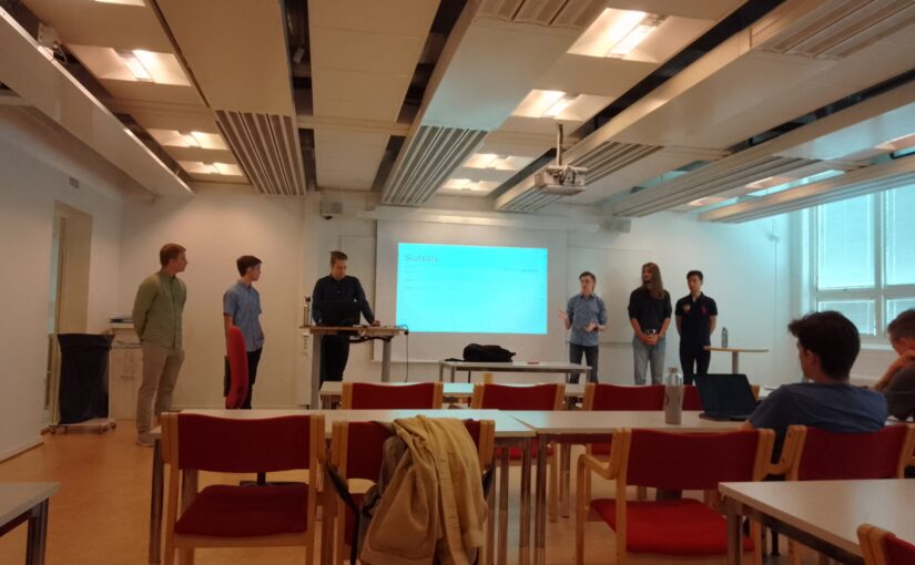
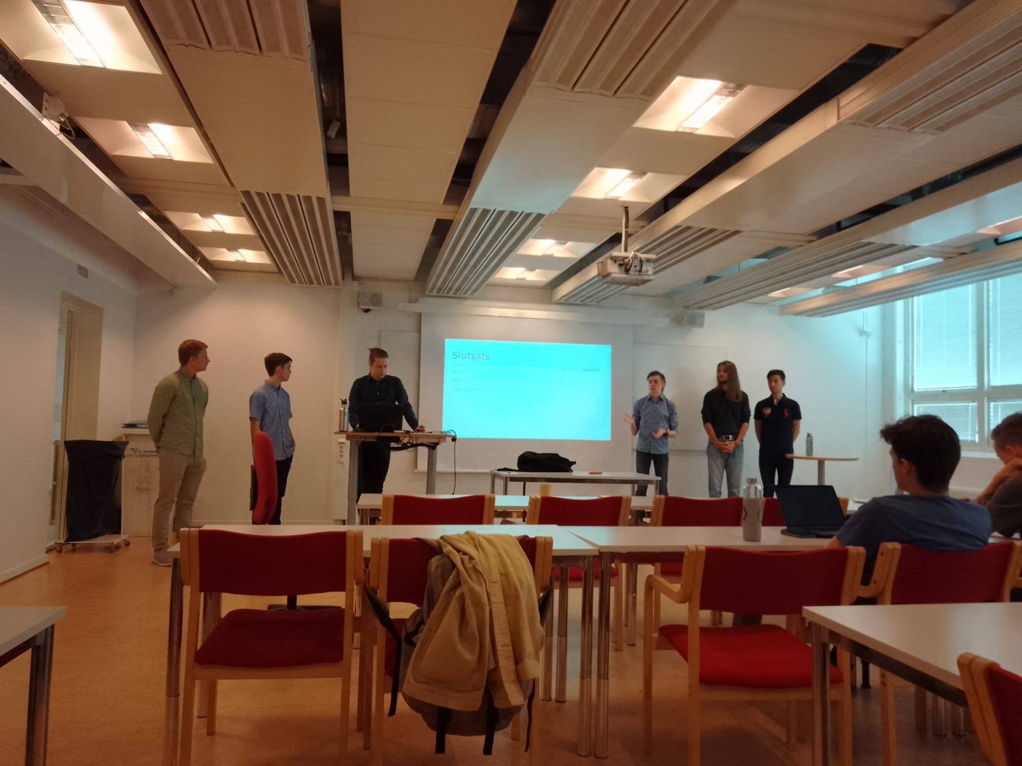
Slide from E. Clément’s presentation. (Image by A. Callegari via Zoom)Bacteria exploring Newtonian and non-Newtonian complex fluids: from behavioral variability to medium assisted tumbling
Eric Clément
PMMH-ESPCI-PSL, Sorbonne University, University Paris-Cité
31 May 2022, 11:00 CET
Understanding the way motile micro-organisms such as bacteria explore their environment is central to many ecological, medical and biotechnological questions. Here, I will present recent advances on the actual spatial exploration process undertaken by flagellated bacteria such as E.coli, undergoing sequences of runs and tumbles, leading to a random-walk. The extreme sensitivity of the motor rotation switch (CCW/CW) to the presence of a phosphorylated protein (CheYP) in its vicinity, leads to a behavioral variability of run-times, characterized by a log-normal distribution [1]. This mechanism prevails in most Newtonian fluids and has important consequences on the residence times at surfaces [2] as well as the large scale transport and dispersion in confined environments [3]. However when the surrounding fluid is a yield-stress fluid, the locally high resistance to penetration takes control of the exploration process and the run persistence time distribution is strongly affected by the mechanical bending of the flagella bundle, hence controlling the spatial diffusivity as well as the onset of a motility barrier.
[1] N. Figueroa-Morales et al., 3D spatial exploration by E.coli echoes motor temporal variability, Phys. Rev. X, 10, 021004 (2020).
[2] G. Junot et al., Run-to-tumble variability controls the surface residence times of E. coli bacteria, to appear in Phys. Rev. Lett. (2022).
[3] N. Figueroa-Morales et al., E.coli “super-contaminates” narrow channels fostered by broad motor switching statistics, Science Advances, 6, eaay0155 (2020).
Kymographs of DNA inside Channel II. (Image by the Authors.)Label-free nanofluidic scattering microscopy of size and mass of single diffusing molecules and nanoparticles
Barbora Špačková, Henrik Klein Moberg, Joachim Fritzsche, Johan Tenghamn, Gustaf Sjösten, Hana Šípová-Jungová, David Albinsson, Quentin Lubart, Daniel van Leeuwen, Fredrik Westerlund, Daniel Midtvedt, Elin K. Esbjörner, Mikael Käll, Giovanni Volpe & Christoph Langhammer
Nature Methods 19, 751–758 (2022)
doi: 10.1038/s41592-022-01491-6
Label-free characterization of single biomolecules aims to complement fluorescence microscopy in situations where labeling compromises data interpretation, is technically challenging or even impossible. However, existing methods require the investigated species to bind to a surface to be visible, thereby leaving a large fraction of analytes undetected. Here, we present nanofluidic scattering microscopy (NSM), which overcomes these limitations by enabling label-free, real-time imaging of single biomolecules diffusing inside a nanofluidic channel. NSM facilitates accurate determination of molecular weight from the measured optical contrast and of the hydrodynamic radius from the measured diffusivity, from which information about the conformational state can be inferred. Furthermore, we demonstrate its applicability to the analysis of a complex biofluid, using conditioned cell culture medium containing extracellular vesicles as an example. We foresee the application of NSM to monitor conformational changes, aggregation and interactions of single biomolecules, and to analyze single-cell secretomes.
August Kälvesten, Richard Blücher, Vilhelm Hedquist, Andreas Bauner, Adam Törnkvist, Eric Dat Le presenting their bachelor thesis. (Photo by A. Callegari.)August Kälvesten, Richard Blücher, Vilhelm Hedquist, Andreas Bauner, Adam Törnkvist, Eric Dat Le defended their Bachelor Thesis at Chalmers University of Technology on 25 May 2025. Congrats!
Title: Can slower predators catch faster swarming prey?
A study of systems where faster swarming prey interact with slower predators through simulation
Title: Kan långsammare rovdjur fånga snabbare svärmande byten?
En undersökning av system där snabbare svärmande byten interagerar med långsammare rovdjur genom simulation
Abstract: This project examined how predators can catch prey in a predator-prey system where the predators have a lower speed than their swarming prey. The investigated factors were the varied angular velocity of prey and predator, complex environment, and several cooperating predators. This was done through simulations based on the Vicsek model where a base model was modified for each of the investigated factors. When varied angular velocity was investigated it was found that the angular velo- city of the predator didn’t have a large effect on the numbers of prey captured, but what had an effect was the angular velocity of the prey. That could be explained by the predators traveling towards the prey head-on and the prey not being able to turn away fast enough. For complex environments, it was shown that an increased radius and number of obstacles in the environment led to increased numbers of prey caught. This is contradictory to the phenomenon in nature and could be explained by limitations in the model. Finally, when many cooperating predators were introduced, it was found that groups of three or four predators were required for prey to be caught. When many predators were introduced, more such groups could be created and therefore capture more prey. Although only three uncountable factors that govern predator-prey systems have been investigated, there are some indications that slower predators can catch faster swarming prey.
Sammandrag: I detta projekt undersöktes hur rovdjur kan fånga byten i ett rovdjur-bytessystem där rovdjuren har lägre fart än dess svärmande byten. De faktorer som undersökts är varierande vinkelhastighet hos byten och rovdjur, komplexa miljöer, och flera sam- arbetande rovdjur. Detta gjordes genom simuleringar baserat på Vicsek-modellen där en basmodell modifierades för varje faktor som undersöktes. Då varierande vin- kelhastighet undersöktes noterades det att rovdjurets vinkelhastighet inte har någon större inverkan på antalet fångade byten, utan det var snarare bytesdjurens vinkel- hastighet som hade störst inverkan. Det kunde förklaras av att rovdjuren lyckades fånga byten då de färdades rakt mot varandra och bytesdjuret inte kunde svänga av tillräckligt snabbt. För komplexa miljöer visades att en ökad radie och antal hinder i miljön ökade antalet fångade byten. Detta var motsägande observerade fenomen i naturen och kunde förklaras av begränsningar i modellen. Slutligen observerades, när flera samarbetande rovdjur undersöktes, att det krävdes grupper av tre eller fyra rovdjur för att byten skall fångas. Då många rovdjur introducerades kunde flera sådana grupper skapas och därför fånga fler byten. Trots att endast tre av de oräkneliga faktorer som styr rovdjur-bytessystem i verkligheten har undersökts kan vissa indikationer finnas på att långsammare rovdjur kan fånga snabbare svärmande byten.
The AnDi Challenge: Objective comparison of methods to decode anomalous diffusion
Giovanni Volpe
18 May 2022, 9:00 (CEST)
Online for: 27th Annual IASBS Meeting on Condensed Matter Physics
IASBS, Zanjan, Iran
18-19 May 2022
Deviations from Brownian motion leading to anomalous diffusion are found in transport dynamics from quantum physics to life sciences. The characterization of anomalous diffusion from the measurement of an individual trajectory is a challenging task, which traditionally relies on calculating the trajectory mean squared displacement. However, this approach breaks down for cases of practical interest, e.g., short or noisy trajectories, heterogeneous behaviour, or non-ergodic processes. Recently, several new approaches have been proposed, mostly building on the ongoing machine-learning revolution. To perform an objective comparison of methods, we gathered the community and organized an open competition, the Anomalous Diffusion challenge (AnDi). Participating teams applied their algorithms to a commonly-defined dataset including diverse conditions. Although no single method performed best across all scenarios, machine learning-based approaches achieved superior performance for all tasks. The discussion of the challenge results provides practical advice for users and a benchmark for developers.
Imaging large neuronal circuits from the Brain to the Gut Gilles Claude Vanwalleghem
4 May 2022, 12:30 CET
As a transparent animal and with powerful light-based tools to monitor the brain, the larval zebrafish offers a perfect window into functioning neural circuits. We can image the whole brain of zebrafish with cellular resolution, as they respond to various stimuli and record the activity of thousands of neurons. I will focus on two recent studies, one in collaboration with optical physicists, using optical tweezers to move otolith in the inner ear and simulate acceleration. We identified several salient response types, and showed the fish can respond to unnatural stimuli. The other used a microfluidics device to apply water flow to the fish and stimulate the lateral line. The fish’s brain could encode the speed, duration and direction of the water flow, but we also showed that the circuit was biased towards one specific direction of flow. Finally, I will briefly present the new focus of my lab, the gut-brain axis is a physiological communication network between the microbiome, enteric and central nervous system. We are using light sheet microscopy to image the activity of the ENS neurons from 3 to 7 days post fertilization fish. We observed that the spontaneous neuronal activity increases from 3 to 5 dpf, before dropping suddenly at day 7.
Bio
I received my PhD in 2012 from the Universite Libre de Bruxelles where I worked on the Trypanosoma brucei parasite. We discovered a key role of Trypanosoma brucei adenylate cyclases in host-pathogen interactions, as well as the mechanisms through which the human APOL1 can trigger the parasite’s death. In 2014, I was awarded an EMBO long-term fellowship, to shift my focus to neuroscience and the use of optogenetics in larval zebrafish. My work since has spanned several sensory modalities in the zebrafish, including optical traps for vestibular stimulation, visual loom responses, auditory processing, and water flow perception.
I am an assistant professor at Aarhus University since October 2021, where I will focus on the gut-brain axis, I am especially interested in the interactions between neurons, bacteria and the immune system.
Medical image segmentation using deep learning.
Hang Zhao
3 May 2022, 11:00
Image segmentation and synthesis of CT image based on deep learning: Deep learning methods for medical image segmentation are hindered by the lack of training data. This thesis aims to develop a method that overcomes this problem. Basic U-net trained on XCAT phantom data was tested first. The segmentation results were unsatisfactory even when artificial quantum noise was added. As a workaround, CycleGAN was used to add tissue textures to the XCAT phantom images by analyzing patient CT images. The generated images were used to train the network. The textures introduced by CycleGAN improved the segmentation, but some errors remained. Basic U-net was replaced with Attention U-net, which further improved the segmentation. More work is needed to fine-tune and thoroughly evaluate the method. The results obtained so far demonstrate the potential of this method for the segmentation of medical images. The proposed algorithms may be used in iterative image reconstruction algorithms in multi-energy computed tomography.
3D Cell nuclei segmentation using digital nuclei phantom and 3D deep learning methods : The analysis of microscopy image is helpful to pathological analysis. Nowadays, deep learning has shown the capabilities of processing the medical imaging data. However, developing deep learning methods in microscopy image analysis can be challenging because of the lack of ground truth and various resolution of microscopy image data. This project aims to build a digital nuclei phantom that simulates the actual microscopy images, including mitotic rate, nucleus size, noise, point spread function, and diverse resolutions. The phantom images were used to train 3D deep neural network for nuclei segmentation. The trained neural network was tested for segmentation on datasets with different resolutions. The neural network successfully performed segmentation on most resolutions in our dataset, and the segmentation results reflect the morphology and density of nuclei in microscopy images. The future work will focus on improving the nuclei phantom to generate more realistic phantom images, thereby further helping with segmentation.
Bio:
My name is Hang, I took my bachelor’s in China on radiophysics, and master’s at Linköping University on biomedical imaging. After I graduate, I joined Karolinska as a research assistant on 3D microscopy image processing. Now, I am working at Linköping university as a full time research engineer, contributing to cardiovascular MR image processing, supervised by Petter Dyverfeldt.
How fundamental physics leads the way to a better understanding of life’s complexity
Timo Betz
Georg August University Göttingen
20 April 2022, 12:30 CEST
Online
Many biological systems rely on fundamental physical principles for their proper function. Here, mechanical processes such as force generation and adaptation of stiffness and viscosity have been very successfully used to explain complex biomedical questions with physical concepts. Such advances have been largely driven by new methods that allow to quantify biological processes and to construct theoretical models with high predictive power. I will present our recent approaches that allow to study active force generation and mobility in different biological systems over several length scales. Starting with active motion of membranes and intracellular particles in oocytes followed by cytosolic fluidification during cell division we will construct a surprisingly general description of active motion inside the cytoplasm. In a further direction, similar principles are used to study the flow of whole tissue. Here we analyze pressure driven outbursts of cancer cells from model tumors, but also the collective motion during zebrafish development that eventually shapes a whole new animal. The tools we use are largely based on continuum mechanics and statistical mechanics, and give deep insights into the physical principles that are exploited by cells and living objects to perform their intriguing function.
Clogging, Dynamics and Reentrant Fluid for Active Matter on Periodic Substrates
Cynthia Reichhardt
Theoretical Division, Los Alamos National Laboratory, Los Alamos, NM, USA
6 April 2022, 16:00 CEST
Online
We explore the interactions between substrate length scales and correlation length scales of run-and-tumble active matter disks. For the case of a Casimir geometry of two plates placed a distance d apart, we show that an effective active-matter-mediated attraction arises due to a geometric shadowing effect [1]. Next we shrink the plates down to columns and consider connections to jamming [2] and clogging effects [3] found in passive granular matter. The active particles are driven with an external force through columns placed in a square periodic array [4]. When the drive is applied along a symmetry direction of the array, we find a clog-free uniform liquid state for low activity, while at higher activity, the density becomes increasingly heterogeneous and an active clogged state emerges in which the mobility is strongly reduced. For driving along non-symmetry or incommensurate directions, there are two different clogging behaviors consisting of a drive dependent clogged state in the low activity thermal limit and a drive independent clogged state at high activity. These regimes are separated by a uniform flowing liquid at intermediate activity. There is a critical activity level above which the thermal clogged state does not occur, as well as an optimal activity level that maximizes the disk mobility. Thermal clogged states are dependent on the driving direction while active clogged states are not [5].
References:
[1] D. Ray, C. Reichhardt, and C.J.O. Reichhardt, Phys. Rev. E 90, 013019 (2014).
[2] J.A. Drocco, M.B. Hastings, C.J.O. Reichhardt, and C. Reichhardt, Phys. Rev. Lett. 95, 088001 (2005).
[3] H. Peter, A. Libal, C. Reichhardt, and C.J.O. Reichhardt, Sci. Rep. 8, 10252 (2018).
[4] C. Reichhardt and C.J.O. Reichhardt, Phys. Rev. E 102, 042616 (2020).
[5] C. Reichhardt and C.J.O. Reichhardt, Phys. Rev. E 103, 062603 (2021).
(Photo by A. Argun.)Giuseppe Pesce starts his employment as a researcher at the Physics Department of the University of Gothenburg on 30th March 2022.
Giuseppe has a PhD degree in Physics from the University of Naples, Italy, where he was working for several years. His field of expertise is laser spectroscopy and optical tweezers used for several experiments, in particular for microrheology and statistical mechanics.
During his employment, Giuseppe will work on a project about optical tweezers combined with deep learning for construction of scalable quantum dots arrays and on automatisation of a double optical tweezers system to stretch biomolecules.