Optical Tweezers: A Comprehensive Tutorial from Calibration to Applications accepted on Advances in Optics and Photonics

Schematic of a bistable potential generated with a double-beam optical tweezers.

Optical Tweezers: A Comprehensive Tutorial from Calibration to Applications
Jan Gieseler, Juan Ruben Gomez-Solano, Alessandro Magazzù, Isaac Pérez Castillo, Laura Pérez García, Marta Gironella-Torrent, Xavier Viader-Godoy, Felix Ritort, Giuseppe Pesce, Alejandro V. Arzola, Karen Volke-Sepulveda & Giovanni Volpe
Advances in Optics and Photonics, 13(1), 74-241 (2021)
doi: https://doi.org/10.1364/AOP.394888
arXiv: 2004.05246

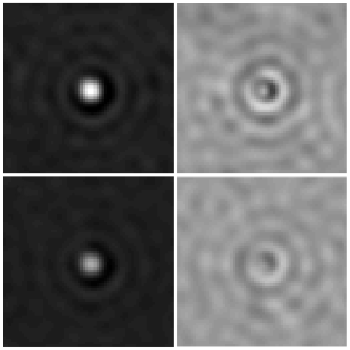
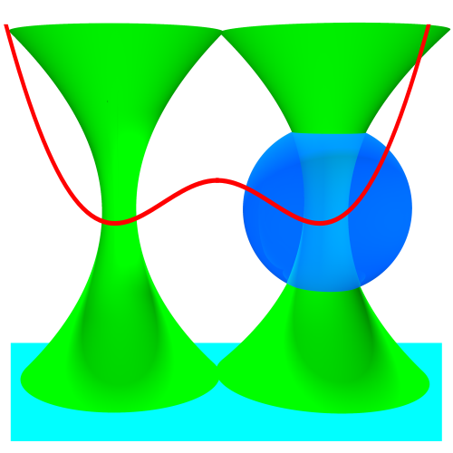
Since their invention in 1986 by Arthur Ashkin and colleagues, optical tweezers have become an essential tool in several fields of physics, spectroscopy, biology, nanotechnology, and thermodynamics. In this Tutorial, we provide a primer on how to calibrate optical tweezers and how to use them for advanced applications. After a brief general introduction on optical tweezers, we focus on describing and comparing the various available calibration techniques. Then, we discuss some cutting-edge applications of optical tweezers in a liquid medium, namely to study single-molecule and single-cell mechanics, microrheology, colloidal interactions, statistical physics, and transport phenomena. Finally, we consider optical tweezers in vacuum, where the absence of a viscous medium offers vastly different dynamics and presents new challenges. We conclude with some perspectives for the field and the future application of optical tweezers. This Tutorial provides both a step-by-step guide ideal for non-specialists entering the field and a comprehensive manual of advanced techniques useful for expert practitioners. All the examples are complemented by the sample data and software necessary to reproduce them.

Quantitative Digital Microscopy with Deep Learning published in Applied Physics Reviews

Particle tracking and characterization in terms of radius and refractive index.

Quantitative Digital Microscopy with Deep Learning
Benjamin Midtvedt, Saga Helgadottir, Aykut Argun, Jesús Pineda, Daniel Midtvedt, Giovanni Volpe
Applied Physics Reviews 8, 011310 (2021)
doi: 10.1063/5.0034891
arXiv: 2010.08260

Video microscopy has a long history of providing insights and breakthroughs for a broad range of disciplines, from physics to biology. Image analysis to extract quantitative information from video microscopy data has traditionally relied on algorithmic approaches, which are often difficult to implement, time consuming, and computationally expensive. Recently, alternative data-driven approaches using deep learning have greatly improved quantitative digital microscopy, potentially offering automatized, accurate, and fast image analysis. However, the combination of deep learning and video microscopy remains underutilized primarily due to the steep learning curve involved in developing custom deep-learning solutions. To overcome this issue, we introduce a software, DeepTrack 2.0, to design, train and validate deep-learning solutions for digital microscopy. We use it to exemplify how deep learning can be employed for a broad range of applications, from particle localization, tracking and characterization to cell counting and classification. Thanks to its user-friendly graphical interface, DeepTrack 2.0 can be easily customized for user-specific applications, and, thanks to its open-source object-oriented programming, it can be easily expanded to add features and functionalities, potentially introducing deep-learning-enhanced video microscopy to a far wider audience.

Optical trapping and critical Casimir forces published in EPJP

Measuring the dynamics of colloids interacting with critical Casimir interaction via blinking optical tweezers: graphical representation of the optical traps.

Optical trapping and critical Casimir forces
Agnese Callegari, Alessandro Magazzù, Andrea Gambassi & Giovanni Volpe
The European Physical Journal Plus (EPJP), 136, 213 (2021)
doi: 10.1140/epjp/s13360-020-01020-4
arXiv: 2008.01537

Critical Casimir forces emerge between objects, such as colloidal particles, whenever their surfaces spatially confine the fluctuations of the order parameter of a critical liquid used as a solvent. These forces act at short but microscopically large distances between these objects, reaching often hundreds of nanometers. Keeping colloids at such distances is a major experimental challenge, which can be addressed by the means of optical tweezers. Here, we review how optical tweezers have been successfully used to quantitatively study critical Casimir forces acting on particles in suspensions. As we will see, the use of optical tweezers to experimentally study critical Casimir forces can play a crucial role in developing nano-technologies, representing an innovative way to realize self-assembled devices at the nano- and microscale.

Intercellular Communication Induces Glycolytic Synchronisation Waves published in PNAS

Intercellular communication induces glycolytic synchronization waves between individually oscillating cells

Intercellular communication induces glycolytic synchronization waves between individually oscillating cells
Martin Mojica-Benavides, David D. van Niekerk, Mite Mijalkov, Jacky L. Snoep, Bernhard Mehlig, Giovanni Volpe, Caroline B. Adiels & Mattias Goksör
PNAS 118(6), e2010075118 (2021)
doi: 10.1073/pnas.2010075118
arXiv: 1909.05187

Metabolic oscillations in single cells underlie the mechanisms behind cell synchronization and cell-cell communication. For example, glycolytic oscillations mediated by biochemical communication between cells may synchronize the pulsatile insulin secretion by pancreatic tissue, and a link between glycolytic synchronization anomalies and type-2 diabetes has been hypotesized. Cultures of yeast cells have provided an ideal model system to study synchronization and propagation waves of glycolytic oscillations in large populations. However, the mechanism by which synchronization occurs at individual cell-cell level and overcome local chemical concentrations and heterogenic biological clocks, is still an open question because of experimental limitations in sensitive and specific handling of single cells. Here, we show how the coupling of intercellular diffusion with the phase regulation of individual oscillating cells induce glycolytic synchronization waves. We directly measure the single-cell metabolic responses from yeast cells in a microfluidic environment and characterize a discretized cell-cell communication using graph theory. We corroborate our findings with simulations based on a kinetic detailed model for individual yeast cells. These findings can provide insight into the roles cellular synchronization play in biomedical applications, such as insulin secretion regulation at the cellular level.

Press release on joint research on intercellular communication mechanism by Biological Physics Lab and Soft Matter Lab

The article Intercellular Communication Induces Glycolytic Synchronisation Waves published in PNAS has been featured in the News of the Faculty of Science of Gothenburg University.

Here the links to the press releases:
Swedish: Forskare har knäckt koden för cellkommunikation
English: Researchers have broken the code for cell communication

Giovanni Volpe is committee member at OSA-OMA 2021

Giovanni Volpe is part of the committee of the conference Optical Manipulation and its Applications (OMA), which is part of the OSA Biophotonics Congress: Optics in the Life Sciences.

Optical Manipulation encompasses all areas of manipulation and measurement using light, from optical manipulation of microparticles to photoactivated materials and optogenetics, emphasizing new and developing application areas in biophysics and biomedicine.

The categories of topics in the conference are Optical Manipulation in Biophysics and Biomedicine, Optical Manipulation Fundamentals, Optical Manipulation Applications, and Alternative Manipulation Techniques.

The conference will be held online 12-16 April 2021.

Online seminar by G. Volpe at Indian Institute of Science Education and Research (IISER), Pune, India

Deep learning for microscopy and optical trapping
Giovanni Volpe
21 January 2021, 16:30 CEST
Online
Invited seminar for Indian Institute of Science Education and Research (IISER), Pune, India

After a brief overview of artificial intelligence, machine learning and deep learning, I will present a series of recent works in which we have employed deep learning for applications in photonics and active matter. In particular, I will explain how we employed deep learning to enhance digital video microscopy, to estimate the properties of anomalous diffusion, to characterize microscopic force fields, to improve the calculation of optical forces, and to characterize nanoparticles. Finally, I will provide an outlook for the application of deep learning in photonics and active matter.

Fast and Accurate Nanoparticle Characterization Using Deep-Learning-Enhanced Off-Axis Holography published in ACS Nano

Phase and amplitude signals from representative particles for testing the performance of the Deep-learning approach

Fast and Accurate Nanoparticle Characterization Using Deep-Learning-Enhanced Off-Axis Holography
Benjamin Midtvedt, Erik Olsén, Fredrik Eklund, Fredrik Höök, Caroline Beck Adiels, Giovanni Volpe, Daniel Midtvedt
ACS Nano 15(2), 2240–2250 (2021)
doi: 10.1021/acsnano.0c06902
arXiv: 2006.11154

The characterisation of the physical properties of nanoparticles in their native environment plays a central role in a wide range of fields, from nanoparticle-enhanced drug delivery to environmental nanopollution assessment. Standard optical approaches require long trajectories of nanoparticles dispersed in a medium with known viscosity to characterise their diffusion constant and, thus, their size. However, often only short trajectories are available, while the medium viscosity is unknown, e.g., in most biomedical applications. In this work, we demonstrate a label-free method to quantify size and refractive index of individual subwavelength particles using two orders of magnitude shorter trajectories than required by standard methods, and without assumptions about the physicochemical properties of the medium. We achieve this by developing a weighted average convolutional neural network to analyse the holographic images of the particles. As a proof of principle, we distinguish and quantify size and refractive index of silica and polystyrene particles without prior knowledge of solute viscosity or refractive index. As an example of an application beyond the state of the art, we demonstrate how this technique can monitor the aggregation of polystyrene nanoparticles, revealing the time-resolved dynamics of the monomer number and fractal dimension of individual subwavelength aggregates. This technique opens new possibilities for nanoparticle characterisation with a broad range of applications from biomedicine to environmental monitoring.

Giovanni Volpe awarded new ERC Consolidator Grant

ERC logo.
Giovanni Volpe has been awarded a new European Research Council (ERC) Consolidator Grant on Wednesday, December 9th 2020.

The title of his project is “Microscopic Active Particles with Embodied Intelligence”.

Active particles and active matter research tries to understand and replicate the characteristics of living microorganisms in artificial systems. Over billions of years of evolution, living organisms have developed complex strategies to survive and thrive. The artificial active particles are still incapable of autonomous information processing.

Giovanni Volpe’s project aims to address three main challenges in the current research on active matter:

  • Make active particles capable of autonomous information processing.
  • Optimize the behavioral strategies of individual active particles.
  • Optimize the interactions between active particles.

Links
Giovanni Volpe awarded new ERC Consolidator Grant
Official press release (ERC): CoG Recipients 2020
14 forskare verksamma i Sverige får ERC Consolidator grant