Intracavity Optical Trapping published in Nature Commun.

Intracavity Optical Trapping

Intracavity optical trapping of microscopic particles in a ring-cavity fiber laser
Fatemeh Kalantarifard, Parviz Elahi, Ghaith Makey, Onofrio M. Maragò, F. Ömer Ilday & Giovanni Volpe
Nature Communications 10, 2683 (2019)
doi: 10.1038/s41467-019-10662-7
arXiv: 1808.07831

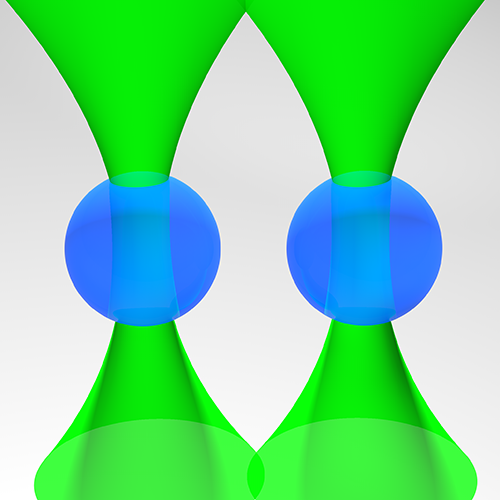
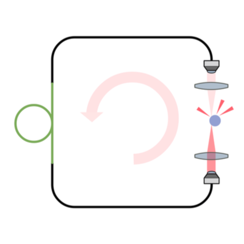
Standard optical tweezers rely on optical forces arising when a focused laser beam interacts with a microscopic particle: scattering forces, pushing the particle along the beam direction, and gradient forces, attracting it towards the high-intensity focal spot. Importantly, the incoming laser beam is not affected by the particle position because the particle is outside the laser cavity. Here, we demonstrate that intracavity nonlinear feedback forces emerge when the particle is placed inside the optical cavity, resulting in orders-of-magnitude higher confinement along the three axes per unit laser intensity on the sample. This scheme allows trapping at very low numerical apertures and reduces the laser intensity to which the particle is exposed by two orders of magnitude compared to a standard 3D optical tweezers. These results are highly relevant for many applications requiring manipulation of samples that are subject to photodamage, such as in biophysics and nanosciences.

Subtypes of Brain Atrophy in Alzheimer’s Disease published in Front. Neurol.

Subtypes of Alzheimer’s disease display distinct network abnormalities extending beyond their pattern of brain atrophy

Subtypes of Alzheimer’s disease display distinct network abnormalities extending beyond their pattern of brain atrophy
Daniel Ferreira, Joana B. Pereira, Giovanni Volpe & Eric Westman
Frontiers in Neurology 10, 524 (2019)
DOI: 10.3389/fneur.2019.00524

Different subtypes of Alzheimer’s disease (AD) with characteristic distributions of neurofibrillary tangles and corresponding brain atrophy patterns have been identified using structural magnetic resonance imaging (MRI). However, the underlying biological mechanisms that determine this differential expression of neurofibrillary tangles are still unknown. Here, we applied graph theoretical analysis to structural MRI data to test the hypothesis that differential network disruption is at the basis of the emergence of these AD subtypes. We studied a total of 175 AD patients and 81 controls. Subtyping was done using the Scheltens’ scale for medial temporal lobe atrophy, the Koedam’s scale for posterior atrophy, and the Pasquier’s global cortical atrophy scale for frontal atrophy. A total of 89 AD patients showed a brain atrophy pattern consistent with typical AD; 30 patients showed a limbic-predominant pattern; 29 patients showed a hippocampal-sparing pattern; and 27 showed minimal atrophy. We built brain structural networks from 68 cortical regions and 14 subcortical gray matter structures for each AD subtype and for the controls, and we compared between-group measures of integration, segregation, and modular organization. At the global level, modularity was increased and differential modular reorganization was detected in the four subtypes. We also found a decrease of transitivity in the typical and hippocampal-sparing subtypes, as well as an increase of average local efficiency in the minimal atrophy and hippocampal-sparing subtypes. We conclude that the AD subtypes have a distinct signature of network disruption associated with their atrophy patterns and further extending to other brain regions, presumably reflecting the differential spread of neurofibrillary tangles. We discuss the hypothetical emergence of these subtypes and possible clinical implications.

Digital Video Microscopy Enhanced by Deep Learning published in Optica

Digital video microscopy enhanced by deep learning

Digital video microscopy enhanced by deep learning
(Cover article)
Saga Helgadottir, Aykut Argun & Giovanni Volpe
Optica 6(4), 506—513 (2019)
doi: 10.1364/OPTICA.6.000506
arXiv: 1812.02653
GitHub: DeepTrack

Single particle tracking is essential in many branches of science and technology, from the measurement of biomolecular forces to the study of colloidal crystals. Standard methods rely on algorithmic approaches; by fine-tuning several user-defined parameters, these methods can be highly successful at tracking a well-defined kind of particle under low-noise conditions with constant and homogenous illumination. Here, we introduce an alternative data-driven approach based on a convolutional neural network, which we name DeepTrack. We show that DeepTrack outperforms algorithmic approaches, especially in the presence of noise and under poor illumination conditions. We use DeepTrack to track an optically trapped particle under very noisy and unsteady illumination conditions, where standard algorithmic approaches fail. We then demonstrate how DeepTrack can also be used to track multiple particles and non-spherical objects such as bacteria, also at very low signal-to-noise ratios. In order to make DeepTrack readily available for other users, we provide a Python software package, which can be easily personalized and optimized for specific applications.

Featured in :
Deep Learning for Particle Tracking”, Optics & Photonics News (December 1, 2019)

Funding:

ERC-founder H2020 European Research Council (ERC) Starting Grant ComplexSwimmers (677511).

Video 1:

Video 2:

Video 3:

Video 4:

Video 5:

Video 6:

Video 7:

Video 8:

Active Colloidal Molecules published in J. Chem. Phys.

Light-controlled Assembly of Active Colloidal Molecules

Light-controlled Assembly of Active Colloidal Molecules
Falko Schmidt, Benno Liebchen, Hartmut Löwen & Giovanni Volpe
Journal of Chemical Physics 150(9), 094905 (2019)
doi: 10.1063/1.5079861
arXiv: 1801.06868

Thanks to a constant energy input, active matter can self-assemble into phases with complex architectures and functionalities such as living clusters that dynamically form, reshape, and break-up, which are forbidden in equilibrium materials by the entropy maximization (or free energy minimization) principle. The challenge to control this active self-assembly has evoked widespread efforts typically hinging on engineering of the properties of individual motile constituents. Here, we provide a different route, where activity occurs as an emergent phenomenon only when individual building blocks bind together in a way that we control by laser light. Using experiments and simulations of two species of immotile microspheres, we exemplify this route by creating active molecules featuring a complex array of behaviors, becoming migrators, spinners, and rotators. The possibility to control the dynamics of active self-assembly via light-controllable nonreciprocal interactions will inspire new approaches to understand living matter and to design active materials.

Funding:

ERC-founder H2020 European Research Council (ERC) Starting Grant ComplexSwimmers (677511).
VR-founder Vetenskapsrådet (Grant No. 2016-03523).
German Research Foundation DFG within LO 418–19-1.

Ordering of binary colloidal crystals by random potentials on ArXiv

Ordering of binary colloidal crystals by random potentials

Ordering of binary colloidal crystals by random potentials
André S. Nunes, Sabareesh K. P. Velu, Iryna Kasianiuk, Denys Kasyanyuk, Agnese Callegari, Giorgio Volpe, Margarida M. Telo da Gama, Giovanni Volpe & Nuno A. M. Araújo
arXiv: 1903.01579

Structural defects are ubiquitous in condensed matter, and not always a nuisance. For example, they underlie phenomena such as Anderson localization and hyperuniformity, and they are now being exploited to engineer novel materials. Here, we show experimentally that the density of structural defects in a 2D binary colloidal crystal can be engineered with a random potential. We generate the random potential using an optical speckle pattern, whose induced forces act strongly on one species of particles (strong particles) and weakly on the other (weak particles). Thus, the strong particles are more attracted to the randomly distributed local minima of the optical potential, leaving a trail of defects in the crystalline structure of the colloidal crystal. While, as expected, the crystalline ordering initially decreases with increasing fraction of strong particles, the crystalline order is surprisingly recovered for sufficiently large fractions. We confirm our experimental results with particle-based simulations, which permit us to elucidate how this non-monotonic behavior results from the competition between the particle-potential and particle-particle interactions.

Controlling Colloidal Dynamics by Critical Casimir Forces published in Soft Matter

Controlling the dynamics of colloidal particles by critical Casimir forces

Controlling the dynamics of colloidal particles by critical Casimir forces
(Back cover article)
Alessandro Magazzù, Agnese Callegari, Juan Pablo Staforelli, Andrea Gambassi, Siegfried Dietrich & Giovanni Volpe
Soft Matter 15(10), 2152—2162 (2019)
doi: 10.1039/C8SM01376D
arXiv: 1806.11403

Critical Casimir forces can play an important role for applications in nano-science and nano-technology, owing to their piconewton strength, nanometric action range, fine tunability as a function of temperature, and exquisite dependence on the surface properties of the involved objects. Here, we investigate the effects of critical Casimir forces on the free dynamics of a pair of colloidal particles dispersed in the bulk of a near-critical binary liquid solvent, using blinking optical tweezers. In particular, we measure the time evolution of the distance between the two colloids to determine their relative diffusion and drift velocity. Furthermore, we show how critical Casimir forces change the dynamic properties of this two-colloid system by studying the temperature dependence of the distribution of the so-called first-passage time, i.e., of the time necessary for the particles to reach for the first time a certain separation, starting from an initially assigned one. These data are in good agreement with theoretical results obtained from Monte Carlo simulations and Langevin dynamics.

Funding:

ERC-founder H2020 European Research Council (ERC) Starting Grant ComplexSwimmers (677511).

Active Matter Influence on Coffee Rings published in Soft Matter

Active Matter Alters the Growth Dynamics of Coffee Rings

Active Matter Alters the Growth Dynamics of Coffee Rings
(Back cover article)
Tuğba Andaç, Pascal Weigmann, Sabareesh K. P. Velu, Erçağ Pinçe, Agnese Callegari, Giorgio Volpe, Giovanni Volpe & Agnese Callegari
Soft Matter 15(7), 1488—1496 (2019)
doi: 10.1039/C8SM01350K
arXiv: 1803.02619

How particles are deposited at the edge of evaporating droplets, i.e. the coffee ring effect, plays a crucial role in phenomena as diverse as thin-film deposition, self-assembly, and biofilm formation. Recently, microorganisms have been shown to passively exploit and alter these deposition dynamics to increase their survival chances under harshening conditions. Here, we show that, as the droplet evaporation rate slows down, bacterial mobility starts playing a major role in determining the growth dynamics of the edge of drying droplets. Such motility-induced dynamics can influence several biophysical phenomena, from the formation of biofilms to the spreading of pathogens in humid environments and on surfaces subject to periodic drying. Analogous dynamics in other active matter systems can be exploited for technological applications in printing, coating, and self-assembly, where the standard coffee-ring effect is often a nuisance.

FORMA – Enhanced Optical Tweezers Calibration published in Nature Commun.

High-Performance Reconstruction of Microscopic Force Fields from Brownian Trajectories

High-Performance Reconstruction of Microscopic Force Fields from Brownian Trajectories
Laura Pérez García, Jaime Donlucas Pérez, Giorgio Volpe, Alejandro V. Arzola & Giovanni Volpe
Nature Communications 9, 5166 (2018)
doi: 10.1038/s41467-018-07437-x
arXiv: 1808.05468

The accurate measurement of microscopic force fields is crucial in many branches of science and technology, from biophotonics and mechanobiology to microscopy and optomechanics. These forces are often probed by analysing their influence on the motion of Brownian particles. Here we introduce a powerful algorithm for microscopic force reconstruction via maximum-likelihood-estimator analysis (FORMA) to retrieve the force field acting on a Brownian particle from the analysis of its displacements. FORMA estimates accurately the conservative and non-conservative components of the force field with important advantages over established techniques, being parameter-free, requiring ten-fold less data and executing orders-of-magnitude faster. We demonstrate FORMA performance using optical tweezers, showing how, outperforming other available techniques, it can identify and characterise stable and unstable equilibrium points in generic force fields. Thanks to its high performance, FORMA can accelerate the development of microscopic and nanoscopic force transducers for physics, biology and engineering.

See also freeware software at 10.6084/m9.figshare.7181888

Featured in:
Optimerad optisk pincett, Forskning.se

Phototactic Robot Tunable by Sensorial Delays published in Phys. Rev. E

Phototactic Robot Tunable by Sensorial Delays

Tuning phototactic robots with sensorial delays (Editors’ suggestion)
Maximilian Leyman, Freddie Ogemark, Jan Wehr & Giovanni Volpe
Physical Review E 98(26), 052606 (2018)
DOI: 10.1103/PhysRevE.98.052606
arXiv: 1807.11765

The presence of a delay between sensing and reacting to a signal can determine the long-term behavior of autonomous agents whose motion is intrinsically noisy.
In a previous work [M. Mijalkov, A. McDaniel, J. Wehr, and G. Volpe, Phys. Rev. X 6, 011008 (2016)], we have shown that sensorial delay can alter the drift and the position probability distribution of an autonomous agent whose speed depends on the illumination intensity it measures. Here, using theory, simulations, and experiments with a phototactic robot, we generalize this effect to an agent for which both speed and rotational diffusion depend on the illumination intensity and are subject to two independent sensorial delays. We show that both the drift and the probability distribution are influenced by the presence of these sensorial delays. In particular, the radial drift may have positive as well as negative sign, and the position probability distribution peaks in different regions depending on the delay.
Furthermore, the presence of multiple sensorial delays permits us to explore the role of the interaction between them.

Stability of Brain Graph Measures published in Sci. Rep.

Stability of graph theoretical
measures in structural brain
networks in Alzheimer’s disease

Stability of graph theoretical measures in structural brain networks in Alzheimer’s disease
Gustav Mårtensson, Joana B. Pereira, Patrizia Mecocci, Bruno Vellas, Magda Tsolaki, Iwona Kłoszewska, Hilkka Soininen, Simon Lovestone, Andrew Simmons, Giovanni Volpe & Eric Westman
Scientific Reports 8, 11592 (2018)
DOI: 10.1038/s41598-018-29927-0

Graph analysis has become a popular approach to study structural brain networks in neurodegenerative disorders such as Alzheimer’s disease (AD). However, reported results across similar studies are often not consistent. In this paper we investigated the stability of the graph analysis measures clustering, path length, global efficiency and transitivity in a cohort of AD (N = 293) and control subjects (N = 293). More specifically, we studied the effect that group size and composition, choice of neuroanatomical atlas, and choice of cortical measure (thickness or volume) have on binary and weighted network properties and relate them to the magnitude of the differences between groups of AD and control subjects. Our results showed that specific group composition heavily influenced the network properties, particularly for groups with less than 150 subjects. Weighted measures generally required fewer subjects to stabilize and all assessed measures showed robust significant differences, consistent across atlases and cortical measures. However, all these measures were driven by the average correlation strength, which implies a limitation of capturing more complex features in weighted networks. In binary graphs, significant differences were only found in the global efficiency and transitivity measures when using cortical thickness measures to define edges. The findings were consistent across the two atlases, but no differences were found when using cortical volumes. Our findings merits future investigations of weighted brain networks and suggest that cortical thickness measures should be preferred in future AD studies if using binary networks. Further, studying cortical networks in small cohorts should be complemented by analyzing smaller, subsampled groups to reduce the risk that findings are spurious.