Stability of Brain Graph Measures published in Sci. Rep.

Stability of graph theoretical
measures in structural brain
networks in Alzheimer’s disease

Stability of graph theoretical measures in structural brain networks in Alzheimer’s disease
Gustav Mårtensson, Joana B. Pereira, Patrizia Mecocci, Bruno Vellas, Magda Tsolaki, Iwona Kłoszewska, Hilkka Soininen, Simon Lovestone, Andrew Simmons, Giovanni Volpe & Eric Westman
Scientific Reports 8, 11592 (2018)
DOI: 10.1038/s41598-018-29927-0

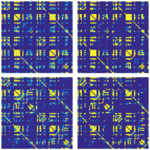
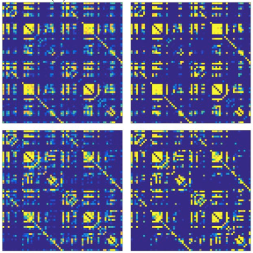
Graph analysis has become a popular approach to study structural brain networks in neurodegenerative disorders such as Alzheimer’s disease (AD). However, reported results across similar studies are often not consistent. In this paper we investigated the stability of the graph analysis measures clustering, path length, global efficiency and transitivity in a cohort of AD (N = 293) and control subjects (N = 293). More specifically, we studied the effect that group size and composition, choice of neuroanatomical atlas, and choice of cortical measure (thickness or volume) have on binary and weighted network properties and relate them to the magnitude of the differences between groups of AD and control subjects. Our results showed that specific group composition heavily influenced the network properties, particularly for groups with less than 150 subjects. Weighted measures generally required fewer subjects to stabilize and all assessed measures showed robust significant differences, consistent across atlases and cortical measures. However, all these measures were driven by the average correlation strength, which implies a limitation of capturing more complex features in weighted networks. In binary graphs, significant differences were only found in the global efficiency and transitivity measures when using cortical thickness measures to define edges. The findings were consistent across the two atlases, but no differences were found when using cortical volumes. Our findings merits future investigations of weighted brain networks and suggest that cortical thickness measures should be preferred in future AD studies if using binary networks. Further, studying cortical networks in small cohorts should be complemented by analyzing smaller, subsampled groups to reduce the risk that findings are spurious.

Disrupted Network Topology in Alzheimer published in Cerebral Cortex

Disrupted Network Topology in Patients with Stable and Progressive Mild Cognitive Impairment and Alzheimer’s Disease

Disrupted Network Topology in Patients with Stable and Progressive Mild Cognitive Impairment and Alzheimer’s Disease
Joana B. Pereira, Mite Mijalkov, Ehsan Kakaei, Patricia Mecocci, Bruno Vellas, Magda Tsolaki, Iwona Kłoszewska, Hilka Soininen, Christian Spenger, Simmon Lovestone, Andrew Simmons, Lars-Olof Wahlund, Giovanni Volpe & Eric Westman, AddNeuroMed consortium, for the Alzheimer’s Disease Neuroimaging Initiative
Cerebral Cortex 26(8), 3476—3493 (2016)
DOI: 10.1093/cercor/bhw128

Recent findings suggest that Alzheimer’s disease (AD) is a disconnection syndrome characterized by abnormalities in large- scale networks. However, the alterations that occur in network topology during the prodromal stages of AD, particularly in patients with stable mild cognitive impairment (MCI) and those that show a slow or faster progression to dementia, are still poorly understood. In this study, we used graph theory to assess the organization of structural MRI networks in stable MCI (sMCI) subjects, late MCI converters (lMCIc), early MCI converters (eMCIc), and AD patients from 2 large multicenter cohorts: ADNI and AddNeuroMed. Our findings showed an abnormal global network organization in all patient groups, as reflected by an increased path length, reduced transitivity, and increased modularity compared with controls. In addition, lMCIc, eMCIc, and AD patients showed a decreased path length and mean clustering compared with the sMCI group. At the local level, there were nodal clustering decreases mostly in AD patients, while the nodal closeness centrality detected abnormalities across all patient groups, showing overlapping changes in the hippocampi and amygdala and nonoverlapping changes in parietal, entorhinal, and orbitofrontal regions. These findings suggest that the prodromal and clinical stages of AD are associated with an abnormal network topology.

Aberrant Cerebral Topology in Early Parkinson published in Human Brain Mapp.

Aberrant cerebral network topology and mild cognitive impairment in early Parkinson’s disease

Aberrant cerebral network topology and mild cognitive impairment in early Parkinson’s disease
Joana B. Pereira, Dag Aarsland, Cedric E. Ginestet, Alexander V. Lebedev, Lars-Olof Wahlund, Andrew Simmons, Giovanni Volpe & Eric Westman
Human Brain Mapping 36(8), 2980—2995 (2015)
DOI: 10.1002/hbm.22822

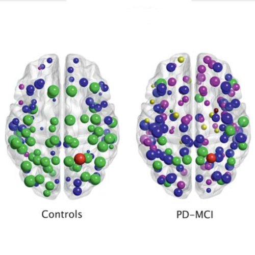
The aim of this study was to assess whether mild cognitive impairment (MCI) is associated with disruption in large-scale structural networks in newly diagnosed, drug-na€ıve patients with Parkin- son’s disease (PD). Graph theoretical analyses were applied to 3T MRI data from 123 PD patients and 56 controls from the Parkinson’s progression markers initiative (PPMI). Thirty-three patients were classified as having Parkinson’s disease with mild cognitive impairment (PD-MCI) using the Movement Disorders Society Task Force criteria, while the remaining 90 PD patients were classified as cognitively normal (PD- CN). Global measures (clustering coefficient, characteristic path length, global efficiency, small-world- ness) and regional measures (regional clustering coefficient, regional efficiency, hubs) were assessed in the structural networks that were constructed based on cortical thickness and subcortical volume data. PD-MCI patients showed a marked reduction in the average correlation strength between cortical and subcortical regions compared with controls. These patients had a larger characteristic path length and reduced global efficiency in addition to a lower regional efficiency in frontal and parietal regions com- pared with PD-CN patients and controls. A reorganization of the highly connected regions in the network was observed in both groups of patients. This study shows that the earliest stages of cognitive decline in PD are associated with a disruption in the large-scale coordination of the brain network and with a decrease of the efficiency of parallel information processing. These changes are likely to signal further cognitive decline and provide support to the role of aberrant network topology in cognitive impairment in patients with early PD.