Flash Talk by F. Schmidt at 729. WE Heraeus Seminar on Fluctuation Induced Forces, Online, 16 February 2022

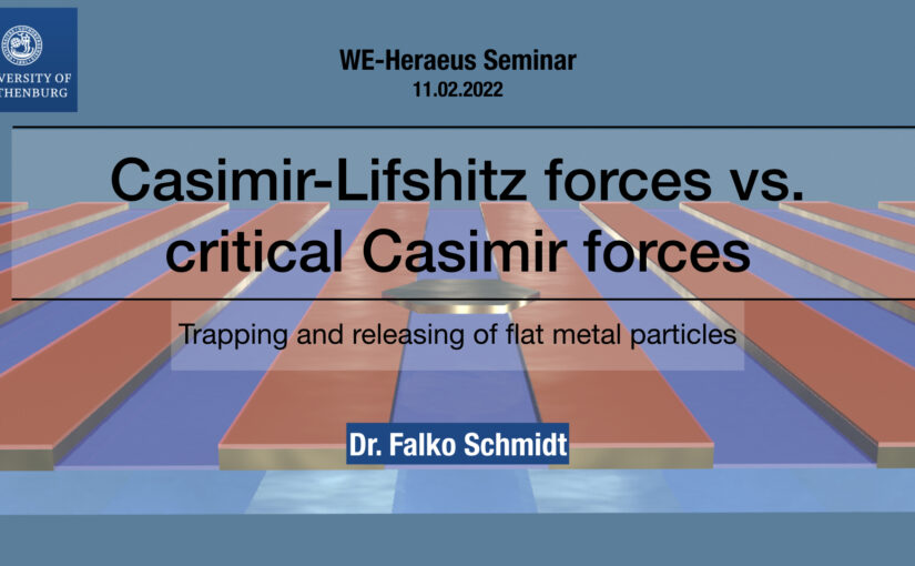
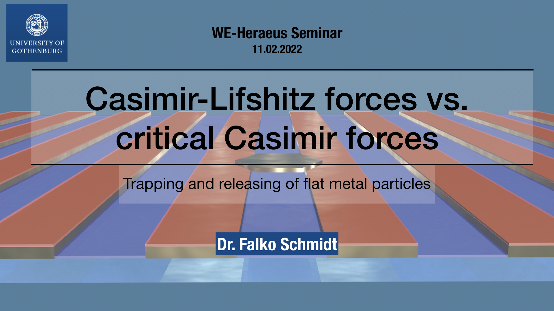
Title slide of the presentation. (Image by F. Schmidt.)
Casimir-Lifshitz forces vs. Critical Casimir forces: Trapping and releasing of flat metallic particles
Falko Schmidt
729. WE-Heraeus Stiftung Seminar on Fluctuation-induced Forces
16 February 2022, 14:50 CET

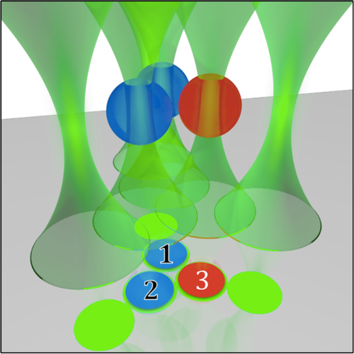
Casimir forces in quantum electrodynamics emerge between microscopic metallic objects because of the confinement of the vacuum electromagnetic fluctuations occuring even at zero temperature. Their generalization at finite temperature and in material media are referred to as Casimir-Lifshitz forces. These forces are typically attractive, leading to the widespread problem of stiction between the metallic parts of micro- and nanodevices. Recently, repulsive Casimir forces have been experimentally realized but their use of specialized materials stills means that the system can not be controlled dynamically and thus limits further implementation to real-world applications. Here, we experimentally demonstrate that repulsive critical Casimir forces, which emerge in a critical binary liquid mixture upon approaching the critical temperature, can be used to prevent stiction due to Casimir-Lifshitz forces. We show that critical Casimir forces can be dynamically tuned via temperature, eventually overcoming Casimir-Lifshitz attraction. We study a microscopic gold flake above a flat gold-coated substrate immersed in a critical mixture. Far from the critical temperature, stiction occurs because of Casimir-Lifshitz forces. Upon approaching the critical temperature, however, we observe the emergence of repulsive critical Casimir forces that are sufficiently strong to counteract stiction. By removing one of the key limitations to their deployment, this experimental demonstration can accelerate the development of micro- and nanodevices for a broad range of applications.

Invited Talk by G. Volpe at UFS Day 10.02.22

DeepTrack 2.0 Logo. (Image from DeepTrack 2.0 Project)

Deep learning for experimental soft matter
Giovanni Volpe
Invited Talk at UFS Day 10.02.22
Online
10 February 2022
14:00 CET

After a brief overview of artificial intelligence, machine learning and deep learning, I will present a series of recent works in which we have employed deep learning for applications in experimental soft matter.

Directed Brain Connectivity Identifies Widespread Functional Network Abnormalities in Parkinson’s Disease published in Cerebral Cortex

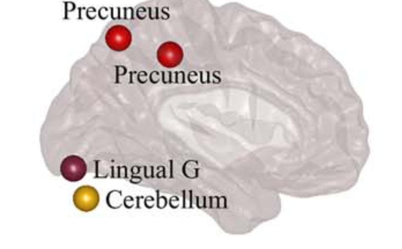
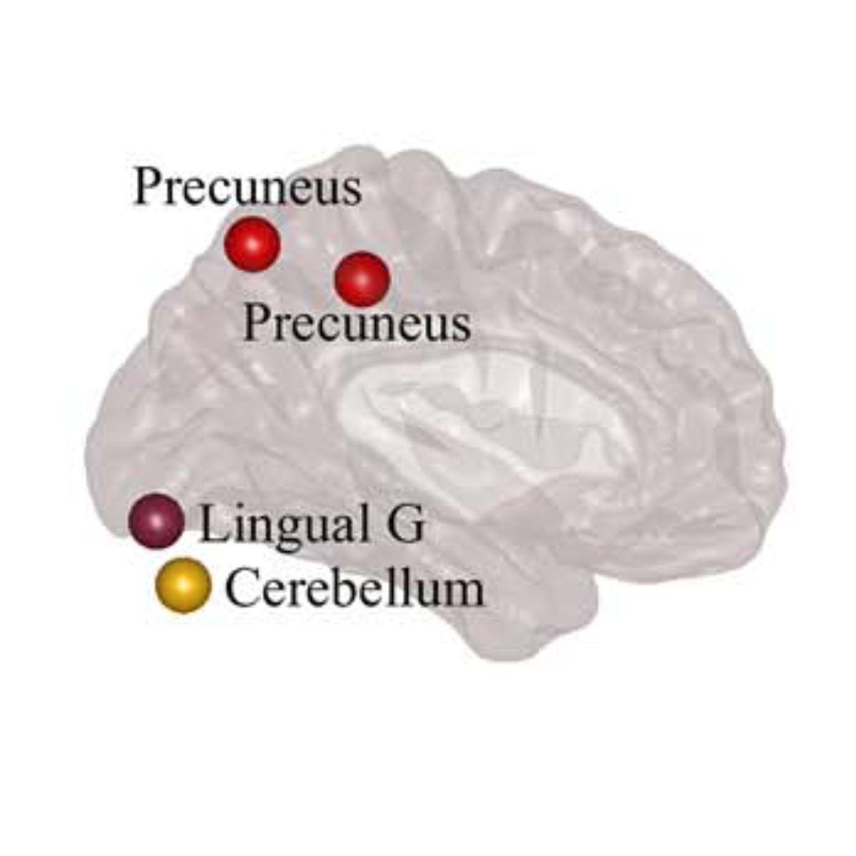
Visual display of the nodes that show significant differences between controls and participants with PD in network measures using the anti-symmetric correlation method. (Image by the Authors.)
Directed Brain Connectivity Identifies Widespread Functional Network Abnormalities in Parkinson’s Disease
Mite Mijalkov, Giovanni Volpe, Joana B Pereira
Cerebral Cortex 32(3), 593–607 (2022)
doi: 10.1093/cercor/bhab237

Parkinson’s disease (PD) is a neurodegenerative disorder characterized by topological abnormalities in large-scale functional brain networks, which are commonly analyzed using undirected correlations in the activation signals between brain regions. This approach assumes simultaneous activation of brain regions, despite previous evidence showing that brain activation entails causality, with signals being typically generated in one region and then propagated to other ones. To address this limitation, here, we developed a new method to assess whole-brain directed functional connectivity in participants with PD and healthy controls using antisymmetric delayed correlations, which capture better this underlying causality. Our results show that whole-brain directed connectivity, computed on functional magnetic resonance imaging data, identifies widespread differences in the functional networks of PD participants compared with controls, in contrast to undirected methods. These differences are characterized by increased global efficiency, clustering, and transitivity combined with lower modularity. Moreover, directed connectivity patterns in the precuneus, thalamus, and cerebellum were associated with motor, executive, and memory deficits in PD participants. Altogether, these findings suggest that directional brain connectivity is more sensitive to functional network differences occurring in PD compared with standard methods, opening new opportunities for brain connectivity analysis and development of new markers to track PD progression.

Keynote Talk by G. Volpe at IUPAP Conference on Condensed Matter Physics and Optics, 20 January 2022


Deep learning for microscopy, optical trapping, and active matter
Giovanni Volpe
Keynote Talk at IUPAP conference on Condensed Matter Physics and Optics
Online
20 January 2022
15:00 PST

After a brief overview of artificial intelligence, machine learning and deep learning, I will present a series of recent works in which we have employed deep learning for applications in photonics and active matter. In particular, I will explain how we employed deep learning to enhance digital video microscopy, to estimate the properties of anomalous diffusion, to characterize microscopic force fields, to improve the calculation of optical forces, and to characterize nanoparticles. Finally, I will provide an outlook for the application of deep learning in photonics and active matter.

Multiplex Connectome Changes across the Alzheimer’s Disease Spectrum Using Gray Matter and Amyloid Data published in Cerebral Cortex

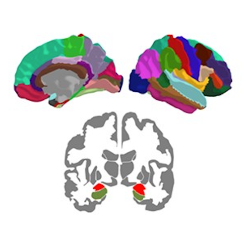
Brain nodes. (Image taken from the article.)
Multiplex Connectome Changes across the Alzheimer’s Disease Spectrum Using Gray Matter and Amyloid Data
Mite Mijalkov, Giovanni Volpe, Joana B Pereira
Anna Canal-Garcia, Emiliano Gómez-Ruiz, Mite Mijalkov, Yu-Wei Chang, Giovanni Volpe, Joana B Pereira, Alzheimer’s Disease Neuroimaging Initiative
Cerebral Cortex, bhab429 (2022)
doi: 10.1093/cercor/bhab429

The organization of the Alzheimer’s disease (AD) connectome has been studied using graph theory using single neuroimaging modalities such as positron emission tomography (PET) or structural magnetic resonance imaging (MRI). Although these modalities measure distinct pathological processes that occur in different stages in AD, there is evidence that they are not independent from each other. Therefore, to capture their interaction, in this study we integrated amyloid PET and gray matter MRI data into a multiplex connectome and assessed the changes across different AD stages. We included 135 cognitively normal (CN) individuals without amyloid-β pathology (Aβ−) in addition to 67 CN, 179 patients with mild cognitive impairment (MCI) and 132 patients with AD dementia who all had Aβ pathology (Aβ+) from the Alzheimer’s Disease Neuroimaging Initiative. We found widespread changes in the overlapping connectivity strength and the overlapping connections across Aβ-positive groups. Moreover, there was a reorganization of the multiplex communities in MCI Aβ + patients and changes in multiplex brain hubs in both MCI Aβ + and AD Aβ + groups. These findings offer a new insight into the interplay between amyloid-β pathology and brain atrophy over the course of AD that moves beyond traditional graph theory analyses based on single brain networks.

Book “Simulation of Complex Systems” published at IOP

Book cover. (From the IOP website.)
The book Simulation of Complex Systems, authored by Aykut Argun, Agnese Callegari and Giovanni Volpe, has been published by IOP in December 2021.

The book is available for the students of Gothenburg University and Chalmers University of Technology through the library service of each institution.
The example codes presented in the book can be found on GitHub.

Links
@ IOP Publishing

@ Amazon.com

Citation 
Aykut Argun, Agnese Callegari & Giovanni Volpe. Simulation of Complex Systems. IOP Publishing, 2022.
ISBN: 9780750338417 (Hardback) 9780750338431 (Ebook).

Comparison of Two-Dimensional- and Three-Dimensional-Based U-Net Architectures for Brain Tissue Classification in One-Dimensional Brain CT published in Frontiers of Computational Neuroscience

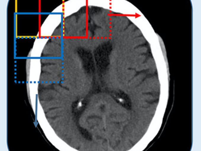
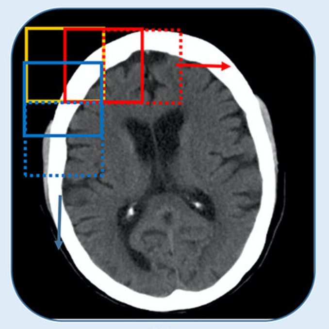
CT is split into smaller patches. (Image by the Authors.)
Comparison of Two-Dimensional- and Three-Dimensional-Based U-Net Architectures for Brain Tissue Classification in One-Dimensional Brain CT
Meera Srikrishna, Rolf A. Heckemann, Joana B. Pereira, Giovanni Volpe, Anna Zettergren, Silke Kern, Eric Westman, Ingmar Skoog and Michael Schöll
Frontiers of Computational Neuroscience 15, 785244 (2022)
doi: 10.3389/fncom.2021.785244

Brain tissue segmentation plays a crucial role in feature extraction, volumetric quantification, and morphometric analysis of brain scans. For the assessment of brain structure and integrity, CT is a non-invasive, cheaper, faster, and more widely available modality than MRI. However, the clinical application of CT is mostly limited to the visual assessment of brain integrity and exclusion of copathologies. We have previously developed two-dimensional (2D) deep learning-based segmentation networks that successfully classified brain tissue in head CT. Recently, deep learning-based MRI segmentation models successfully use patch-based three-dimensional (3D) segmentation networks. In this study, we aimed to develop patch-based 3D segmentation networks for CT brain tissue classification. Furthermore, we aimed to compare the performance of 2D- and 3D-based segmentation networks to perform brain tissue classification in anisotropic CT scans. For this purpose, we developed 2D and 3D U-Net-based deep learning models that were trained and validated on MR-derived segmentations from scans of 744 participants of the Gothenburg H70 Cohort with both CT and T1-weighted MRI scans acquired timely close to each other. Segmentation performance of both 2D and 3D models was evaluated on 234 unseen datasets using measures of distance, spatial similarity, and tissue volume. Single-task slice-wise processed 2D U-Nets performed better than multitask patch-based 3D U-Nets in CT brain tissue classification. These findings provide support to the use of 2D U-Nets to segment brain tissue in one-dimensional (1D) CT. This could increase the application of CT to detect brain abnormalities in clinical settings.

CORDIS News article on ComplexSwimmers

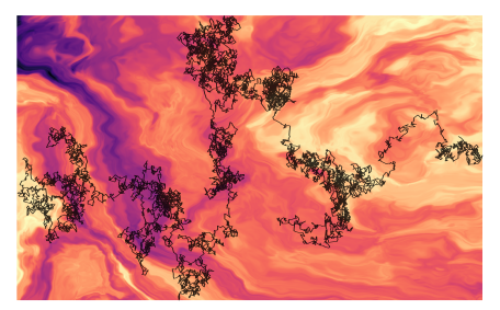
An illustration of anomalous diffusion. (Image by Gorka Muñoz-Gil.)

CORDIS, the Community Research and Development Information Service of the European Commission, recently covered Giovanni Volpe’s ComplexSwimmers ERC-StG grant in a news:
Throwing down the scientific gauntlet to assess methods for anomalous diffusion.
The article highlights the joint results obtained by three EU-backed research projects (NOQIA, OPTOlogic and ComplexSwimmers) dealing with anomalous diffusion.

Raman Tweezers for Tire and Road Wear Micro- and Nanoparticles Analysis published in Environmental Science: Nano

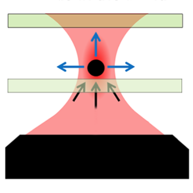
Optical beam focused into the liquid: the tire particles are pushed away from the laser focus.

Raman Tweezers for Tire and Road Wear Micro- and Nanoparticles Analysis
Pietro Giuseppe Gucciardi, Gillibert Raymond, Alessandro Magazzù, Agnese Callegari, David Bronte Ciriza, Foti Antonino, Maria Grazia Donato, Onofrio M. Maragò, Giovanni Volpe, Marc Lamy de La Chapelle & Fabienne Lagarde
Environmental Science: Nano 9, 145 – 161 (2022)
ChemRxiv: https://doi.org/10.33774/chemrxiv-2021-h59n1
doi: https://doi.org/10.1039/D1EN00553G

Tire and Road Wear Particles (TRWP) are non-exhaust particulate matter generated by road transport means during the mechanical abrasion of tires, brakes and roads. TRWP accumulate on the roadsides and are transported into the aquatic ecosystem during stormwater runoffs. Due to their size (sub-millimetric) and rubber content (elastomers), TRWP are considered microplastics (MPs). While the amount of the MPs polluting the water ecosystem with sizes from ~ 5 μm to more than 100 μm is known, the fraction of smaller particles is unknown due to the technological gap in the detection and analysis of < 5 μm MPs. Here we show that Raman Tweezers, a combination of optical tweezers and Raman spectroscopy, can be used to trap and chemically analyze individual TWRPs in a liquid environment, down to the sub-micrometric scale. Using tire particles mechanically grinded from aged car tires in water solutions, we show that it is possible to optically trap individual sub-micron particles, in a so-called 2D trapping configuration, and acquire their Raman spectrum in few tens of seconds. The analysis is then extended to samples collected from a brake test platform, where we highlight the presence of sub-micrometric agglomerates of rubber and brake debris, thanks to the presence of additional spectral features other than carbon. Our results show the potential of Raman Tweezers in environmental pollution analysis and highlight the formation of nanosized TRWP during wear.

Featured in:
University of Gothenburg > News and Events: New technology enables the detection of microplastics from road wear
Phys.org > News > Nanotechnology:New technology enables the detection of microplastics from road wear
Nonsologreen > Green: Le Raman-tweezers per la guerra alle nanoplastiche che inquinano fiumi e mari

Invited Presentation by G. Volpe at FiO LS, 4 November 2021

DeepTrack 2.0 Logo. (Image from DeepTrack 2.1 Project)
DeepTrack 2.1: A Framework for Deep Learning for Microscopy
Giovanni Volpe
Invited Presentation at Frontiers in Optics + Laser Science
Online
4 November 2021
4:00 PM

We present DeepTrack 2.0, a software to design, train, and validate deep-learning solutions for digital microscopy. We demonstrate it for applications from particle localization, tracking, and characterization, to cell counting and classification, to virtual staining.

Link: FTh6A.3