News

Presentation by M. Selin at SBE congress, 30 June 2023

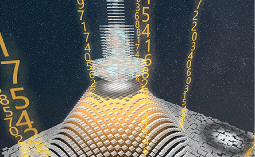
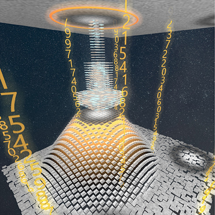
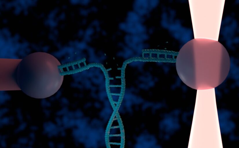
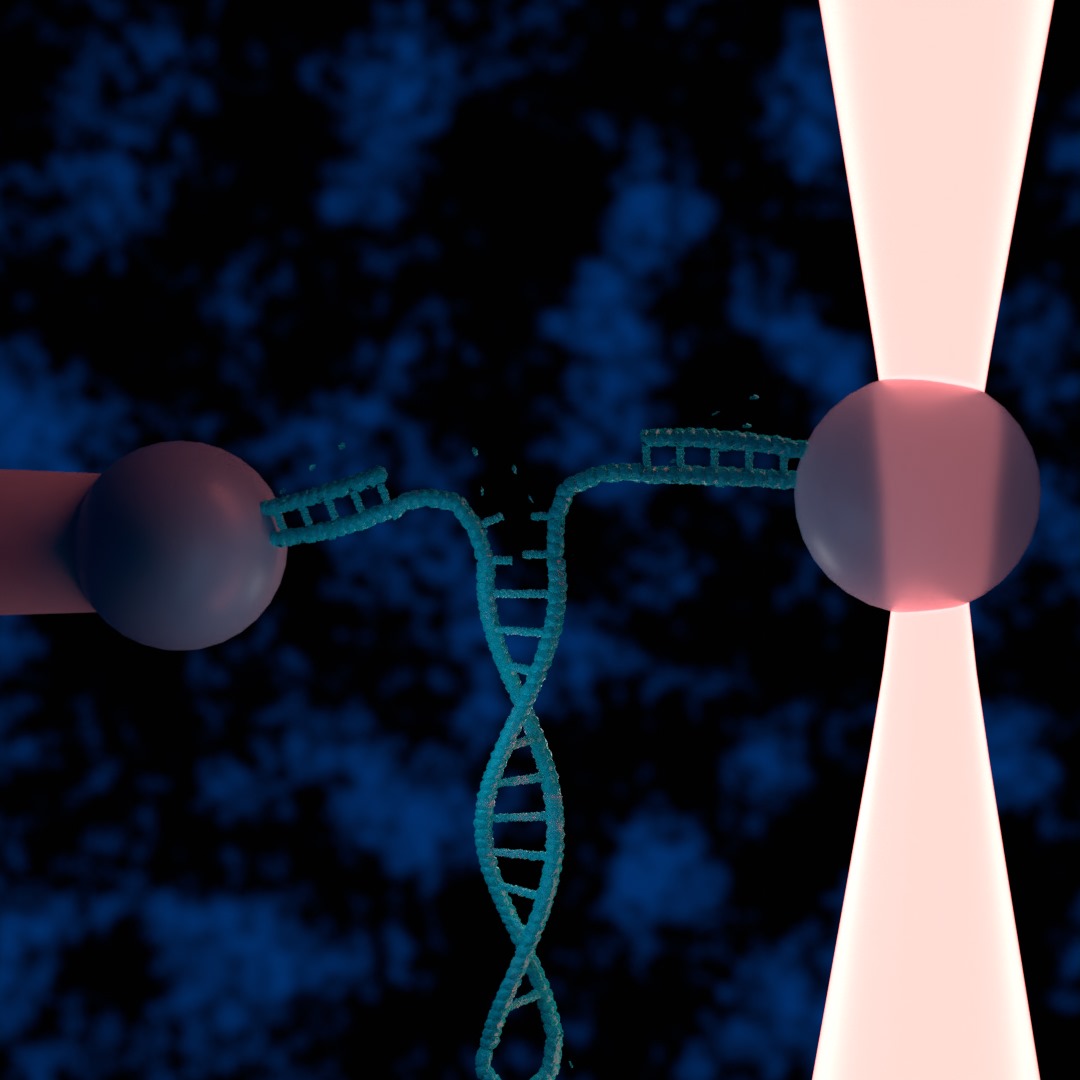
Illustration of a DNA hairpin being unzipped by an optical tweezers. (Illustration by M. Selin.)
Automating optical tweezers experiments using deep learning and custom electronics
Martin Selin
30 June 2023, 13:00 CEST

Optical tweezers are powerful tools for manipulating and studying the mechanical properties of single biomolecules, such as DNA. However, conducting such experiments manually is both time-consuming and labor-intensive limiting the amount of data collectable. In this work, we present a method to automate optical tweezers with the use of deep learning applying it to DNA pulling experiments.

A typical DNA pulling experiment can be divided into three main steps, each of which we have automated. The first is positioning of a bead in a micropipette(or secondary optical trap), second is connecting DNA of a another optically trapped bead with the bead in the micropipette and lastly the stretching of the DNA by moving the trapped bead while monitoring the force.

We have used deep learning, in particular a unet, to track beads and identify important features in the sample such as the micropipette. Combining this with realtime feedback allows the system to both trap beads and carefully position trap beads.

We demonstrate the viability of our method by stretching lambda DNA, showing human like reliability in performing the experiments. We expect our method to find use in the study of small biomolecules enabling more and faster data collection as well as longer running experiments.
Presentation by J. Pineda at the XVII International Congress of the Spanish Biophysical Society, Castelldefels, 30 June 2023

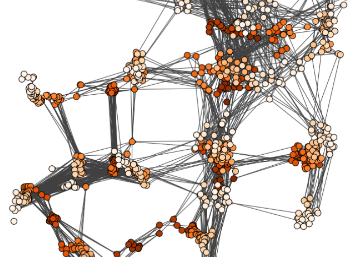
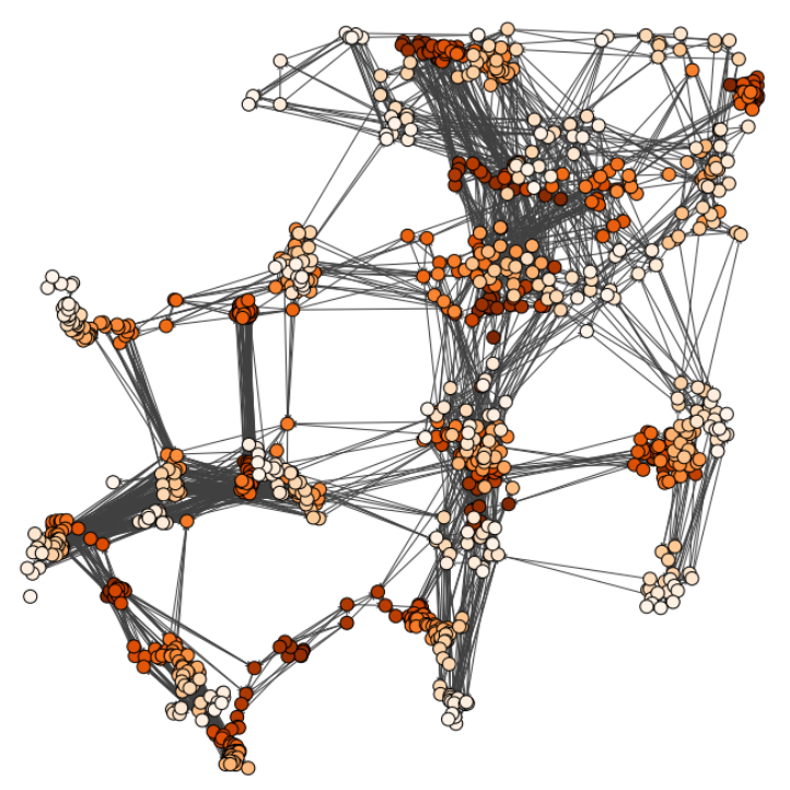
Input graph structure including a redundant number of edges. (Image by J. Pineda.)
MAGIK: Microscopic motion analysis through graph inductive knowledge
Jesús Pineda

Characterizing dynamic processes in living systems provides essential information for advancing our understanding of life processes in health and diseases and for developing new technologies and treatments. In the past two decades, optical microscopy has undergone significant developments, enabling us to study the motion of cells, organelles, and individual molecules with unprecedented detail at various scales in space and time. However, analyzing the dynamic processes that occur in complex and crowded environments remains a challenge. This work introduces MAGIK, a deep-learning framework for the analysis of biological system dynamics from time-lapse microscopy. MAGIK models the movement and interactions of particles through a directed graph where nodes represent detections and edges connect spatiotemporally close nodes. The framework utilizes an attention-based graph neural network (GNN) to process the graph and modulate the strength of associations between its elements, enabling MAGIK to derive insights into the dynamics of the systems. MAGIK provides a key enabling technology to estimate any dynamic aspect of the particles, from reconstructing their trajectories to inferring local and global dynamics. We demonstrate the flexibility and reliability of the framework by applying it to real and simulated data corresponding to a broad range of biological experiments.

Date: 30 June 2023
Time: 12:30
Event: XVII International Congress of the Spanish Biophysical Society

Invited Talk by G. Volpe at XVII Congress of the Spanish Biophysical Society, Castelldefels, Spain, 30 June 2023

(Image by A. Argun)
Deep Learning for Imaging and Microscopy
Giovanni Volpe
XVII Congress of the Spanish Biophysical Society, Castelldefels, Spain, 30 June 2023
Date: 30 June 2023
Time: 11:00

Invited Talk by G. Volpe at Active Matter at Surfaces and in Complex Environments, Dresden, 20 June 2023

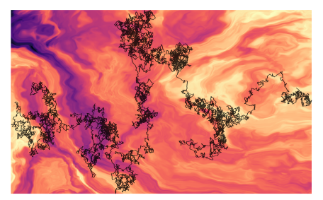
Illustration of anomalous diffusion. (Image by G. Muñoz-Gil)
The anomalous diffusion challenge 2
Giovanni Volpe
Active Matter at Surfaces and in Complex Environments, Dresden, Germany
Date: 20 June 2023
Time: 15:30

Presentation by H. Bachimanchi at XVII International Congress of the Spanish Biophysical Society, Castelldefels, 30 June 2023

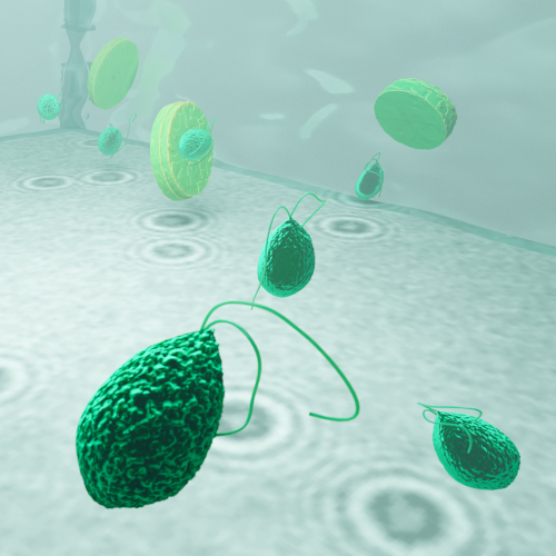
Planktons imaged under a holographic microscope. (Illustration by J. Heuschele.)
Bringing microplankton into focus: Deep learning meets holographic microscopy
Harshith Bachimanchi
30 June 2023, 12:40 CEST

The marine microbial food web plays a central role in the global carbon cycle. However, our mechanistic understanding of the ocean is biased toward its larger constituents, while rates and biomass fluxes in the microbial food web are mainly inferred from indirect measurements and ensemble averages. Yet, resolution at the level of the individual microplankton is required to advance our understanding of the microbial food web. Here, we demonstrate that, by combining holographic microscopy with deep learning, we can follow microplanktons throughout their lifespan, continuously measuring their three-dimensional position and dry mass. The deep-learning algorithms circumvent the computationally intensive processing of holographic data and allow rapid measurements over extended time periods. This permits us to reliably estimate growth rates, both in terms of dry mass increase and cell divisions, as well as to measure trophic interactions between species such as predation events. The individual resolution provides information about selectivity, individual feeding rates, and handling times for individual microplanktons. The method is particularly useful to detail the rates and routes of organic matter transfer in micro-zooplankton, the most important and least known group of primary consumers in the oceans. Studying individual interactions in idealized small systems provides insights that help us understand microbial food webs and ultimately larger-scale processes. We exemplify this by detailed descriptions of micro-zooplankton feeding events, cell divisions, and long-term monitoring of single cells from division to division.

The article related to this presentation can be found at the following link: Microplankton life histories revealed by holographic microscopy and deep learning.

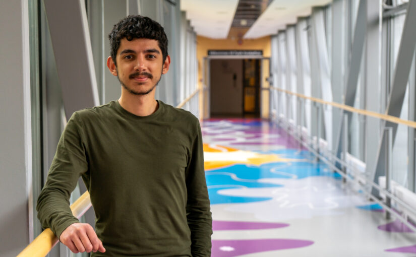
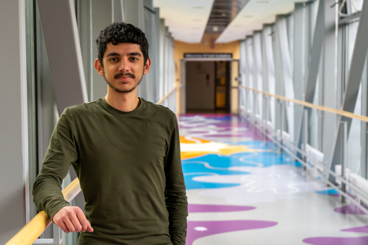
Emir Erdem joins the Soft Matter Lab

(Photo by A. Ciarlo.)
Emir Erdem joined the Soft Matter Lab on June 17, 2023.

Emir is an undergraduate student at the Mechanical Engineering Department of Bilkent University in Ankara, Turkey.

During his time at the Soft Matter Lab, he will be working on the modeling of red blood cells with geometrical optics.

He is supported by an Erasmus Traineeship scholarship. He will stay in our lab till September 13, 2023.

Poster by G. Wang at DINAMO 2023, Svolvær, 13 June 2023

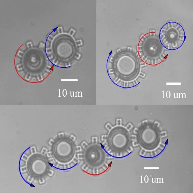
Light-driven micromachines. (Image by G. Wang.)
Nanophotonic encoding of light-driven micromachines
Gan Wang, Marcel Rey, Mahdi Shanei, Kunli Xiong, Einstom Engay, Mikael Käll, and Giovanni Volpe
Date: 13 June 2023
Time: 21:00 (CEST)

On-chip micromotors hold significant application potential in various fields, including cells, microfluidic manipulation, and the micro integration of machines. .The driving mechanism plays a crucial role in the design of micromotors. Currently, various methods such as static electricity, light, magnetism, chemical energy, and mechanical conduction are utilized for this purpose. Optics, in particular, offers distinct advantages including precise control, addressability, non-contact operation, and compatibility with diverse liquid environments. However, existing optically actuated on-chip motors necessitate high energy input, resulting in phototoxicity concerns and hindrances to large-scale manipulation. Furthermore, achieving precise control over speed and direction remains challenging, along with difficulties in establishing coupling among multiple devices.

Christian Rutgersson defended his Master Thesis on 9 June 2023. Congrats!

Christian Rutgersson defended his Master thesis in Complex Adaptive Systems at Chalmers University of Technology on 9 June 2023 at 17:00. Congrats!

Title: Characterizing Active Matter Particle Systems with Graph Neural Networks

Abstract:
Biological systems often have self-organizing properties. On the microscopic scale, the self-organization may be driven by constituents that generate their own motility by expending energy. Systems made up of such constituents, that self-propel, are termed active matter. Via this definition, it is clear that it is of importance to understand the rules that govern the microscopic constituents of an active matter in order to understand the active matter itself. Even though active matter is a topic of interest today, finding good methods of qualitatively and quantitatively characterizing the constituents of active matter remains an issue. Coincidentally, different types of artificial neural networks (ANN) have, in recent years, been used increasingly in research settings with great success. One such network is called the graph neural network (GNN). As the name suggests, this network is specifically designed to work with graphs as input data. Graphs can act as a useful representation of a system of particles, including active matter systems. Therefore, this project aims to characterize the forces that underpin an active matter system consisting of interacting particles that also have an active component, using a special type of GNN called message passing network (MPN). This was done by simulating the active matter using a Python code written from scratch, and training the MPN with standard machine learning algorithms. In the end, the simulations were found to give rise to characteristic active matter phenomena, and the MPN was able to correctly predict the force dynamics of a particle in the given active matter system.

Supervisors: Miguel Ruiz Garcia, Carlo Manzo, Jesus Pineda Castro, Giovanni Volpe
Examiner: Giovanni Volpe
Opponent: Ludvig Lindahl

Place: Nexus
Time: 9 June, 2023, 12:00

Gideon Jägenstedt defended his Master Thesis on 8 June 2023. Congrats!

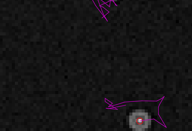
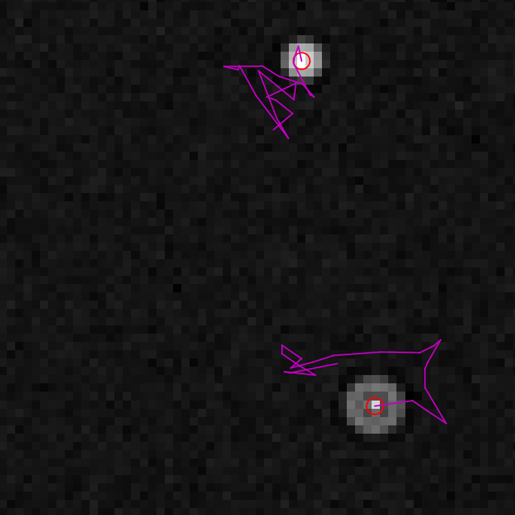
Tracking of two simulated particles over 20 frames using an object-focused variational autoencoder. (Image by G. Jägenstedt.)
Gideon Jägenstedt defended his Master Thesis on 8 June 2023 at 11:00. Congrats!

Title: End-to-End Object Tracking on Simulated Microscopy Video

Abstract:
Object tracking in microscopy time-lapse videos is currently mostly done in two steps often using two neural network models, first the images are segmented in order to detect each object within each time frame and extract their centroids using one neural network model. A selected set of properties on the centroids are then used as an input to a second neural network that creates the temporal trajectories by linking the centroids over a sequence of frames.

This work proposes a novel method to combine the object detection step and the
linking step which should, in theory, create better linking in time since the combined model has access to not only a set of properties but the complete image of the centroids. Two different architectures of a combined model were tested, one supervised model based on graph neural networks (GNN) and one unsupervised model based on a variational autoencoder (VAE).

The supervised GNN-based model did not succeed in predicting the position of the centroids, but it showed promise in linking the centroids between frames. Therefore, the VAE-based model was developed that uses the same approach for linking. The VAE-based model resulted in a mean absolute error of under 0.002 on its detection placement, a detection miss-rate of 2.69 %, and an F1-score of 81.2 % when linking trajectories on simulated data.

Supervisor: Jesus Pineda Castro
Examiner: Giovanni Volpe
Opponent: Mirja Granfors

Place: PJ
Time: 8 June, 2023, 11:00