How morphology affects optical trapping of red blood cells in health and disease published in Biomedical Optics Express

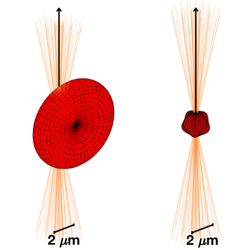
 Left: trapping of a healthy (biconcave) red blood cell. Right: trapping of an echinocyte-like cell with membrane protrusions. In the representation of the trapping (focused) beam, only rays originating from points close enough to the propagation axis are drawn. More peripheral rays are omitted. (Image from the manuscript.)
How morphology affects optical trapping of red blood cells in health and disease
Emir Erdem, Gökberk Kabacaoğlu, Giovanni Volpe, Agnese Callegari, and Luca Biancofiore
Biomedical Optics Express 17, 5355-5372 (2026)
doi: 10.1364/BOE.609102

Optical tweezers are widely used to manipulate microscopic particles and biological cells through optical forces and torques, yet how cell shape controls trapping response has not been fully elucidated. Here, we develop a computational framework to investigate single-beam optical trapping of red blood cells with physiologically and pathologically relevant deformed morphologies. Representative cell shapes are generated parametrically and analyzed using static displacement and rotation tests, together with Brownian dynamics simulations. We show that all considered morphologies remain laterally and orientationally confined in a single-beam trap, while axial localization is weaker by pure optical means and is experimentally realized by the balance between the optical force and the buoyancy-corrected cell weight. Cell geometry strongly affects translational and rotational stiffness, preferred orientation, and Brownian confinement. In particular, shape-induced anisotropy produces distinct dynamical signatures, indicating that optical trapping measurements may be sensitive to morphological alterations beyond simple changes in cell size or volume. These results establish a quantitative connection between red-blood-cell morphology, trap stiffness, and dynamics, providing a computational framework for interpreting morphology-dependent optical trapping experiments.
Presentation by E. Erdem, 4 October 2023

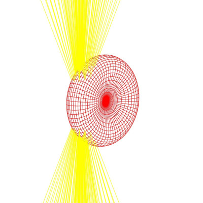
Schematic of a red blood cell in a focused optical beam. (Image by E. Erdem.)
Optical trapping of red blood cells and different geometrical shapes
Emir Erdem

Red Blood Cells (RBC), also known as erythrocyts, are essential cells that are present in the blood of every vertebrate. Because of their hemoglobin protein content, they carry oxygen to the cells and perform a vital function. Due to their complex shapes, behavior of cells like RBCs under optical forces are not fully been discovered. In this study, the behavior of RBCs as well as other shapes under optical trap are simulated using OTGO which is a numeric toolbox utilizing geometrical optics approximation for optical calculations. As a result of the simulations, it is observed that the RBC aligns itself in a vertical configuration, parallel to the incident beam propagating towards the cell from below. Conducted static analysis showed that it is possible to stably trap a RBC in all three dimensions. The center of the trap is near the edge of the cell, where the thickness is larger. After the analysis on RBC, how well different geometrical shapes can optically be trapped are investigated by integrating different shapes modeled by spherical harmonics to OTGO. A similar static analysis is conducted on a dumbbell shape and its trapping effectiveness is compared with an ellipsoid. A dumbbell shape can effectively be trapped in the horizontal plane similar to an ellipsoid, but in the light propagation direction, it is more challenging to trap the shape and it requires modifications on optical properties of the setup. The aim of this study after this point is to optimize the optical force calculations by training a neural network model and to apply flow conditions to cells.

Emir Erdem joins the Soft Matter Lab

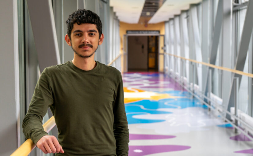
(Photo by A. Ciarlo.)
Emir Erdem joined the Soft Matter Lab on June 17, 2023.

Emir is an undergraduate student at the Mechanical Engineering Department of Bilkent University in Ankara, Turkey.

During his time at the Soft Matter Lab, he will be working on the modeling of red blood cells with geometrical optics.

He is supported by an Erasmus Traineeship scholarship. He will stay in our lab till September 13, 2023.