Falko Schmidt, Agnese Callegari, Abdallah Daddi-Moussa-Ider, Battulga Munkhbat, Ruggero Verre, Timur Shegai, Mikael Käll, Hartmut Löwen, Andrea Gambassi and Giovanni Volpe
Nature Physics 19, 271-278 (2023)
arXiv: 2202.10926
doi: 10.1038/s41567-022-01795-6
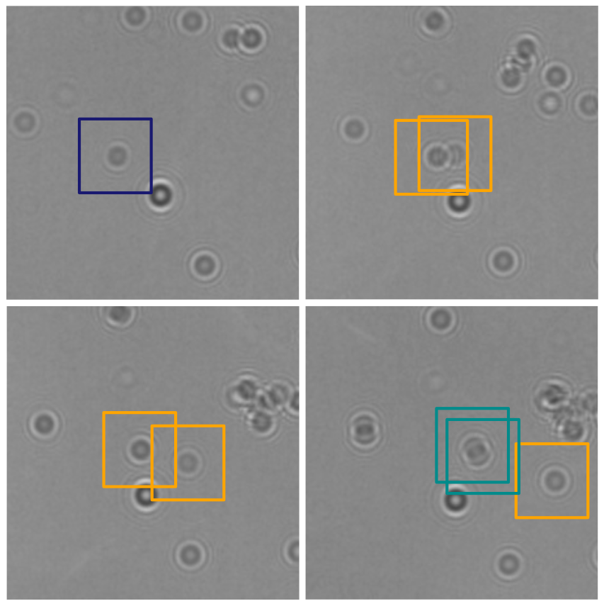
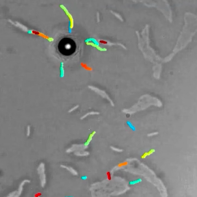
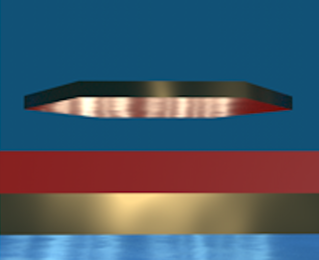
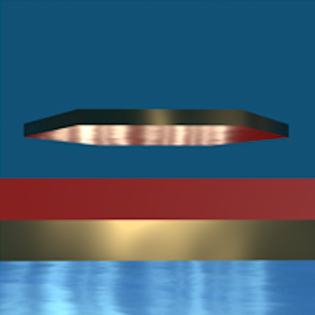
Casimir forces in quantum electrodynamics emerge between microscopic metallic objects because of the confinement of the vacuum electromagnetic fluctuations occurring even at zero temperature. Their generalization at finite temperature and in material media are referred to as Casimir-Lifshitz forces. These forces are typically attractive, leading to the widespread problem of stiction between the metallic parts of micro- and nanodevices. Recently, repulsive Casimir forces have been experimentally realized but their reliance on specialized materials prevents their dynamic control and thus limits their further applicability. Here, we experimentally demonstrate that repulsive critical Casimir forces, which emerge in a critical binary liquid mixture upon approaching the critical temperature, can be used to actively control microscopic and nanoscopic objects with nanometer precision. We demonstrate this by using critical Casimir forces to prevent the stiction caused by the Casimir-Lifshitz forces. We study a microscopic gold flake above a flat gold-coated substrate immersed in a critical mixture. Far from the critical temperature, stiction occurs because of dominant Casimir-Lifshitz forces. Upon approaching the critical temperature, however, we observe the emergence of repulsive critical Casimir forces that are sufficiently strong to counteract stiction. This experimental demonstration can accelerate the development of micro- and nanodevices by preventing stiction as well as providing active control and precise tunability of the forces acting between their constituent parts.