John is a master student in Physics at the University of Gothenburg.
During his time at the Soft Matter Lab, he will investigate the light-controlled self-organisation of active particles.
John is a master student in Physics at the University of Gothenburg.
During his time at the Soft Matter Lab, he will investigate the light-controlled self-organisation of active particles.
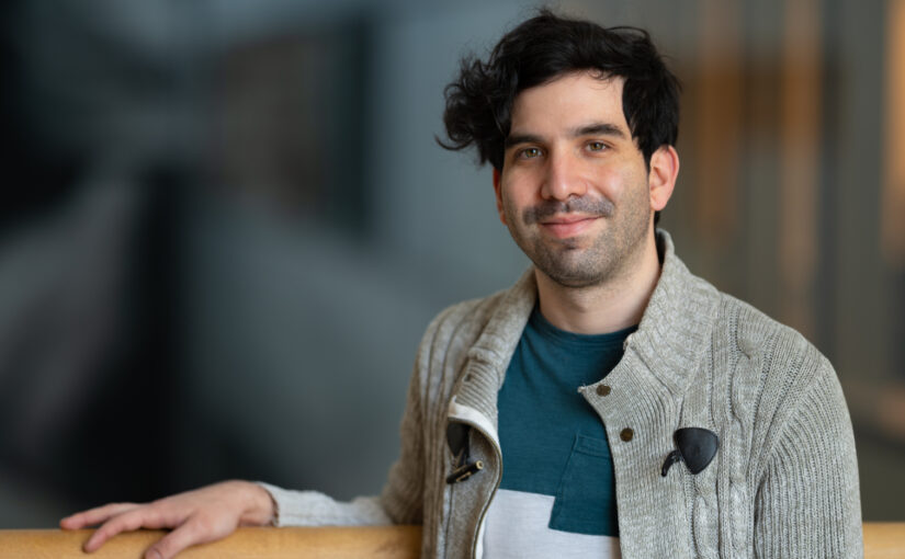
Hari Prakash has a Master degree in Advanced Robotics from Queen Mary University of London, United Kingdom.
In his PhD, he will focus on the development of soft robots to study collective emergent behaviours in soft active matter systems.
Meera Srikrishna, Rolf A. Heckemann, Joana B. Pereira, Giovanni Volpe, Anna Zettergren, Silke Kern, Eric Westman, Ingmar Skoog and Michael Schöll
Frontiers of Computational Neuroscience 15, 785244 (2022)
doi: 10.3389/fncom.2021.785244
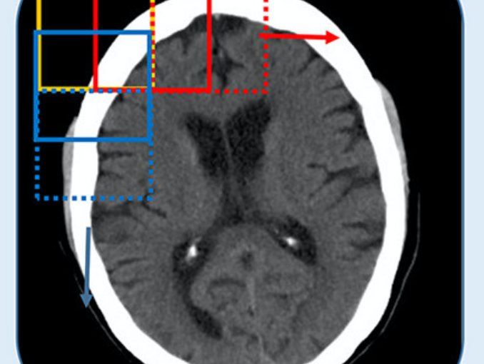
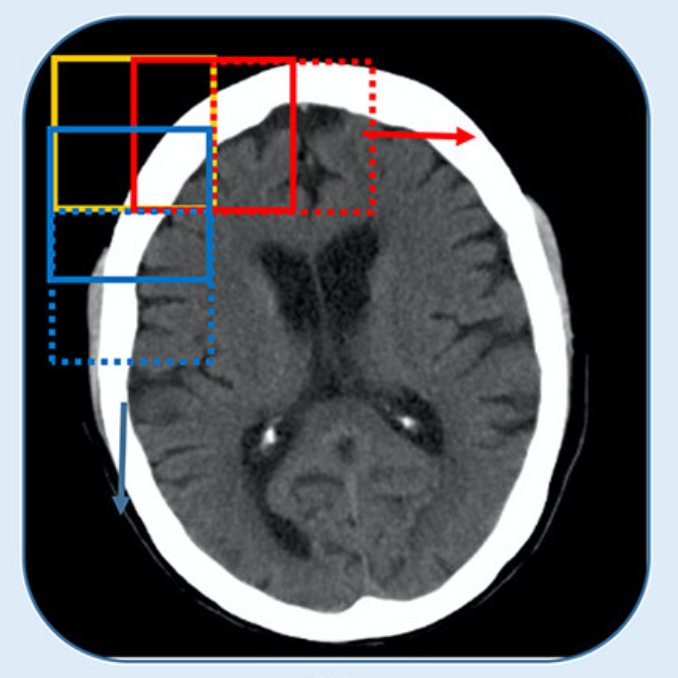
Brain tissue segmentation plays a crucial role in feature extraction, volumetric quantification, and morphometric analysis of brain scans. For the assessment of brain structure and integrity, CT is a non-invasive, cheaper, faster, and more widely available modality than MRI. However, the clinical application of CT is mostly limited to the visual assessment of brain integrity and exclusion of copathologies. We have previously developed two-dimensional (2D) deep learning-based segmentation networks that successfully classified brain tissue in head CT. Recently, deep learning-based MRI segmentation models successfully use patch-based three-dimensional (3D) segmentation networks. In this study, we aimed to develop patch-based 3D segmentation networks for CT brain tissue classification. Furthermore, we aimed to compare the performance of 2D- and 3D-based segmentation networks to perform brain tissue classification in anisotropic CT scans. For this purpose, we developed 2D and 3D U-Net-based deep learning models that were trained and validated on MR-derived segmentations from scans of 744 participants of the Gothenburg H70 Cohort with both CT and T1-weighted MRI scans acquired timely close to each other. Segmentation performance of both 2D and 3D models was evaluated on 234 unseen datasets using measures of distance, spatial similarity, and tissue volume. Single-task slice-wise processed 2D U-Nets performed better than multitask patch-based 3D U-Nets in CT brain tissue classification. These findings provide support to the use of 2D U-Nets to segment brain tissue in one-dimensional (1D) CT. This could increase the application of CT to detect brain abnormalities in clinical settings.
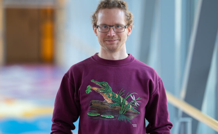
Mathias is a master student in Physics at the University of Gothenburg.
During his time at the Soft Matter Lab, he will focus on the simulation of an intracavity optical tweezers with the help of neural networks.
Angelo is a master student in Complex Adaptive Systems at Chalmers University of Technology.
During his time at the Soft Matter Lab, he will investigate the behaviour of active matter via experiments with toy robots (HEXBUG nano®).
CORDIS, the Community Research and Development Information Service of the European Commission, recently covered Giovanni Volpe’s ComplexSwimmers ERC-StG grant in a news:
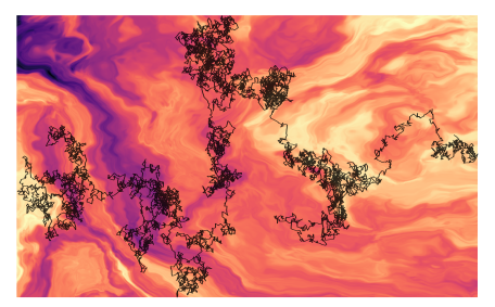
Throwing down the scientific gauntlet to assess methods for anomalous diffusion.
The article highlights the joint results obtained by three EU-backed research projects (NOQIA, OPTOlogic and ComplexSwimmers) dealing with anomalous diffusion.
Thermoplasmonic Tweezers: Probing single-molecules and more
G. V. Pavan Kumar
IISER, Pune, India.
24 November 2021
Online
In this presentation, we will discuss two specific issues: How to perform single-molecule surface enhanced Raman scattering (SERS) in an optothermal trap? and how to design optothermal fields to trap and interrogate molecules and colloids in a fluid?
In recent years, performing SERS in optical traps has emerged as an important development in nano- and bio-photonics. To this end, tweezer techniques based on surface-plasmons facilitate deeper optical potentials at sub-wavelength scales, and simultaneously provide enhanced electric and optothermal fields. In this
presentation, we will discuss various strategies developed in my laboratory to perform single-molecule SERS in optical and plasmonic tweezer platforms. Specifically, we will highlight some thermoplasmonic effects and directionality aspects of the tweezer platforms in metallic thin film and some plasmonic nano-architectures.
Short bio:
G.V. Pavan Kumar is an associate professor of physics at the Indian Institute of Science Education and Research (IISER), Pune, India.
He obtained his PhD from JNCASR, Bangalore. Subsequently he was a postdoctoral fellow at ICFO-Barcelona and Purdue University, before joining IISER in 2010.
His current research interests are optical, optothermal and nanophotonic forces and their utility in probing single molecules and soft-matter systems at micro and nanoscale.
To this end, his lab has been interfacing optical tweezer platforms with a variety of optical spectroscopy and microscopy tools.
He blogs on topics related to science: https://backscattering.wordpress.com/
Raman Tweezers for Tire and Road Wear Micro- and Nanoparticles Analysis
Pietro Giuseppe Gucciardi, Gillibert Raymond, Alessandro Magazzù, Agnese Callegari, David Bronte Ciriza, Foti Antonino, Maria Grazia Donato, Onofrio M. Maragò, Giovanni Volpe, Marc Lamy de La Chapelle & Fabienne Lagarde
Environmental Science: Nano 9, 145 – 161 (2022)
ChemRxiv: https://doi.org/10.33774/chemrxiv-2021-h59n1
doi: https://doi.org/10.1039/D1EN00553G
Tire and Road Wear Particles (TRWP) are non-exhaust particulate matter generated by road transport means during the mechanical abrasion of tires, brakes and roads. TRWP accumulate on the roadsides and are transported into the aquatic ecosystem during stormwater runoffs. Due to their size (sub-millimetric) and rubber content (elastomers), TRWP are considered microplastics (MPs). While the amount of the MPs polluting the water ecosystem with sizes from ~ 5 μm to more than 100 μm is known, the fraction of smaller particles is unknown due to the technological gap in the detection and analysis of < 5 μm MPs. Here we show that Raman Tweezers, a combination of optical tweezers and Raman spectroscopy, can be used to trap and chemically analyze individual TWRPs in a liquid environment, down to the sub-micrometric scale. Using tire particles mechanically grinded from aged car tires in water solutions, we show that it is possible to optically trap individual sub-micron particles, in a so-called 2D trapping configuration, and acquire their Raman spectrum in few tens of seconds. The analysis is then extended to samples collected from a brake test platform, where we highlight the presence of sub-micrometric agglomerates of rubber and brake debris, thanks to the presence of additional spectral features other than carbon. Our results show the potential of Raman Tweezers in environmental pollution analysis and highlight the formation of nanosized TRWP during wear.
Featured in:
University of Gothenburg > News and Events: New technology enables the detection of microplastics from road wear
Phys.org > News > Nanotechnology:New technology enables the detection of microplastics from road wear
Nonsologreen > Green: Le Raman-tweezers per la guerra alle nanoplastiche che inquinano fiumi e mari
Giovanni Volpe
Invited Presentation at Frontiers in Optics + Laser Science
Online
4 November 2021
4:00 PM
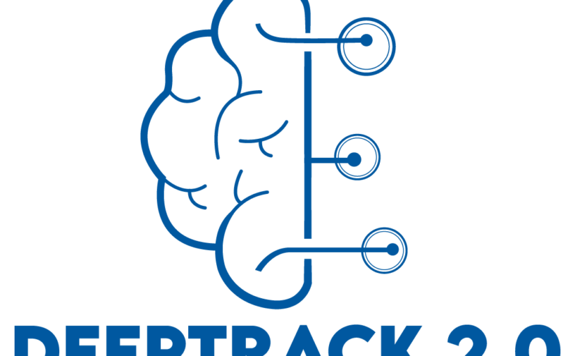
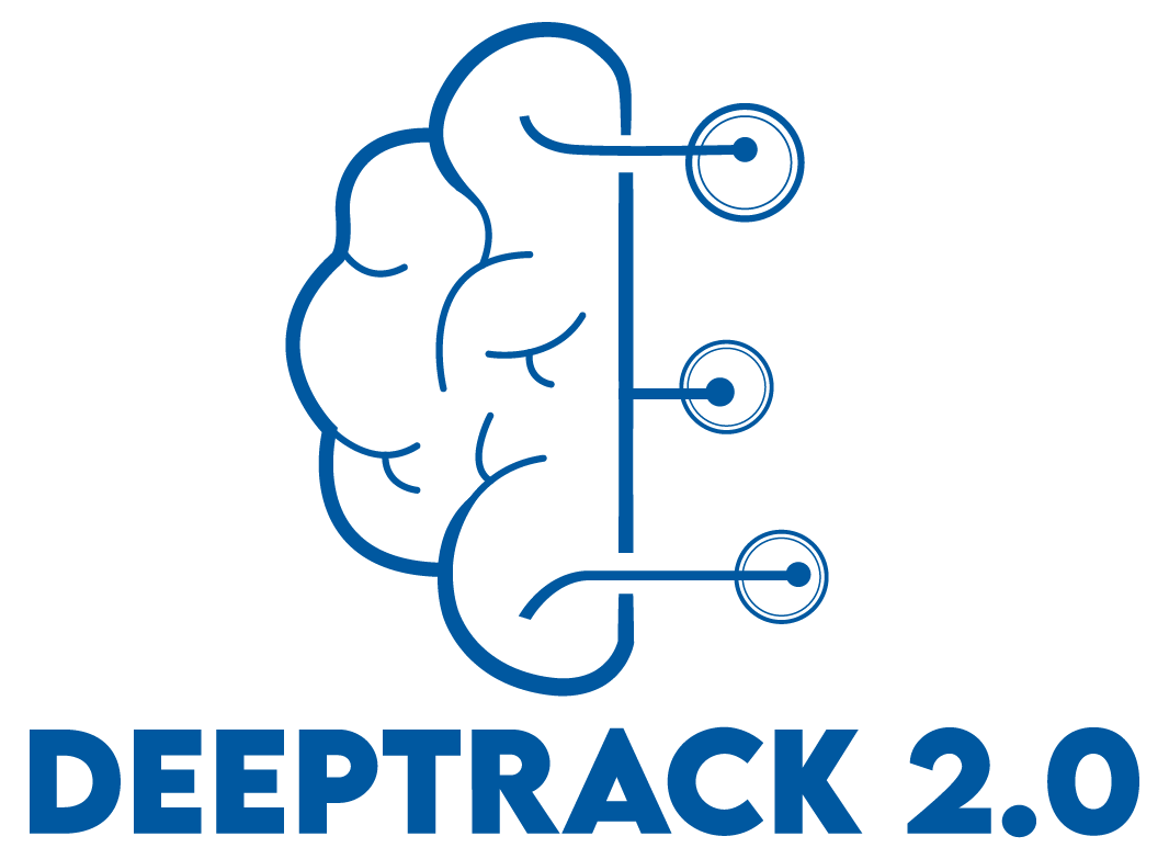
We present DeepTrack 2.0, a software to design, train, and validate deep-learning solutions for digital microscopy. We demonstrate it for applications from particle localization, tracking, and characterization, to cell counting and classification, to virtual staining.
Link: FTh6A.3
The article Objective comparison of methods to decode anomalous diffusion has been featured in the News of the University of Gothenburg.
The study, published in Nature Communications and co-written by researchers at the Soft Matter Lab of the Department of Physics at the University of Gothenburg, originates from the AnDi Challenge, a competition co-organised by Giovanni Volpe with researchers from University of Vic – Central University of Catalunya, Institute of Photonic Sciences in Barcelona, University of Potsdam, and Valencia Polytechnic University.
The challenge was held during March–November 2020 and consisted of three main tasks concerning anomalous exponent inference, model classification, and trajectory segmentation. The goal was to provide an objective assessment of the performance of methods to characterise anomalous diffusion from single trajectories.
Here the links to the press releases:
English: A scientific competition led to improved methods for analysing the diffusion of particles.
Swedish: En vetenskaplig tävling ledde till förbättrade metoder för att analysera diffusion av partiklar.